The Health Education Assets Library (HEAL) is a collection of over 22,000 freely available digital materials for health sciences education. The collection is now housed at the University of Utah J. Willard Marriott Digital Library.
1 - 25 of 2
| Title | Description | Subject | Collection | ||
|---|---|---|---|---|---|
| 1 |
 |
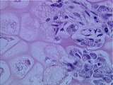
Bone / Cartilage | Between the bone spicules can be seen various bone cell types, including osteoblasts and osteoprogenitor cells. The left side of this image shows hypertrophied chondrocyte lacunae. UCLA Histology Collection. | Bone; Cartilage; osteoblasts, osteoprogenitor | UCLA Histology |
| 2 |
 |
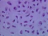
Cells and Organelles | Cartilage in the newborn rat knee. Here each cell is surrounded in a hole or lacuna, created by the preparation method; these in turn are embedded in extracellular matrix (ECM). The round, dark structures are nuclei. The cytoplasm of the cells contains reddish-staining glycogen, a storage form of gl... | Cartilage | UCLA Histology |
1 - 25 of 2
