The Health Education Assets Library (HEAL) is a collection of over 22,000 freely available digital materials for health sciences education. The collection is now housed at the University of Utah J. Willard Marriott Digital Library.
TO
1 - 25 of 14
| Title | Description | Subject | Collection | ||
|---|---|---|---|---|---|
| 1 |
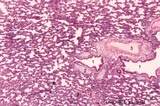 |
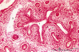
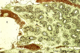
Late terminal sac period of developing lung (human, fetus, low magnification) | Stain: Hematoxylin and eosin. Cross-sections of a bronchus (1) with cartilage rings (2) closely associated with pulmonary arteries (3). The alveolar appearance is evident, and the open spaces represent future alveolar duct systems as well as alveolar sacs. The cellular septa are still thick due to n... | Lung development; Terminal sac period | Poja Histology Collection - Respiratory System Subset |
| 2 |
 |
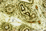
Pseudoglandular - canalicular period of developing lung (human, fetus) | Stain: Azan. Part of a bronchus (1) with young cartilage (2). The hyaline cartilage presents solitary chondrocytes embedded in the blue intercellular substance surrounded by a perichondrium (3). Note that the bronchus as well as the bronchial tubes (4) show an apical position of the epithelial nucle... | Lung development; Pseudoglandular period; Canalicular period; Mesenchyme | Poja Histology Collection - Respiratory System Subset |
| 3 |
 |
Pseudoglandular period of developing lung (human, embryo) | Stain: Trichrome (Goldner). Cross-sectioned future bronchial tubes (1) of varying sizes, note the apical position of the nuclei with light-stained basal parts (glycogen). The surrounding mesenchyme condenses (↓) around the epithelium and in the neighbourhood small blood vessels (*) are present. At... | Lung development; Pseudoglandular period; Bronchial tubes; Mesenchyme | Poja Histology Collection - Respiratory System Subset |
| 4 |
 |
Pseudoglandular period of developing lung (human, embryo) | Stain: Trichrome (Goldner). Cross-sectioned future bronchial tubes (1) of varying sizes, note the apical position of the nuclei with light-stained basal parts (glycogen). The surrounding mesenchyme becomes condensed (↓) around the epithelium and in between small blood vessels (*) are present. | Lung development; Pseudoglandular period; Bronchial tubes; Mesenchyme | Poja Histology Collection - Respiratory System Subset |
| 5 |
 |
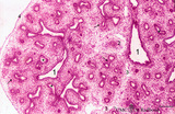
Pseudoglandular period of developing lung (human, embryo, low magnification) | Stain: Hematoxylin and eosin. The two future lung lobes contain many cross-sectioned future bronchial tubes (1). Note in these epithelial cells the apical position of the nuclei with light-stained basal parts (glycogen). The surrounding mesenchyme becomes more condensed (↓) around the epithelium a... | Lung development; Visceral pleura; Pseudoglandular period; Bronchial tubes; Mesenchyme | Poja Histology Collection - Respiratory System Subset |
| 6 |
 |
Pseudoglandular - canalicular period of developing lung (human, fetus) | Stain: Azan. Terminal budding and elongation of the future bronchial tree: branching of a bronchial tubes (1) within an islet where the mesenchyme condenses (4). A large blood vessel (2) as well as a lymph vessel (3) are recognizable. Note in the mesenchyme formation of blood capillaries in the vici... | Lung development; Pseudoglandular period ; Canalicular period; Mesenchyme | Poja Histology Collection - Respiratory System Subset |
| 7 |
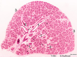 |
Pseudoglandular-canalicular period of developing lung (human, fetus, low magnification) | Stain: Azan. A future lung lobe with cross-sectioned larger bronchi (1) in close association with islets of future smaller bronchial tubes (*). Around the larger bronchi several cartilage structures (↓) are already present. Note that the mesenchyme around the tubes within the islets becomes more c... | Lung development; Visceral pleura; Pseudoglandular period; Canalicular period; Bronchial tubes; Mesenchyme | Poja Histology Collection - Respiratory System Subset |
| 8 |
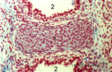 |
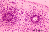
Pseudoglandular - canalicular period of developing lung (human, fetus) | Stain: Azan. Location of a cartilagineous ring (1) between two cross-sectioned bronchi (2). Note in the bronchial epithelial cells the apical position of the epithelial nuclei and the light-stained basal part (↑) containing glycogen. The young hyaline cartilage presents solitary chondrocytes embed... | Lung development; Pseudoglandular period; Canalicular period; Mesenchyme | Poja Histology Collection - Respiratory System Subset |
| 9 |
 |
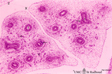
Pseudoglandular period of developing lung (human, embryo, low magnification) | Stain: Hematoxylin and eosin. Cross-sectioned future bronchial tubes (1), the surrounding mesenchyme becomes more condensed around the epithelium. The mesoderm of the future visceral pleura (2) as well as the future parietal pleura (3) and (4) indicates pleural cavity. The cartilagineous spinal colu... | Lung development; Visceral pleura; Parietal pleura; Pseudoglandular period; Bronchial tubes; Mesenchyme | Poja Histology Collection - Respiratory System Subset |
| 10 |
 |
Pseudoglandular period of developing lung (human, embryo, low magnification) | Stain: Trichrome (Goldner). Cross-sectioned future bronchial tubes (1) of varying sizes. Note the apical position of the nuclei in these epithelial cells with light-stained basal parts (glycogen). The surrounding mesenchyme becomes more condensed (↓) around the epithelium and in between numerous ... | Lung development; Visceral pleura; Pseudoglandular period; Bronchial tubes; Mesenchyme | Poja Histology Collection - Respiratory System Subset |
| 11 |
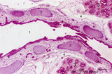 |
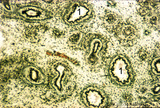
Pseudoglandular - canalicular period of developing lung (human, fetus) | Stain: Azan. Longitudinal section through a large bronchus (1) with cartilagineous rings (2). At (3) developing glandular structures in islets of bronchial tubes surrounded by condensed mesenchyme. At (4) lymph vessels. | Lung development; Pseudoglandular period; Canalicular period; Mesenchyme; Bronchial tubes | Poja Histology Collection - Respiratory System Subset |
| 12 |
 |
Pseudoglandular period of developing lung (human, embryo) | Stain: Hematoxylin and eosin. Two cross-sectioned future bronchial tubes. Note the basal position of the nuclei with light-stained apical cytoplasm (↓, glycogen). The surrounding mesenchyme condenses (1) around the epithelium and developing capillaries and small blood vessels (*) are present. Futu... | Lung development; Visceral pleura; Pseudoglandular period; Bronchial tubes; Mesenchyme | Poja Histology Collection - Respiratory System Subset |
| 13 |
 |
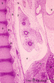
Terminal sac period of developing lung (human, fetus) | Stain: Hematoxylin and eosin. The transition of a terminal bronchiolus (1) into two future respiratory bronchioli (2), present as dilated spaces (saccules derived from the primitive respiratory channels, hence the name terminal sac period). The surrounding cellular tissue is composed of developing p... | Lung development; Terminal sac period; Respiratory bronchioli; Mesenchyme | Poja Histology Collection - Respiratory System Subset |
| 14 |
 |
Pseudoglandular - canalicular period of developing lung (human, fetus) | Stain: Azan. Part of a future lung lobe with cross-sectioned larger bronchi (1) in close association with islets of future smaller bronchial tubes (*). Note the branching of the bronchial tubes within an islet. The mesenchyme within the islets of future tubes becomes more condensed (↓). Blood vess... | Lung development; Visceral pleura; Pseudoglandular period; Canalicular period; Mesenchyme | Poja Histology Collection - Respiratory System Subset |
1 - 25 of 14