The Health Education Assets Library (HEAL) is a collection of over 22,000 freely available digital materials for health sciences education. The collection is now housed at the University of Utah J. Willard Marriott Digital Library.
TO
1 - 25 of 8
| Title | Description | Subject | Collection | ||
|---|---|---|---|---|---|
| 1 |
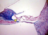 |
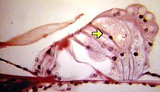
Cochlea, axons leaving basilar membrane | Cochlea, axons leaving basilar membrane. Photograph. Multimedia. | Cochlea; Basilar membrane; Sense organs; Ear; Anatomy; Axons | Slice of Life |
| 2 |
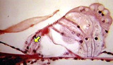 |
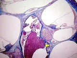
Inner hair cell above | Inner hair cell above. Cochlea. Photograph. Multimedia. | Hair cells, inner; Cochlea; Ear; Sense organs; Anatomy | Slice of Life |
| 3 |
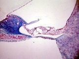 |
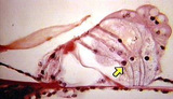
Organ of corti | Organ of corti. Cochlea. Photograph. Multimedia. | Organ of corti; Cochlea; Sense organs; Ear; Anatomy | Slice of Life |
| 4 |
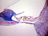 |
Outer pilar cell tunnel of corti | Outer pilar cell tunnel of corti. Cochlea. Photograph. Multimedia. | Cochlea; Sense organs; Ear; Anatomy | Slice of Life |
| 5 |
 |
Outer hairs cell 3 | Outer hair cell 3. Cochlea. Photograph. Multimedia. | Hair Cells, Outer; Cochlea; Ear; Sense Organs; Anatomy | Slice of Life |
| 6 |
 |
Cochlear nerve within cochlea | Cochlear nerve within cochlea. Photograph. Multimedia. | Cochlear Nerve; Ear; Sense Organs; Cochlea; Anatomy | Slice of Life |
| 7 |
 |
Organ of corti outer supporting cells | Organ of corti outer supporting cells. Cochlea. Photograph. Multimedia. | Organ of Corti; Labyrinth Supporting Cells; Cochlea; Ear; Sense Organs; Anatomy | Slice of Life |
| 8 |
 |
Ear | This image shows one section through the coiled cochlear canal. The scala vestibuli and scala tympani are filled with perilymph, and the scala media is filled with endolymph. Also visible is a section of the spiral ganglion and the large acoustic nerve. The cochlea is encased in bone. UCLA Histology... | Cochlea; Ear | UCLA Histology |
1 - 25 of 8