Home
Browse
Ask Us
Chat
Harmful Language Statement
Log in
NOVEL - Neil R. Miller Collection
Advanced Search
About
The Neil R. Miller Collection covers a broad range of neuro-ophthalmologic conditions and syndromes in videos, images, and lecture presentations.
Year
1996
1997
1998
1999
2000
2001
2002
2003
2004
2005
2006
2007
2008
2009
2010
2011
2012
2013
2014
2015
2016
2017
2018
2019
2020
2021
2022
2023
2024
TO
1996
1997
1998
1999
2000
2001
2002
2003
2004
2005
2006
2007
2008
2009
2010
2011
2012
2013
2014
2015
2016
2017
2018
2019
2020
2021
2022
2023
2024
Type
Image
640
Image/MovingImage
135
Text
1
Format
image/jpeg
680
video/mp4
136
application/pdf
8
Collection
NOVEL - Neil R. Miller Collection
824
Filters:
Collection:
"ehsl_novel_nrm"
551
-
575
of
824
<
18
19
20
21
22
23
24
25
26
27
>
Gallery view
Number of results to display per page
10
25
50
100
200
Sort by Relevance
Sort by Title A-Z
Sort by Title Z-A
Sort by Date Ascending
Sort by Date Descending
Sort by Last Modified Ascending
Sort by Last Modified Descending
Title
Date
Type
551
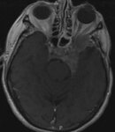
Extensive Skull Base Meningioma That Caused Left Optic Disc Cupping
Image
552
External Appearance and Ocular Motility Disorder in Patient With Myotonic Dystrophy Type 1
2024-06
Image/MovingImage
553
External Appearance of the Lid Crease Incision Site Used to Access the Superior Ophthalmic Vein for Transvenous Embolization of a Dural or Direct Carotid-cavernous Sinus Fistula
2024-07-10
Image
554
Eyelid Myokymia
2024-05
Image/MovingImage
555
Eyelid Myokymia
2024-05
Image/MovingImage
556
Familial Cavernomas
2024-06-28
Image
557
Familial Cavernomas (SWI)
2024-06-28
Image
558
Focal Area of Atrophy in a Diabetic Patient who Previously had an Attack of Nonarteritic Anterior Ischemic Optic Neuropathy
2024-06
Image
559
Fundus Appearance in a Patient With Oculopalatal Tremor
2024-06
Image/MovingImage
560
Giant Intracavernous Aneurysm
2024-07-10
Image
561
Giant Intracavernous Aneurysm
2024-07-10
Image
562
Heimann-Bielschowsky Phenomenon
2024-05
Image/MovingImage
563
Hemifacial Spasm
2024-05
Image/MovingImage
564
Hemifacial Spasm
2024-05
Image/MovingImage
565
Hemifacial Spasm (Seesaw)
2024-05
Image/MovingImage
566
Horner Syndrome
2024-05
Image/MovingImage
567
Hydrocephalus Caused by a Large Pineal Region Mass
2024-07
Image
568
Hydrocephalus Causing Downward Compression and Displacement of the Optic Chiasm
2024-07
Image
569
Hydrocephalus Causing Downward Compression of the Optic Chiasm by an Enlarged Third Ventricle
2024-07
Image
570
Hydrocephalus from a Large Posterior Fossa Mass
2024-07
Image
571
Hydrocephalus from Large Posterior Fossa Mass
2024-07
Image
572
Hydrocephalus from Large Posterior Fossa Mass
2024-07
Image
573
Hydrocephalus from Large Posterior Fossa Mass
2024-07
Image
574
Hyrocephalus with Downward Compression of the Optic Chiasm by an Enlarged Third Ventricle
2024-07
Image
575
Inflammatory Cells in the Infarcted Region and Surrounding Area of an Optic Nerve from a Patient with Recent Nonarteritic Anterior Ischemic Optic Neuropathy
2024-06
Image
551
-
575
of
824
<
18
19
20
21
22
23
24
25
26
27
>