The Health Education Assets Library (HEAL) is a collection of over 22,000 freely available digital materials for health sciences education. The collection is now housed at the University of Utah J. Willard Marriott Digital Library.
TO
1 - 25 of 23
| Title | Description | Subject | Collection | ||
|---|---|---|---|---|---|
| 1 |
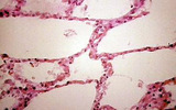 |
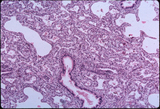
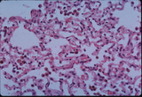
Lung, normal | Lung, normal | Respiratory System; Lung | Slice of Life |
| 2 |
 |
Hyaline membrane formation, lung | Hyaline membrane formation, lung | Respiratory System; Lung | Slice of Life |
| 3 |
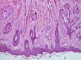
 |
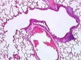
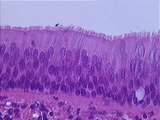
Emboli, lima bean, lungs | Emboli, lima bean, lungs | Respiratory System; Lung; Embolism | Slice of Life |
| 4 |
 |
Respiratory System | Bronchus-associated lymphoid tissue BALT consists of submucosal lymphoid tissue in intermediate and small airways. They are part of the diffuse mucosa-associated lymphoid tissue (MALT) also found in many other tissues. These structures are normal, but similar collections in lung alveolar tissue are ... | Lung; lymphoid tissue; Respiratory System | UCLA Histology |
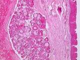
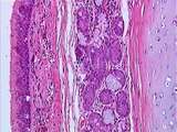
| 5 |
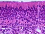
 |
Respiratory System | In this image, a respiratory bronchiole (with intermittent alveoli) branches into alveolar ducts (walled by continuous alveoli). Alveolar sacs and alveoli can also be seen. UCLA Histology Collection. | Lung; respiratory bronchiole; Respiratory System | UCLA Histology |
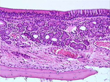
| 6 |
 |
Respiratory System | In this small bronchus, the respiratory epithelium is less tall, with a thick basal lamina. The submucosa contains connective tissue, smooth muscle, and the mixed glands made up of serous and mucous components. UCLA Histology Collection. | epithelium; Lung; respiratory epithelium; Respiratory System | UCLA Histology |
| 7 |
 |
Respiratory System | Observe the narrowing of this small bronchus (with cartilage plates) into a bronchiole, terminal bronchiole, then respiratory bronchiole (with intermittent alveoli). A branch of the pulmonary artery is also visible. UCLA Histology Collection. | Lung; Respiratory System | UCLA Histology |
| 8 |
 |
Respiratory System | A macrophage rests on the alveolar wall. It is difficult to differentiate between type I pneumocytes and endothelial cells lining the alveolar capillaries. The type II pneumocytes, which produce surfactant, have large round nuclei with prominent nucleoli. UCLA Histology Collection. | Lung; pneumocytes; Respiratory System | UCLA Histology |
| 9 |
 |
Respiratory System | In this image the transition between olfactory epithelium on the right and respiratory epithelium on the left can be seen. Bowman's glands and bone are also visible. UCLA Histology Collection. | Nose; olfactory epithelium; respiratory epithelium; Respiratory System | UCLA Histology |
| 10 |
 |
Respiratory System | This image from the underside of the epiglottis shows large amounts of mixed glands in the submucosa. Here the epithelium changes to respiratory epithelium (or something approaching it). Again note the elastic cartilage. UCLA Histology Collection. | respiratory epithelium; Respiratory System; Epiglottis | UCLA Histology |
| 11 |
 |
Respiratory System | It can be difficult to distinguish between the three cell types in the olfactory epithelium. The lowest layer of nuclei belongs to the basal cells, the middle layer holds the olfactory cell nuclei, and the uppermost layer of nuclei beongs to the sustentacular cells sustentacular cells. Find the duct... | Nose; olfactory epithelium; Respiratory System | UCLA Histology |
| 12 |
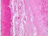 |
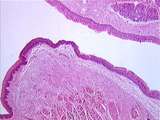
Respiratory System | Stratified squamous epithelium covers much of the surface of the epiglottis, and there is a connective tissue layer which separates the epithelium from the perichondrium of the elastic cartilage. UCLA Histology Collection. | Epiglottis; Respiratory System; Stratified squamous epithelium | UCLA Histology |
| 13 |
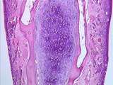 |
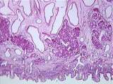
Respiratory System | Here we can see the lamellar bone and hyaline cartilage of the median septum of the nose. At this magnification it is hard to distinguish between respiratory epithelium (lower 1/3rd, with pale goblet cells) and olfactory epithelium (upper 2/3rds). UCLA Histology Collection. | hyaline cartilage; median septum; Nose; Respiratory System | UCLA Histology |
| 14 |
 |
Respiratory System | Underneath the olfactory epithelium is the submucosa, which contains Bowman's glands. The ducts of Bowman's glands carry watery secretions to the epithelial surface, where odorant molecules are dissolved and detected. Branches of the olfactory nerve and bone of the nasal septum is visible in this ar... | Bowman's glands; Nose; olfactory epithelium; Respiratory System | UCLA Histology |
| 15 |
 |
Respiratory System | Fetal lung showing developing airways and alveoli. UCLA Histology Collection. | Fetal lung; Respiratory System | UCLA Histology |
| 16 |
 |
Respiratory System | The outside of the nares or nostrils is covered by skin. The dermis contains sebaceous glands associated with the hair follicles. Red strips of skeletal muscle are also visible. UCLA Histology Collection. | Nares; Respiratory System | UCLA Histology |
| 17 |
 |
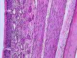
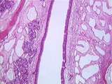
Respiratory System | The respiratory epithelium, together with the basal lamina, constitutes the mucosa. The submucosa contains rather dense connective tissue, blood vessels, and mixed glands which are made up of serous and mucous components. Next can be found the perichondrium, which covers the C-shaped hyaline cartila... | hyaline cartilage; respiratory epithelium; Respiratory System; trachea | UCLA Histology |
| 18 |
 |
Respiratory System | At a higher power, the respiratory epithelium can be seen to consist of pseudostratified columnar ciliated cells, goblet cells and basal cells; the basal cells divide and differentiate to become columnar cells. The basal lamina separates the epithelium from the submucosal connective tissue. UCLA His... | pseudostratified columnar ciliated cells; respiratory epithelium; Respiratory System; trachea | UCLA Histology |
| 19 |
 |
Respiratory System | A small number of alveolar macrophages is found in alveolar spaces of normal healthy lungs. They are part of the lung's defense mechanism. UCLA Histology Collection. | alveolar spaces; Lung; macrophage; Respiratory System | UCLA Histology |
| 20 |
 |
Respiratory System | The vocal cords consist of a vestibular fold or false vocal cord, found above a space, the laryngeal ventricle. The false vocal cords are covered by respiratory epithelium, under which can be found mixed glands. The edge of the true vocal cord is covered by stratified squamous epithelium, and its ce... | Respiratory System; vocal cords | UCLA Histology |
| 21 |
 |
Respiratory System | In the posterior nose, air must traverse rather narrow slits with underlying venous sinuses, creating a heat-exchange system for inspired air. Mixed glands can be seen under the respiratory epithelium. UCLA Histology Collection. | nares; Respiratory System; venous sinuses | UCLA Histology |
| 22 |
 |
Respiratory System | Identify an alveolus and a small blood vessel. Macrophages are usually found resting on the thin alveolar wall. UCLA Histology Collection. | Lung; Respiratory System | UCLA Histology |
| 23 |
 |
Respiratory System | Deeper within the nasal cavity can be found respiratory epithelium, mixed serous/mucous glands and a network of venous sinuses, useful for warming inspired air. UCLA Histology Collection. | Nares; nasal cavity; Respiratory System; venous sinuses | UCLA Histology |
1 - 25 of 23