The Health Education Assets Library (HEAL) is a collection of over 22,000 freely available digital materials for health sciences education. The collection is now housed at the University of Utah J. Willard Marriott Digital Library.
TO
Filters: Collection: "ehsl_heal" Subject: "cortex"
1 - 25 of 10
| Title | Description | Subject | Collection | ||
|---|---|---|---|---|---|
| 1 |
 |
Lymph node (mouse) | Stain: Hematoxylin & eosin. For comparison with the human lymph node a survey of a small rodent lymph node is demonstrated. Basically the same divisions are found: (1) a cortex (outer cortex zone, nodular cortex or superficial cortex) covered with a (2) thin capsule and subcapsular sinus; a paracor... | follicle; hilum; medulla; cortex | Poja Histology Collection - Lymphatic Tissues and Organs Subset |
| 2 |
 |
Lymph node (human) | Stain: Azan. Left and right: survey of lymph nodes, compare the individual differences between the nodes. The pictures display the cortex (1) with capsule (3), and the medulla (2). The cortex consists of (secondary) follicles displaying a clear germinal centre (4). The paracortex (T cell area) is ... | follicle; paracortex; cortex; germinal center | Poja Histology Collection - Lymphatic Tissues and Organs Subset |
| 3 |
 |
Scheme of lymph node (human) | Lymph node: A. survey; B. detail subcapsular (or marginal) sinus 1. adipose tissue; 2. capsule; 3. afferent lymph vessel with valves (B); 4. subcapsular (or marginal) sinus; 5. germinal centre of a secondary lymphatic nodule (or follicle); 6. crescent or mantle zone indicated by cap of per... | cortex; scheme; paracortex; germinal center | Poja Histology Collection - Lymphatic Tissues and Organs Subset |
| 4 |
 |
Afferent lymph vessel in lymph node (human) | Stain: Azan. Left (A) and right (B): part of the cortex of a lymph node with the capsule (1) and subcapsular (or marginal) sinus (2) filled with lymphocytes. Left (A): (3) show perpendicularly localized reticular cells and fibres (blue) in the sinus. (4) indicate afferent lymph vessel with valves ... | lymph vessel; subcapsular sinus; follicle; cortex | Poja Histology Collection - Lymphatic Tissues and Organs Subset |
| 5 |
 |
Cortex of lymph node (human) | Stain: Azan. The cortex of the lymph node comprises the (secondary) follicles with germinal centres (1) and corona or mantle zone (2), all filled with B lymphocytes. The paracortical area (3) below the follicles largely comprises T cells. Afferent lymph vessels enter via the capsule (6) into the m... | cortex; paracortex | Poja Histology Collection - Lymphatic Tissues and Organs Subset |
| 6 |
 |
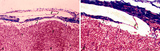
Thymus - Young Thymus | Young thymus, showing cortex and portions of the medulla. Note a Hassals corpuscle in the medulla. ( This is a diagnostic feature of the thymus.) UCLA Histology Collection. | cortex; lymphoid organ | UCLA Histology |
| 7 |
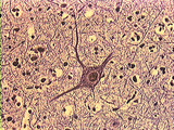 |
Central Nervous System | Pyramidal cells of the cortex have several dendrites and one axon emanating for the cell body. The cell body has a nucleus with a nucleolus. The dendrites are branched and taper as they leave the cell body. One slender axon can be seen emerging from the cell body. Darkly stained nuclei of neuroglial... | Brain; Central Nervous System; cortex; pyramidal cell | UCLA Histology |
| 8 |
 |
Central Nervous System | Higher power examination of a small pyramidal cell. At this magnification the dendritic spines are easily observed. These spines are specializations that increase the surface area of dendrites. Spines are preferential sites for synaptic contact. Large neurons may receive up to 100,000 synapses via t... | Brain; Central Nervous System; cortex; dendritic spines; pyramidal cell | UCLA Histology |
| 9 |
 |
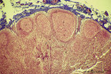
Thymus | The connective tissue capsule of the thymus sends septa into the organ, dividing it into lobules. Each lobule is further divided into cortex and medulla. Cortex stains darkly because it contains numerous lymphocytes embedded in a meshwork of epithelial reticular cells. The Hassal's corpuscle in the ... | cortex; medulla; Thymus | UCLA Histology |
| 10 |
 |
Central Nervous System | Fibrous astrocytes of the cerebrum. The stellate body gives off numerous radial processes which can extend to surround a capillary. Note the long protoplasmic process marked in this section. UCLA Histology Collection. | astrocytes; Brain; Central Nervous system; cerebrum; cortex | UCLA Histology |
1 - 25 of 10