The Health Education Assets Library (HEAL) is a collection of over 22,000 freely available digital materials for health sciences education. The collection is now housed at the University of Utah J. Willard Marriott Digital Library.
TO
1 - 25 of 13
| Title | Description | Subject | Collection | ||
|---|---|---|---|---|---|
| 1 |
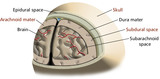 |
Potential Dural Spaces (Labeled) | Potential dural spaces. | Brain; Arachnoid Mater; Epidural Space; Skull; Dura Mater; Subdural Space; Subarachnoid Space | Royal College of Surgeons in Ireland Illustrations |
| 2 |
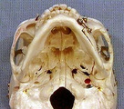 |
Skull ventral surface | Skull ventral surface. Other or model. Photograph. Multimedia. | Skull; Musculoskeletal system; Anatomy | Slice of Life |
| 3 |
 |
Skull ventral surface | Skull ventral surface. Other or model. Photograph. Multimedia. | Skull; Musculoskeletal System; Anatomy | Slice of Life |
| 4 |
 |
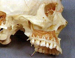
Palate, skull, closeup | Palate, skull, closeup. Photograph. Multimedia. | Palate; Skull; Musculoskeletal system; Anatomy | Slice of Life |
| 5 |
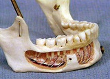 |
Mandible, inferior alveolar nerve | Mandible, inferior alveolar nerve. Painted skull model. Other or model. Photograph. Multimedia. | Mandibular Nerve; Skull; Mandible; Central Nervous System; Anatomy | Slice of Life |
| 6 |
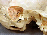 |
Mastoid process, mastoid air cells, temporal bone | Mastoid process, mastoid air cells, temporal bone. Lateral view skull, closeup. Other or model. Photograph. Multimedia. | Mastoid; Temporal Bone; Skull; Musculoskeletal System; Anatomy | Slice of Life |
| 7 |
 |
Orbit, close-up | Orbit, close-up | Musculoskeletal System; Skull; Orbit; Models, Anatomic | Slice of Life |
| 8 |
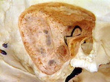 |
Mastoid air cells | Mastoid air cells | Musculoskeletal System; Skull; Mastoid; Semicircular Canals; Models, Anatomic | Slice of Life |
| 9 |
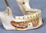 |
Inferior alveolar nerve&vessels | Inferior alveolar nerve&vessels. Mandible, dissected and prepared. Other or model, labeled. Photograph. Multimedia. | Mandible; Skull; Musculoskeletal System; Anatomy | Slice of Life |
| 10 |
 |
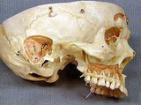
Maxilla and skull lateral view | Maxilla and skull lateral view | Musculoskeletal System; Skull; Maxilla; Models, Anatomic | Slice of Life |
| 11 |
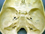 |
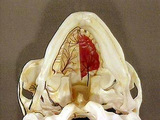
Skull and sella turcica, view of base of skull from interior. Good foramina | Skull and sella turcica, view of base of skull from interior. Good foramina. Other or model. Photograph. Multimedia. | Skull; Sella turcica; Musculoskeletal system; Anatomy | Slice of Life |
| 12 |
 |
Bone | At a later stage of the developing skull, note the differences in cell density and organization between the immature or woven bone and the mature or lamellar bone. Often a cementing line separates the two types of bone. UCLA Histology Collection. | bone; lamellar bone; Skull | UCLA Histology |
| 13 |
 |
Bone | Intramembranous bone formation. In the fetal skull, plates of bone are laid down in the mesenchymal field. This image shows woven or immature bone, which tends to be more cellular than lamellar or adult bone. Osteoprogenitor cells differentiate into osteoblasts, which produce the osteoid or uncalcif... | bone; Intramembranous bone formation; Skull | UCLA Histology |
1 - 25 of 13