Home
Browse
Ask Us
Chat
Harmful Language Statement
Log in
Advanced Search
Year
1990
1991
1992
1993
1994
1995
1996
1997
1998
1999
2000
2001
2002
2003
2004
2005
2006
2007
2008
2009
2010
2011
2012
2013
2014
2015
2016
2017
2018
2019
2020
2021
2022
2023
2024
TO
1990
1991
1992
1993
1994
1995
1996
1997
1998
1999
2000
2001
2002
2003
2004
2005
2006
2007
2008
2009
2010
2011
2012
2013
2014
2015
2016
2017
2018
2019
2020
2021
2022
2023
2024
Type
Image
188
Text
50
Image/MovingImage
13
Format
image/jpeg
182
application/pdf
56
video/mp4
13
Collection
Honors Theses Closed Archive
1
NOVEL
4
NOVEL - Andrew Lee Collection
6
NOVEL - Emory Eye Center
1
NOVEL - Jonathan D. Trobe Collection
1
NOVEL - Journal of Neuro-Ophthalmology
3
NOVEL - NANOS Annual Meeting
40
NOVEL - Neil R. Miller Collection
188
NOVEL - Walsh and Hoyt Textbook Selec...
3
Theses & Dissertations
3
UScholar Works
1
More
Filters:
Subject:
"Imaging"
176
-
200
of
251
<
2
3
4
5
6
7
8
9
10
11
>
Gallery view
Number of results to display per page
10
25
50
100
200
Sort by Relevance
Sort by Title A-Z
Sort by Title Z-A
Sort by Date Ascending
Sort by Date Descending
Sort by Last Modified Ascending
Sort by Last Modified Descending
Title
Date
Type
Setname
176
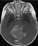
Large Posterior Fossa Mass with Brainstem Compression
2024-07
Image
ehsl_novel_nrm
177
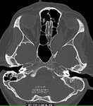
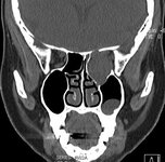
Left Orbital and Retro-orbital Cholesterol Granuloma
2024-07
Image
ehsl_novel_nrm
178
Left Orbital Invasion by a Large Cholesterol Granuloma
2024-07
Image
ehsl_novel_nrm
179
Left Orbital Invasion by Cholesterol Granuloma
2024-07
Image
ehsl_novel_nrm
180
Left Orbital Invasion by Large Cholesterol Granuloma
2024-07
Image
ehsl_novel_nrm
181
Left Orbital Invasion by Large Cholesterol Granuloma
2024-07
Image
ehsl_novel_nrm
182
Left orbital invasion by large cholesterol granuloma
2024-07
Image
ehsl_novel_nrm
183
Left-sided Intracavernous Aneurysm
2024-07-10
Image
ehsl_novel_nrm
184
Limitations of Neuroimaging
2008-03-13
Text
ehsl_novel_nam
185
Magnetic Resonance Imaging of Superior Oblique Muscle Atrophy in Trochlear Nerve Schwannoma
2013-02-12
Text
ehsl_novel_nam
186
Magnetic Resonance Imaging of the Human Lateral Geniculate Body
1990-02-04
Text
ehsl_novel_nam
187
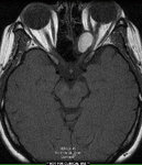
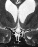
Marked Compression and Downward Displacement of the Chiasm by an Enlarged Third Ventricle in a Patient with Hydrocephalus
2024-07
Image
ehsl_novel_nrm
188
Metamaterials and their applications in imaging
2017
Text
ir_etd
189
Modern Imaging of Optic Disc Drusen
2021
Text
ehsl_novel_novel
190
MR Imaging of the Cavernous Sinus
1990-02-07
Text
ehsl_novel_nam
191
MRI Diagnosis of Clival Cancer and Sixth Nerve Palsy
2023-03
Text
ehsl_novel_jno
192
MRI Findings in Giant Cell Arteritis
2022
Text
ehsl_novel_eec
193
MRI in Neuro-ophthalmology
2019-03
Image/MovingImage
ehsl_novel_lee
194
MRI Showing Extensive White Matter Disease in a Patient with CADASIL
2024-07-10
Image
ehsl_novel_nrm
195
MRI Showing Extensive White Matter Disease in a Patient with CADASIL
2024-07-10
Image
ehsl_novel_nrm
196
MRI T1, Non-contrast Sagittal Image of Large Parietal-occipital Cavernoma
2024-07-10
Image
ehsl_novel_nrm
197
MRI, Axial T2 Image, Showing Large Left Parietal-occipital Cavernoma
2024-07-10
Image
ehsl_novel_nrm
198
MRI, T1 Axial, Following Gadolinium Administration, Showing Large, Left Parietal-occipital Cavernoma
2024-07-10
Image
ehsl_novel_nrm
199
MRI, T1 Image After Contrast Injection, Shows a Lesion in the Midbrain that Caused a Benedikt Syndrome
2024-07-10
Image
ehsl_novel_nrm
200
MRI, T2 Axial Image, Showing Midbrain Lesion Causing a Benedikt Syndrome
2024-07-10
Image
ehsl_novel_nrm
176
-
200
of
251
<
2
3
4
5
6
7
8
9
10
11
>