The Emory Eye Center Neuro-Ophthalmology Collection contains a variety of lectures, videos and images relating to the discipline of neuro-ophthalmology created by faculty at Emory University in Atlanta, GA.
NOVEL: https://novel.utah.edu/
TO
1 - 200 of 93
| Title | Description | Creator | ||
|---|---|---|---|---|
| 1 |
 |
Anatomy of the Ocular Fundus | A review of normal features of the ocular fundus. Fundus photography using various techniques illustrate anatomic features of the ocular fundus. - Figure 1 : A) Color fundus photograph of the left optic disc and peripapillary retina showing a normal optic disc, retinal arteries, retinal veins, and... | Devin D. Mackay, MD; Valérie Biousse, MD |
| 2 |
 |
Anatomy of the Trigeminal Nerve | MRI and CT scan imaging of the trigeminal nerve and its 3 divisions. Figure 1 : trigeminal nerve. Overview Figure 2 : trigeminal nuclei Figure 3 : trigeminal root. Cisternal segment. Figure 3A. FIESTA axial image through midpons. Figure 3B. T2 coronal image through prepontine cistern Figure 4 : trig... | Samuel Bidot, MD; Amit M. Saindane, MD; Valérie Biousse, MD |
| 3 |
 |
Anterior and Posterior Scleritis | A case of anterior and posterior scleritis secondary to idiopathic orbital inflammation, also known as orbital pseudotumor. Various imaging modalities are included to demonstrate optic disc edema, macular edema, and fluid in tenon's capsule which may be seen in posterior scleritis. Figure 1 : Exter... | Joshua Levinson, MD; Valérie Biousse, MD |
| 4 |
 |
Artifact from Incomplete Orbital Fat Suppression on Magnetic Resonance Imaging | Orbital fat has short relaxation times that results in a hyperintense appearance on T1-weighted magnetic resonance imaging (MRI). Fat suppressed T1 MRI sequences are needed to remove the fat signal and better visualize the orbital anatomy, including the optic nerve. Contrast can be used with fat sup... | Matthew Boyko, MD; Valérie Biousse, MD |
| 5 |
 |
Bilateral Lens Subluxation in Marfan Syndrome | This is a case of known Marfan syndrome with bilateral progressive visual loss. The ocular examination showed bilateral lens dislocation. Figure 1a: Typical superonasal lens subluxation in both eyes Figure 1b: The arrows show the inferior edges of the lenses Figure 2: Optical section of the lenses u... | Rabih Hage, MD; Valérie Biousse, MD |
| 6 |
 |
Bilateral Optic Disc Edema from Hypertensive Retinopathy | A 29 year-old woman was assessed for 2 weeks of headaches and 4 days of blurred vision in both eyes. Her blood pressure was 225/135. Her examination showed: best-corrected visual acuity: 20/25 OD, 20/30 OS; pupils equal and reactive without relative afferent pupillary defect; intraocular pressures 1... | Benjamin I. Meyer, MD; Valérie Biousse, MD |
| 7 |
 |
Cavernous Sinus Meningioma Extending into the Orbital Apex | Septic left cavernous sinus and superior ophthalmic vein thrombosis, secondary to left maxillary tooth abscess. MRI characteristics Figure 1 : MRI Orbits (Coronal T2 with fat suppression) : Left periorbital edema (increased T2 signal, yellow arrows) extends inferiorly along the premalar tissues to t... | Supharat Jariyakosol, MD; Valérie Biousse, MD |
| 8 |
 |
Central Retinal Artery Occlusion with Cilioretinal Artery Sparing | Central retinal artery occlusion with sparing of the cilioretinal artery Figure 1 : Fundus photographs show retinal whitening in the right eye, with sparing of the perfused retina in the distribution of the cilioretinal artery (arrows); the left eye has a normal funduscopic appearance. Figure 2 : Mo... | Supharat Jariyakosol, MD; Valérie Biousse, MD |
| 9 |
 |
Cerebral Arterial Vascularization | Arteries of the neck and brain as seen on a CT Angiogram. Figure 1 : Overview. Figure 1A. Anterior view. Figure 1B. Lateral view. Figure 2 : Internal carotid artery. Segmentation. Figure 3 : Internal carotid artery and vertebral arteries. Extracranial part. Posterolateral view. Figure 4 : Internal c... | Samuel Bidot, MD; Valérie Biousse, MD |
| 10 |
 |
Cerebral Venous Sinus Thrombosis with Papilledema | A case of superior sagittal sinus, right transverse sinus and right sigmoid sinus thrombosis, presenting with increased intracranial pressure (headaches, bilateral sixth palsy and papilledema). Figure 1 : Disc photos of the right and left eyes demonstrating bilateral disc edema. Figure 2 : Non-contr... | Supharat Jariyakosol, MD; Valérie Biousse, MD |
| 11 |
 |
Cerebral Venous Vascularization | MRV and CTV scan imaging of the brain veins. Figure 1 : Overview. MRV with contrast Figure 1A. Right postero-lateral view. Figure 1B. Sagittal view. Figure 2 : Dural sinuses. Superior endocranial view. CTV. Figure 3 : Dural sinuses. Sagittal endocranial view. CTV. Figure 4 : Dural sinuses. Right an... | Samuel Bidot, MD; Amit M. Saindane, MD; Valérie Biousse, MD |
| 12 |
 |
Checkboard Visual Field Loss Due to Bilateral Homonymous Hemianopsias | A 70-year-old man with vascular risk factors was seen for assessment of sudden visual field loss in both eyes. His examination showed: visual acuity: 20/20 OD and 20/25 OS; pupils: equal with no relative afferent pupillary defect; color vision: 14/14 plates correct OU; anterior segment exam: normal ... | Rahul A. Sharma, MD, MPH; Valérie Biousse, MD |
| 13 |
 |
Choroidal Hypoperfusion Defect in Giant Cell Arteritis | Here, we present a case of a 62 year-old male with vision loss in the right eye, headaches, and neck/shoulder/temporal pain, found to have choroidal hypoperfusion and diagnosed with giant cell arteritis (GCA). In combination with anterior ischemic optic neuropathy and cotton wool spots, choroidal hy... | Nithya Shanmugam; Michael Dattilo; Valerie Biousse |
| 14 |
 |
Choroidal Infarction in Giant Cell Arteritis | An 80-year-old Caucasian woman presented with a 10-day history of headaches, intermittent binocular diplopia, and jaw pain. Temporal artery biopsy confirmed a diagnosis of giant cell arteritis. Examination showed characteristic large area of choroidal ischemia that is well-known to be associated wit... | Wael A. Alsakran, MD; Andre Aung, MD; Valérie Biousse, MD |
| 15 |
 |
Choroidal Neovascular Membrane in Chronic Papilledema | A 21-year-old woman with papilledema from idiopathic intracranial hypertension developed a peripapillary choroidal neovascular membrane (PCNVM) complicating untreated chronic papilledema 10 years later. | George Alencastro; Valerie Biousse |
| 16 |
 |
Classic Pathology Findings in Giant Cell Arteritis | An 80-year-old Caucasian woman presented with a 10 day history of headaches, intermittent binocular diplopia, and jaw pain. Temporal artery biopsy confirmed a diagnosis of giant cell arteritis. Pathology findings were classic for giant cell arteritis with numerous inflammatory cells in the tunica me... | Andre Aung, MD; Corrina Azarcon, MD; Wael A. Alsakran, MD; Valérie Biousse, MD |
| 17 |
 |
Clinical Features of Neuroretinitis | A 13-year-old girl was seen for assessment of blurred vision and optic disc edema in her right eye. Her examination showed: best-corrected visual acuity of hand motion OD and 20/25 OS; pupils: no relative afferent pupil defect; color vision: 0/14 plates OD and 14/14 plates OS; humphrey visual fields... | Rahul A. Sharma, MD, MPH; Jason H. Peragallo, MD; Valérie Biousse, MD |
| 18 |
 |
Coexisting Thyroid Eye Disease and Myasthenia Gravis | A case of coexisting thyroid orbitopathy and myasthenia gravis. External photographs of the eyes and eyelids, as well as images from an MRI of the orbits, are included. Figure 1 : External photograph of eyes showing right lid retraction and left upper lid ptosis. Figure 2 : External photograph of ... | Devin D. Mackay, MD; Valérie Biousse, MD |
| 19 |
 |
Colloid Cyst Hydrocephalus | This is a case of colloid cyst of the third ventricle complicated by severe hydrocephalus, raised intracranial pressure and papilledema. Figure 1: Fundus photographs demonstrating bilateral optic nerve head edema Figure 2a and 2b: T1-weighted axial brain MRI without contrast: Dilation of both later... | Rabih Hage, MD; Valérie Biousse, MD |
| 20 |
 |
Compressive Optic Neuropathy from Cavernous Hemangioma | A 42-year-old man was seen for assessment of progressive blurring of vision in his left eye (OS) over several weeks. His examination showed a left optic neuropathy: visual acuity: 20/20 OD and 20/20-2 OS; pupils: 1+ left relative afferent pupillary defect; color vision: 14/14 plates correct OU. Ther... | Rahul A. Sharma, MD, MPH; Aaron M. Yeung, MD; Valérie Biousse, MD |
| 21 |
 |
Cotton Wool Spots in Giant Cell Arteritis | This is a case of cotton wool spots in a patient with temporal artery-biopsy proven temporal arteritis.; ; A 66-year-old woman presents with isolated painless vision loss related to a left optic neuropathy in her left eye. She denies systemic symptoms to suggest giant cell arteritis.; Her examinatio... | Rahul A. Sharma, MD, MPH; Valérie Biousse, MD |
| 22 |
 |
Dilated Episcleral Vessels from Carotid Cavernous Fistula | 51 year white woman with a 6 months history of chronic right eye redness, periorbital swelling and progressive proptosis. She was seen by multiple providers and treated for dry eye and conjunctivitis. Her examination showed normal visual acuity, color vision and pupils. There was an intraocular pres... | Amani Alzayani, MD; Valérie Biousse, MD; Ling Chen Chien, MD |
| 23 |
 |
Direct Carotid-Cavernous Sinus Fistula | A 40-year-old man presented with decreased vision and redness in his left eye. He had a significant trauma to the left side of his face about one year ago, but did not seek medical attention. External examination showed significant proptosis of the left eye (Figure 1) and conjunctival injection and ... | Jonathan A. Micieli, MD; Valérie Biousse, MD |
| 24 |
 |
Enophthalmos from Breast Cancer Metastasis to the Orbit | Right painful ophthalmoplegia with right enophthalmos secondary to breast cancer metastasis to the right orbit. | Valérie Biousse, MD |
| 25 |
 |
Fourth Nerve Schwannoma | This is a case of IVth cranial nerve schwannoma, showing an enhancement in the subarachnoid space consistent with the clinical presentation. Figure 1a : T1-weighted axial brain MRI Figure 1b : T1-weighted axial brain MRI : magnification of the brainstem Figure 1c : T1-weighted axial brain MRI : cr... | Rabih Hage, MD; Valérie Biousse, MD |
| 26 |
 |
Fundus Autofluorescence | The retinal pigment epithelium (RPE) has many important functions including phagocytosis of the photoreceptor outer segments. The metabolism of the photoreceptor outer segments leads to the formation of lipofuscin. Disease states and potentially increased oxidative damage can lead to the buildup of ... | Jonathan A. Micieli, MD; Valérie Biousse, MD |
| 27 |
 |
Funduscopic Findings of Acute Central Retinal Artery Occlusion | A 59-year-old man was referred for assessment acute vision loss in the right eye. His examination showed: best-corrected visual acuity: light perception OD, 20/20 OS; pupils: Relative afferent pupillary defect OD; color vision: unable to visualize control plate OD, 14/14 OS correct Ishihara plates. ... | David B. Enfield, MD; Valérie Biousse, MD |
| 28 |
 |
Ganglion Cell Layer Analysis by Optical Coherence Tomography (OCT) | A normal optical coherence tomography (OCT) of the macula is shown (Figure 1) and the various layers of the retina are labelled (Figure 2). The cell bodies of retinal ganglion cells (RGC) are located in the ganglion cell layer (GCL) of the retina and mostly synapse in the lateral geniculate nucleus ... | Jonathan A. Micieli, MD; Valérie Biousse, MD |
| 29 |
 |
Geniculate Nucleus Metastasis with Homonymous Sectoranopia | This is a case of multiple brain metastases in the setting of bladder cancer complicated with right homonymous horizontal sectoranopia. Figure 1: Pet-scan showing liver (yellow arrows) and kidneys (red arrow) metastases Figure 2: Goldmann Visual Fields: Right homonymous horizontal sectoranopia Figu... | Rabih Hage, MD; Valérie Biousse, MD |
| 30 |
 |
Giant Cell Arteritis: Temporal Artery Anatomy and Histology | Gross anatomy and histology of the normal superficial temporal artery.; Histopathology of the superficial temporal artery involved by active and healed GCA; Summary of the main histopathologic findings in GCA | Samuel Bidot, MD; Valérie Biousse, MD |
| 31 |
 |
Homonymous Hemianopia Secondary to an Intracranial Bleed from an Arteriovenous Malformation | This case demonstrates a homonymous hemianopia resulting from hemorrhage secondary to a ruptured intracranial arteriovenous malformation (AVM), providing grounds for illustration and discussion of the correlations between localization of this lesion on cerebral imaging and resultant visual field and... | Lauren Hudson, MD, PhD; Valérie Biousse, MD |
| 32 |
 |
Incipient Non-Arteritic Anterior Ischemic Optic Neuropathy (NAION) | A 61-year old white man with hypertension, diabetes, and dyslipidema was seen in neuro-ophthalmology consultation for asymptomatic right optic disc edema. He had a small, crowded optic disc in the left eye known as a "disc-at-risk" (Figure 1). He had normal visual function including normal 24-2 SITA... | Jonathan A. Micieli, MD; Valérie Biousse, MD |
| 33 |
 |
Incipient Non-Arteritic Anterior Ischemic Optic Neuropathy (NAION) Evolving to Symptomatic NAION | A 54-year old woman with hypertension was seen in neuro-ophthalmology consultation for asymptomatic left optic disc edema. She had a small, crowded optic disc in the right eye known as a "disc-at-risk" (Figure 1). Her visual function including 24-2 SITA-Fast Humphrey visual fields were normal in bot... | Jonathan A. Micieli, MD; Valérie Biousse, MD |
| 34 |
 |
Internal Carotid Artery / Posterior Communicating Artery Aneurysm with Third Nerve Palsy Shown on CT Angiogram | Internal Carotid Artery / Posterior Communicating Artery Aneurysm with Third Nerve Palsy Shown on CT Angiogram ; anatomic description of vascular and bony findings on the CTA. - Figure 1 : 51 year-old man complaining of painful binocular diplopia. Orange arrows indicate the direction of gaze. In p... | Samuel Bidot, MD; Amit M. Saindane, MD; Valérie Biousse, MD |
| 35 |
 |
Internuclear Ophthalmoplegia (INO) | A 67-year-old man with a known history of heart failure and atrial fibrillation developed binocular horizontal diplopia in right gaze after cardiac catheterization. His examination showed normal afferent visual function, full ocular movement of the right eye, and slow adducting saccades in the left ... | Wael A. Alsakran, MD; Valérie Biousse, MD |
| 36 |
 |
Interpreting Ocular Fundus Photographs: a brief guide | Brief guide for interpreting ocular fundus photographs. | Gabriele Berman, MD; Sachin Kedar, MD; Nancy J. Newman, MD; Valérie Biousse, MD |
| 37 |
 |
Iris Transillumination | Single case of iris transillumination in a patient with albinism. Figure 1 : Anterior segment photograph demonstrating reddish hue to iris in albinism Figure 2 : Slit lamp photograph with retroillumination demonstrating iris transillumination Figure 3 : Slit lamp photograph with retroillumination... | Jason Peragallo, MD; Valérie Biousse, MD |
| 38 |
 |
Junctional Scotoma from a Sellar Mass | This is a case of a 55-year-old woman presenting with gradual painless vision loss in both eyes. Although visual acuity was 20/20 in both eyes, there was a left relative afferent pupillary defect and diffuse pallor of both optic nerves (Figure 1). Visual fields (24-2 SITA-Fast) showed a temporal def... | Jonathan A. Micieli, MD; Valérie Biousse, MD |
| 39 |
 |
Large Frontal Meningioma with Mass Effect and Increased Intracranial Pressure | This is a case of frontal meningioma presenting with raised intracranial pressure and bilateral papilledema responsible for visual loss. Figure 1: Goldmann visual field of the left eye. In the right eye, there was no response to the V4e. The visual field is severely constricted in the left eye. Fig... | Rabih Hage, MD; Valérie Biousse, MD |
| 40 |
 |
Large Right Hypophyseal Aneurysm Causing a Junctional Scotoma | Right, multi-lobulated superior hypophyseal artery aneurysm measuring 1.6 x 1.2 x 2.2 cm with 6 mm neck causing a right junctional scotoma . Images from a brain CT with contrast, a brain CT angiography with contrast, cerebral angiogram, Humphrey visual fields and ocular fundus photographs are includ... | Laurel N. Vuong, MD; Valérie Biousse, MD |
| 41 |
 |
Large Sellar and Suprasellar Mass (Pituitary Macroadenoma) With Left Third Nerve Palsy and Left Optic Tract Compression | A case of a large sellar and suprasellar pituitary macroadenoma with an associated left third nerve palsy and left optic tract compression. Images from an MRI of the brain with contrast illustrate the imaging characteristics and extent of the tumor. Figure 1 : Humphrey Visual Fields (24-2 SITA-Fast)... | Devin D. Mackay, MD; Valérie Biousse, MD |
| 42 |
 |
Left RAPD | Video clip displaying pupillary examination and RAPD measurement. | Valérie Biousse, MD |
| 43 |
 |
Malignant Hypertension With Bilateral Optic Nerve Edema | This is a case of malignant hypertension and severe hypertensive retinopathy. A 30-year-old woman with headache and vision loss in the left eye was found to have a markedly elevated blood pressure of 205/100. CT head without contrast showed acute hemorrhage in the right temporal-occipital junction a... | Rahul A. Sharma, MD, MPH; Michael Dattilo, MD, PhD; Valérie Biousse, MD |
| 44 |
 |
Metastatic Ovarian Cancer to the Left Occipital Lobe With Complete Right Homonymous Hemianopia | A case of metastatic ovarian cancer to the left occipital lobe with a complete right homonymous hemianopia. Humphrey visual fields as well as images from an MRI of the brain are included. Figure 1 : Humphrey visual fields showing a complete right homonymous hemianopia Figure 2 : MRI brain T1 axial... | Devin D. Mackay, MD; Valérie Biousse, MD |
| 45 |
 |
MRI Characteristics of Cavernous Sinus and Superior Ophthalmic Vein Septic Thrombosis | Septic left cavernous sinus and superior ophthalmic vein thrombosis, secondary to left maxillary tooth abscess. MRI characteristics. Figure 1 : MRI Orbits (Coronal T2 with fat suppression) : Left periorbital edema (increased T2 signal, yellow arrows) extends inferiorly along the premalar tissues to ... | Devin D. Mackay, MD; Valérie Biousse, MD |
| 46 |
 |
MRI Findings in Giant Cell Arteritis | Case 1. An 80-year-old Caucasian woman presented with a 10-day history of headaches, intermittent binocular diplopia, and jaw pain. Temporal artery biopsy confirmed a diagnosis of giant cell arteritis. MRI with contrast showed enhancement of bilateral optic nerve sheaths in addition to enhancement o... | Wael A. Alsakran, MD; Andre Aung, MD; Valérie Biousse, MD |
| 47 |
 |
MRI Findings in Gliomatosis Cerebri | Single case of gliomatosis cerebri identified on mulltiple MRI images pre- and post-contrast administration with axial and coronal views. Figure 1 : Axial brain MRI (T2) - Diffusely thickened and T2 hyperintense area (yellow arrows) centered within the genu of the corpus callosum with extension into... | Jason Peragallo, MD; Valérie Biousse, MD |
| 48 |
 |
MRI Findings in Optic Nerve and Chiasmal Glioma in Childhood | Multiple cases of optic pathway gliomas are demonstrated using MRI imaging. The optic pathway gliomas are identified at multiple points in the optic pathways. Figure 1 : Patient 1: Axial orbital MRI (T1 postcontrast with fat suppression) showing thickening and enhancement of the right optic nerve a... | Jason Peragallo, MD; Valérie Biousse, MD |
| 49 |
 |
Myelin Oligodendrocyte Glycoprotein (MOG) - Antibody Optic Neuritis | This is an illustrated guide to the clinical diagnosis of myelin oligodendrocyte glycoprotein (MOG)-antibody optic neuritis. Myelin oligodendrocyte glycoprotein (MOG) is a glycoprotein on the surface of myelin and is found exclusively in the central nervous system (CNS). MOG likely mediates a comple... | Bryce Buchowicz, MD; Valérie Biousse, MD |
| 50 |
 |
Neurosarcoidosis | This is an illustrated guide to the clinical diagnosis of neurosarcoidosis. Sarcoidosis is a chronic systemic inflammatory disorder characterized by non-caseating granulomas. It can affect multiple organ systems including the lungs, skin, orbit, and brain. When there is central nervous system (CNS) ... | Bryce Buchowicz, MD; Valérie Biousse, MD |
| 51 |
 |
Non-Arteritic Anterior Ischemic Optic Neuropathy (NAION) With Segmental Optic Disc Edema | A 75-year old white woman with hypertension and diabetes presented with a 1 week history of vision loss in the right eye. Dilated fundus examination revealed superior segmental optic disc edema in the right eye and a small, crowded optic disc in the left eye known as a "disc-at-risk" (Figure 1). Int... | Jonathan A. Micieli, MD; Valérie Biousse, MD |
| 52 |
 |
Nonfunctiong Pituitary Adenoma with Chiasmal Compression | This is a case of large non-functioning pituitary adenoma with mass effect on the optic chiasm inducing loss of optic nerve fibers and subsequent visual field. Figure 1: Fundus photographs demonstrating bilateral temporal optic nerve head pallor Figure 2: Humphrey visual fields demonstrating a bitem... | William Pearce, MD; Valérie Biousse, MD |
| 53 |
 |
Normal Retinal Anatomy | Normal posterior vitreous, retinal and chroroidal anatomy (pictures, fluorescein angiography and optical coherence tomography). Figure 1: Normal fundus photograph of the left eye o a : Optic disc and fovea o b : Foveal reflex in young patients o c : Macular and foveal areas share the same center o d... | Rabih Hage, MD; Valérie Biousse, MD |
| 54 |
 |
Occipital Hemorrhagic Infarction Secondary to Bacterial Endocarditis-Congruent Homonymous Scotomatous Hemianopic Defect | MRI features of occipital hemorrhage secondary to bacterial endocarditis Figure 1 : Humphrey visual fields showing a congruent homonymous scotomatous hemianopic defect Figure 2 : axial gradient echo T2*w brain MRI Figure 3 : axial T1w and T2w brain MRI Figure 4 : axial postcontrast T1w brain MRI | Samuel Bidot, MD; Amit M. Saindane, MD; Valérie Biousse, MD |
| 55 |
 |
Occipital Infarction with Incomplete Congruent Homonymous Hemianopia | CT appearance of a remote occipital infarction. Congruent homonymous hemianopia. | Samuel Bidot, MD; Amit M. Saindane, MD; Valérie Biousse, MD |
| 56 |
 |
Occipital Pyogenic Abscess with Homonymous Hemianopia | This is a case of right occipital abscess with a left homonymous hemianopia. Number of Figures and legend for each: 8 figures Figure 1: Humphrey visual fields: Dense left homonymous hemianopia Figure 2: T2-weighted axial MRI : Round, hyperintense lesion (yellow arrow) in the right occipital lobe sur... | Rabih Hage, MD; Valérie Biousse, MD |
| 57 |
 |
Ocular Fundus Examination | Review of various techniques of ocular fundus examination, including direct ophthalmoscopy, binocular indirect ophthalmoscopy, slit lamp binocular indirect ophthalmoscopy, and fundus photography. Advantages and disadvantages of each technique are discussed. | Devin D. Mackay, MD; Valérie Biousse, MD |
| 58 |
 |
Ocular Hypoperfusion from Severe Internal Carotid Artery Stenosis | 68 year-old man complaining of mildly decreased vision OD with fluctuation of vision throughout the day. Fluorescein angiography shows delayed choroidal and retinal fillings, suggesting hypoperfusion of the right eye. | Samuel Bidot, MD; Amit M. Saindane, MD; Valérie Biousse, MD |
| 59 |
 |
Optic Disc Edema and Pseudoedema | A presentation covering how to approach optic disc edema, including clinical characteristics and the distinction of pseudoedema. | Rahul A. Sharma, MD, MPH; Valérie Biousse, MD |
| 60 |
 |
Optic Disc Melanocytoma | A 26-year-old woman was seen for assessment of an asymptomatic optic nerve abnormality in her left eye. Her examination showed normal afferent visual function: visual acuity: 20/30 OD and 20/25 OS; PH 20/20 OU; pupils: no relative afferent pupil defect; color vision: 14/14 plates correct OU. Humphre... | Rahul A. Sharma, MD, MPH; Valérie Biousse, MD |
| 61 |
 |
Optic Disc Pseudoedema Due to Leber Hereditary Optic Neuropathy | A 23-year-old woman developed sequential painless central vision loss in both eyes (right eye 5 months ago and left eye 2 months ago). Her examination showed bilateral optic neuropathies: visual acuity: 20/300 eccentrically OU (no improvement with pinhole); pupils: equal and reactive with no relativ... | Rahul A. Sharma, MD, MPH; Valérie Biousse, MD |
| 62 |
 |
Optic Nerve Head Granuloma from Sarcoidosis | We present a case of optic neuropathy with optic nerve head granuloma as the presenting sign of neurosarcoidosis. Initial patient presentation was notable for a chronic cough and worsening of vision in the left eye. Subsequent imaging revealed multiple brain lesions and the presence of a subcarinal ... | Nithya Shanmugam; Valerie Biousse |
| 63 |
 |
Optic Nerve Hypoplasia | This is an illustrated guide to the clinical diagnosis of optic nerve hypoplasia. Optic nerve hypoplasia (ONH) is the most common congenital optic nerve anomaly, with an estimated incidence of 1 in 2287 live births. It may present unilaterally or bilaterally. It is seen in isolation or in associati... | Rahul A. Sharma, MD, MPH; Valérie Biousse, MD |
| 64 |
 |
Optic Nerve Sheath Meningioma | This is a case of an optic nerve sheath meningioma (ONSM) in a 56-year-old woman who presented with gradual, painless vision loss in her left eye. Optic disc photos at presentation showed temporal pallor of the left optic nerve (Figure 1) and Cirrus optical coherence tomography (OCT) of the retinal ... | Jonathan A. Micieli, MD; Valérie Biousse, MD |
| 65 |
 |
Optical Coherence Tomography of the Retinal Nerve Fiber Layer | A normal optical coherence tomography (OCT) of the macula is shown highlighting the position of a single retinal ganglion cell and its axon in the retinal nerve fiber layer (Figure 1). The topographical relationship of retinal ganglion cells in the retina to the visual field and position in the ante... | Jonathan A. Micieli, MD; Valérie Biousse, MD |
| 66 |
 |
Orbital and Cavernous Sinus Involvement by Herpes Zoster Ophthalmicus | A single case of the effects of herpes zoster is demonstrated using external photographs and MRI imaging. The effects demonstrated include the typical dermatomal rash as well as extraocular muscle invovlement and cavernous sinus involvement. Figure 1 : External photograph of dermatomal rash and s... | Jason Peragallo, MD; Valérie Biousse, MD |
| 67 |
 |
Osseous Anatomy of the Optic Canals | Anatomic study of the optic canals using 3D reconstruction of CT scan images. Figure 1 : Orbital canal seen through the orbit Figure 2 : Optic canal seen from the intracranial side (1) Figure 3 : Optic canal seen from the intracranial side (2) Figure 4 : Optic canal : axial plane Figure 5 : Optic c... | Samuel Bidot, MD; Amit M. Saindane, MD; Valérie Biousse, MD |
| 68 |
 |
Posterior Cerebral Artery Infarction from Vertebral Artery Dissection | Right posterior cerebral artery ischemic infarction due to post traumatic (martial arts) left vertebral artery dissection with resulting right PCA occlusion. Left homonymous hemianopia due to right occipital lobe infarction and left hemisensory loss due to right thalamic infarction. Imaging of the a... | Kristen Hudson, MD; Valérie Biousse, MD |
| 69 |
 |
Posterior Circulation Infarctions Secondary to a Post Traumatic Vertebral Artery Dissection | A case of a young man with a vertebral artery dissection that caused multiple posterior circulation brain infarcts. Images from an MRI of the brain, digital subtraction angiography, and Humphrey visual fields are included. Figure 1 : Humphrey visual fields showed a right homonymous hemianopia with ... | Devin D. Mackay, MD; Valérie Biousse, MD |
| 70 |
 |
Previous Branch Retinal Artery Occlusion | This is a typical case of an old branch retinal artery occlusion in a 64 year old woman presenting with persistent monocular vision loss. She had sudden onset of painless vision loss in the inferior field of her left eye approximately one year prior. Her past medical history was significant for atri... | Benson S. Chen, MBChB FRACP; Valérie Biousse, MD |
| 71 |
 |
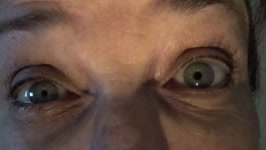
Pulsatile Proptosis from Sphenoid Wing Hypoplasia in Neurofibromatosis Type 1 | Clinical and radiologic features of greater wing sphenoid hypoplasia in the setting of neurofibromatosis type 1. Figure 1 : slit lamp examination showing Lisch nodules; Figure 2 : orbit CT scan (1); Figure 3 : orbit CT scan (2) with annotations. For visual examples of this disorder, please see the... | Samuel Bidot, MD; Amit M. Saindane, MD; Valérie Biousse, MD |
| 72 |
 |
Pulsatile Proptosis Profile | See the PowerPoint description for Pulsatile Proptosis From Sphenoid Wing Hypoplasia in Neurofibromatosis Type 1 at: http://content.lib.utah.edu/cdm/ref/collection/ehsl-eec/id/64 See also Pulsatile Proptosis Full Face video: http://content.lib.utah.edu/cdm/ref/collection/ehsl-eec/id/14 | Valérie Biousse, MD |
| 73 |
 |
Radiologic Appearance of Unilateral Sphenoid Wing Hypoplasia in Neurofibromatosis Type I | MRI features of greater wing sphenoid hypoplasia in the setting of neurofibromatosis type 1. - Figure 1 : Orbital MRI with contrast showing right greater sphenoid wing hypoplasia. The lack of bone tissue leads to herniation of the right temporal lobe into the orbit, pushing forward the orbital conte... | Samuel Bidot, MD; Amit M. Saindane, MD; Valérie Biousse, MD |
| 74 |
 |
Rathke's Cleft Cyst Apoplexy with Junctional Scotoma | MRI features of Rathke's cleft cyst apoplexy. - Figure 1 : Humphrey visual fields at initial presentation - Figure 2 : Brain MRI without contrast at initial presentation - Figure 3 : Brain MRI with contrast at initial presentation - Figure 4 : Postoperative Humphrey visual fields | Samuel Bidot, MD; Amit M. Saindane, MD; Valérie Biousse, MD |
| 75 |
 |
Retrograde Trans-Synaptic Degeneration from a Longstanding Occipital Lobe Tumor | This is an illustrated guide that (1) discusses the localization of paracentral homonymous hemianopic scotomatous visual field defects and (2) discusses the concept of trans-synaptic retrograde degeneration. A 43-year-old woman was assessed for longstanding blurred vision in both eyes. Her examinati... | Rahul A. Sharma, MD, MPH; Valérie Biousse, MD |
| 76 |
 |
Right Lateral Mediullary Syndrome (Wallenberg Syndrome) With Lateropulsion and Ocular Tilt Reaction | A 55-year old man presented with acute onset right-sided facial numbness, left-sided body numbness, vertigo, right ptosis, and binocular vertical diplopia. External examination showed right ptosis and miosis indicating a right Horner syndrome (Figure 1). He had gaze-evoked nystagmus only on right g... | Jonathan A. Micieli, MD; Valérie Biousse, MD |
| 77 |
 |
Right Occipital Arteriovenous Malformation presenting as a Migraineous Visual Aura | Migraine with visual aura is a distinct entity from a migraineous visual aura. A migraine with visual aura is characterized by a visual aura that does not prefer either visual field and accompanies a headache. The mechanism of the visual aura is via cortical spreading depression. A migraineous visua... | Nithya Shanmugam; Fernando Labella; Valerie Biousse |
| 78 |
 |
Second Order Horner Syndrome Revealing Metastatic Squamous Cell Carcinoma | Horner Syndrome is secondary to a lesion of the ipsilateral sympathetic pathway and is associated with ptosis, miosis, and anhidrosis. Here, we present a unique presentation of metastatic squamous cell carcinoma in a patient with a right-sided Horner Syndrome (second order). We also highlight the di... | Nithya Shanmugam; Michael Dattilo; Valerie Biousse |
| 79 |
 |
Second Order Horner Syndrome Revealing Metastatic Squamous Cell Carcinoma (video) | Horner Syndrome is secondary to a lesion of the ipsilateral sympathetic pathway and is associated with ptosis, miosis, and anhidrosis. Here, we present a unique presentation of metastatic squamous cell carcinoma in a patient with a right-sided Horner Syndrome (second order). We also highlight the di... | Nithya Shanmugam; Michael Dattilo; Valerie Biousse |
| 80 |
 |
Sellar Aneurysm with Chiasmal Compression | This is a case of aneurysm of the internal carotid artery, invading the sella and complicated by chiasmal compression and bitemporal hemianopia. Figure 1 : Humphrey visual fields (gray scale and pattern deviations) Figure 2a : T1-weighted axial brain MRI (1): well defined circular intracerebral mass... | Rabih Hage, MD; Valérie Biousse, MD |
| 81 |
 |
Sequential Non-Arteritic Anterior Ischemic Optic Neuropathy (NAION) | A 68-year old woman with hypertension, obstructive sleep apnea and obesity was seen in neuro-ophthalmology consultation for vision loss in the right eye. She had right optic disc edema with a small optic disc hemorrhage a small, crowded optic disc in the left eye known as a "disc-at-risk" (Figure 1)... | Jonathan A. Micieli, MD; Valérie Biousse, MD |
| 82 |
 |
Sturge-Weber Syndrome | A case of Sturge-Weber syndrome (Encephalotrigeminal angiomatosis) with angiomas that involve the leptomeninges, and the skin of the ipsilateral hemiface, associated with congenital glaucoma in the same eye. Various illustrations are included to demonstrate the port wine stain, enlarged optic nerve ... | Supharat Jariyakosol, MD; Valérie Biousse, MD |
| 83 |
 |
Superior Segmental Optic Nerve Hypoplasia | This is a case of superior segmental optic nerve hypoplasia in a woman with a history of maternal diabetes. A 25 year-old woman noticed a visual field defect in her right eye. Her examination showed: visual acuity: 20/20 OD, 20/20 OS; pupils: trace relative afferent pupillary defect OD; color visi... | Naa Naamuah M. Tagoe, MBChB, FWACS, FGCS; Rahul A. Sharma, MD, MPH; Valérie Biousse, MD; Nancy J. Newman, MD |
| 84 |
 |
Terson Syndrome and Subarachnoid Hemorrhage | A case of Terson syndrome resulting with subarachnoid hemorrhage and right vitreous hemorrhage resulting from a left pericallosal artery aneurysm. Figure 1 : External photograph of right eye demonstrates blunted red reflex secondary to vitreous hemorrhage Figure 2 : External photograph of left eye d... | Joshua Levinson, MD; Valérie Biousse, MD |
| 85 |
 |
Terson Syndrome With Cranial Nerve 3 Palsy Due to Subarachnoid Hemorrhage from Arteriovenous Malformation and Aneurysmal Rupture | A case of Terson syndrome due to AVM and posteral cerebral aneurysm. The patient developed a left CN3 palsy due to hematoma involving the left midbrain. Figure 1 : External photograph of right eye demonstrates blunted red reflex secondary to vitreous hemorrhage Figure 2 : External photograph of lef... | Joshua Levinson, MD; Valérie Biousse, MD |
| 86 |
 |
Toxic Retinopathy: Deferoxamine Toxicity | Number of Figures and legend for each: 6 figures Figure 1: Goldmann perimetry showing large cecocentral scotomas in both eyes Figure 2: Fundus photograph of the right eye demonstrating hypopigmentation of the peripapillary and perifoveal retinal pigment epithelium (RPE) with subfoveal yellow lesions... | Will Pearce, MD; Valérie Biousse, MD |
| 87 |
 |
Toxoplasmic Chorioretinitis with Unilateral Disc Edema | A 53-year-old man had a history of high myopia and a seronegative spondyloarthropathy treated with immunosuppressive agents. He presented with mild, painless vision loss in his right eye. His examination showed findings of a right anterior optic neuropathy: visual acuity: 20/20 OD (right eye), 20/20... | Rahul A. Sharma, MD, MPH; Nancy J. Newman, MD; Valérie Biousse, MD |
| 88 |
 |
Typical Idiopathic Intracranial Hypertension: Optic Nerve Appearance and Brain MRI Findings | A 24-year old African American woman was referred for bilateral optic disc edema that was incidentally noted on a routine eye examination. She had excellent visual function and dilated examination showed bilateral optic disc edema with peripapillary wrinkles in the right eye and pseudodrusen in the ... | Jonathan A. Micieli, MD; Valérie Biousse, MD |
| 89 |
 |
Typical Idiopathic Optic Neuritis | This is a case of a typical optic neuritis in a 41-year-old woman presenting with vision loss and pain with eye movements in the right eye. Optic disc photos at presentation showed subtle hyperemia in the right eye (Figure 1) and optical coherence tomography (OCT) of the retinal nerve fiber layer (R... | Jonathan A. Micieli, MD; Valérie Biousse, MD |
| 90 |
|
Vertical Diplopia Secondary to Skew Deviation With Ocular Tilt Reaction With Multiple Posterior Fossa Metastases | This is a case of multiple brain metastases in the posterior fossa resulting in a skew deviation. Figure 1 : Photograph of the patient demonstrating a spontaneous right head tilt. The patient's head is tilted toward his right shoulder to suppress his diplopia Figure 2 : Ocular movements : There is a... | Rabih Hage, MD; Valérie Biousse, MD; Jason Peragallo, MD |
| 91 |
 |
Vitreomacular Traction Syndrome (VMTS) | Vitreomacular traction (VMT) syndrome is characterized by an abnormal vitreous adherence to the macula due to incomplete posterior vitreous detachment (PVD). The resulting traction produces morphological changes of the macula. Patients may complain of decreased visual acuity, photopsia, micropsia, o... | Kirstyn Taylor; Sachin Kedar, MD |
| 92 |
 |
Vitreopapillary Traction | A 64-year-old woman was referred for bilateral optic disc edema. Examination of her optic nerves showed indistinct margins at the nasal aspect of both eyes (Figure 1). Humphrey 24-2 SITA-Fast visual fields showed non-specific depressed points in both eyes (Figure 2). Optical coherence tomography (... | Jonathan A. Micieli, MD; Valérie Biousse, MD |
| 93 |
 |
Vitreopapillary Traction Syndrome (VPT) | Vitreopapillary traction (VPT) is characterized by abnormal vitreous adherence to the optic disc due to a fibrocellular membrane or incomplete posterior vitreous detachment (PVD). Traction on the optic disc can result in elevation of the disc, blurred margins, and rarely peripapillary hemorrhage. Pa... | Kirstyn Taylor; Sachin Kedar, MD |
1 - 200 of 93