The Health Education Assets Library (HEAL) is a collection of over 22,000 freely available digital materials for health sciences education. The collection is now housed at the University of Utah J. Willard Marriott Digital Library.
TO
1 - 25 of 3
| Title | Description | Subject | Collection | ||
|---|---|---|---|---|---|
| 1 |
 |
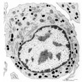
Neutrophilic myelocyte | Scheme electron microscopy. From CFU-S (colony forming units-spleen) stem cells arise CFU-GM (colony forming unit-granulocyte/monocyte) stem cells. The latter divide by mitoses and differentiate via promyeloblasts and myeloblasts into myelocytes (the last proliferative stage). The neutrophilic myelo... | Blood; Bone Marrow; Electron microscopy; Neutrophilic myelocyte; Neutrophilic granulocyte; Primary granule; Secondary granule; Azurophilic granule; Lysosome | Poja Histology Collection - Blood & Bone Marrow Subset |
| 2 |
 |
Pro-megakaryocyte | Scheme electron microscopy. The pro-megakaryocyte (20-80 m) develops from a basophilic megakaryoblast (up to 50 m) that originates from a committed stem cell. The large pro-megakaryocyte starts with the early synthesis of the characteristic granules of platelets. Apart from a bilobed nucleus (1) and... | Blood; Bone Marrow; Electron microscopy; Pro-megakaryocyte; Megakaryocyte | Poja Histology Collection - Blood & Bone Marrow Subset |
| 3 |
 |
Myeloblast in bone marrow smear (human) | Stain: May-Grnwald-Giemsa (MGG). The myeloblast (1) shows a very transparent nucleus and several distinct nucleoli. The slightly basophilic cytoplasm is limited to a small rim in contrast to a promyelocyte. No granules are yet visible. (2) small lymphocyte. | Blood; Bone Marrow; Myeloblast; Lymphocyte | Poja Histology Collection - Blood & Bone Marrow Subset |
1 - 25 of 3
