The Health Education Assets Library (HEAL) is a collection of over 22,000 freely available digital materials for health sciences education. The collection is now housed at the University of Utah J. Willard Marriott Digital Library.
TO
Filters: Collection: "ehsl_heal" Subject: "Eye"
1 - 25 of 16
| Title | Description | Subject | Collection | ||
|---|---|---|---|---|---|
| 1 |
 |
Fovea macula and optic disc | Fovea macula and optic disc. Photograph. Multimedia. | Optic Disk; Sense Organs; Eye; Anatomy | Slice of Life |
| 2 |
 |
Iris muscular layer | Iris muscular layer. Labeled. Photograph. Multimedia. | Iris; Eye; Sense Organs; Anatomy | Slice of Life |
| 3 |
 |
Optic disc, fundus, normal | Optic disc, fundus, normal. Photograph. Multimedia. | Eye; Optic Disk; Fundus Oculi; Sense Organs; Anatomy | Slice of Life |
| 4 |
 |
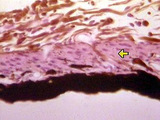
Eye | The iris extends forward from its junction with the ciliary body. The double-layered epithelium of the ciliary body continues onto the posterior surface of the iris. Iris color is determined by the number of melanocytes in the stroma. Note the iris root and the iris epithelium. UCLA Histology Collec... | Eye; iris | UCLA Histology |
| 5 |
 |
Central Nervous System | Although the optic nerve (CN II) appears grossly as a peripheral nerve, it is actually a white matter tract of the CNS. It is surrounded by meninges: dura mater, arachnoid mater, and the pia mater. There is a subarachnoid space filled with CSF. A distinguishing feature of the optic nerve is its cent... | arachnoid mater; Brain; Central Nervous System; dura mater; Eye; meninges; Optic Nerve CN II; pia mater | UCLA Histology |
| 6 |
 |
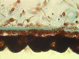
Eye | The corneal epithelium is multi-layered and its continuously proliferating cells change their shape from columnar to squamous as they are displaced from a thick, specialized extracellular matrix called Bowman's membrane. The stromal cells are called keratocytes and they synthesize and organize the h... | cornea; Eye | UCLA Histology |
| 7 |
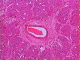 |
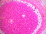
Central Nervous System | Higher power magnification of the optic nerve at the level of the central artery where the nerve is composed of axonic bundles. Note the typical appearance of an artery with endothelial cells and a layer of smooth muscle. A layer of connective tissue surrounds the artery and radiates trabeculae betw... | Brain; Central Nervous System; Eye; optic nerve | UCLA Histology |
| 8 |
 |
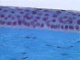
Epithelium | Note that the epithelium is simple in simple squamous epithelium because there is only one layer of cells. UCLA Histology Collection. | cornea; epithelium; Eye; simple squamous epithelium | UCLA Histology |
| 9 |
 |
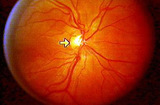
Eye | Optic nerve showing the optic disc (blind spot with no retina) and the central artery. UCLA Histology Collection. | Eye; optic nerve; test | UCLA Histology |
| 10 |
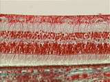 |
Eye | This slide provides a good view of the Mller glial cell processes. Their end feet rest on the inner limiting membrane of the retina and their microvilli extend external to the outer limiting membrane of the retina. UCLA Histology Collection. | Eye; retina | UCLA Histology |
| 11 |
 |
Eye | Further enlargement reveals the anterior lens capsule, area of proliferation (mitotic figures) and area of elongation. The developing ciliary epithelium (double layered) is seen in the top right corner. UCLA Histology Collection. | Eye; lens | UCLA Histology |
| 12 |
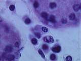 |
Connective Tissue | Plasma cells can also be found in the lacrimal gland of the eye. Note the classic clock face nucleus produced by clumps of heterochromatin. UCLA Histology Collection. | Connective Tissue; Eye; lacrimal gland | UCLA Histology |
| 13 |
 |
Eye | The posterior layer of the iris epithelium is heavily pigmented and the anterior layer has reduced pigment when compared to the epithelium of the ciliary body. The anterior portion of each anterior layer cell is modified to collectively form the dilator muscle of the pupil (under sympathetic innerva... | Eye; iris | UCLA Histology |
| 14 |
|
Connective Tissue | At a higher power of the corneal stroma, the flattened fibroblast nuclei and collagen bundles are easily seen. UCLA Histology Collection. | Connective Tissue; cornea; Eye | UCLA Histology |
| 15 |
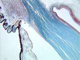 |
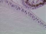
Eye | The limbus is the corneal-scleral junction. Present in this region is the conjunctival fornix, where the corneal epithelium is reflected onto the inner surface of the eyelid. Major anatomical features include the trabecular meshwork and Schlemm's canal, a major drainage site for aqueous humor from t... | Eye; limbus | UCLA Histology |
| 16 |
|
Connective Tissue | Plasma cells can also be found in the lacrimal gland of the eye. Note the classic clock face nucleus produced by clumps of heterochromatin. UCLA Histology Collection. | Connective Tissue; Eye; lacrimal gland | UCLA Histology |
1 - 25 of 16