Home
Browse
Ask Us
Chat
Harmful Language Statement
Log in
NOVEL - Neil R. Miller Collection
Advanced Search
About
The Neil R. Miller Collection covers a broad range of neuro-ophthalmologic conditions and syndromes in videos, images, and lecture presentations.
Year
1996
1997
1998
1999
2000
2001
2002
2003
2004
2005
2006
2007
2008
2009
2010
2011
2012
2013
2014
2015
2016
2017
2018
2019
2020
2021
2022
2023
2024
TO
1996
1997
1998
1999
2000
2001
2002
2003
2004
2005
2006
2007
2008
2009
2010
2011
2012
2013
2014
2015
2016
2017
2018
2019
2020
2021
2022
2023
2024
Type
Image
601
Image/MovingImage
135
Text
1
Format
image/jpeg
642
video/mp4
135
application/pdf
8
Collection
NOVEL - Neil R. Miller Collection
785
Filters:
Collection:
"ehsl_novel_nrm"
576
-
600
of
785
<
19
20
21
22
23
24
25
26
27
28
>
Gallery view
Number of results to display per page
10
25
50
100
200
Sort by Relevance
Sort by Title A-Z
Sort by Title Z-A
Sort by Date Ascending
Sort by Date Descending
Sort by Last Modified Ascending
Sort by Last Modified Descending
Title
Date
Type
576
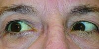
Left Relative Afferent Pupillary Defect in a Patient With Completely Normal Visual Acuity and Visual Field but Who Had a Contralateral Fourth Nerve Palsy Due to a Glioma Involving the Dorsal Midbrain
2024-06
Image/MovingImage
577
Left-sided Intracavernous Aneurysm
2024-07-10
Image
578
Macrosaccadic Oscillations in a Patient With a Cerebellar Mass
2024-06
Image/MovingImage
579
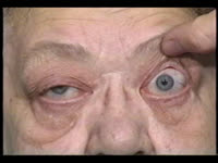
Magnified Fundus Photo of an Optic Disc With No Central Cup (Disc at Risk)
2005-10-24
Image
580
Marked Compression and Downward Displacement of the Chiasm by an Enlarged Third Ventricle in a Patient with Hydrocephalus
2024-07
Image
581
Marked Dilation of the Left Superior Ophthalmic Vein in a Patient with a High-flow Carotid-cavernous Sinus Fistula
Image
582
Marked Left Eye Proptosis, Chemosis, and Arterialization of Conjunctival and Episcleral Vessels in a Patient with a Direct (Traumatic) Carotid-cavernous Sinus Fistula
2024-07-10
Image
583
Miller Fisher Syndrome
2024-05
Image/MovingImage
584
Minimal Residual Right Sixth Nerve Palsy with Resolution of Right Proptosis, Chemosis, and Redness of the Eye After Successful Occlusion of an Ipsilateral Carotid-cavernous Sinus Fistula Using a Transcutaneous Inferior Ophthalmic Vein Approach
Image
585
Monocular Downbeat Nystagmus and Internuclear Ophthalmoplegia
2024-05
Image/MovingImage
586
Monocular Saccadic Pulses
2024-05
Image/MovingImage
587
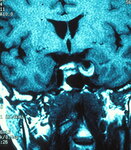
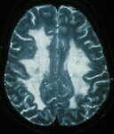
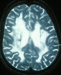
MRI Showing Extensive White Matter Disease in a Patient with CADASIL
2024-07-10
Image
588
MRI Showing Extensive White Matter Disease in a Patient with CADASIL
2024-07-10
Image
589
MRI T1, Non-contrast Sagittal Image of Large Parietal-occipital Cavernoma
2024-07-10
Image
590
MRI, Axial T2 Image, Showing Large Left Parietal-occipital Cavernoma
2024-07-10
Image
591
MRI, T1 Axial, Following Gadolinium Administration, Showing Large, Left Parietal-occipital Cavernoma
2024-07-10
Image
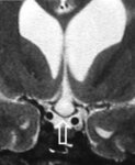
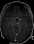
592
MRI, T1 Image After Contrast Injection, Shows a Lesion in the Midbrain that Caused a Benedikt Syndrome
2024-07-10
Image
593
MRI, T2 Axial Image, Showing Midbrain Lesion Causing a Benedikt Syndrome
2024-07-10
Image
594
Multiple Cerebellar Cavernomas in a Patient with Familial Cavernomas who had a Hemorrhage from a Chiasmal Angioma (Chiasmal Apoplexy)
2024-06-28
Image
595
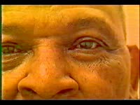
Myasthenia Gravis (Cogan Lid Twitch)
2024-05
Image/MovingImage
596
Myasthenia Gravis (Cogan Lid Twitch)
2024-05
Image/MovingImage
597
Myasthenia Gravis (Enhancement of Ptosis)
2024-05
Image/MovingImage
598
Neonatal Benign Upgaze Paresis
2024-05
Image/MovingImage
599
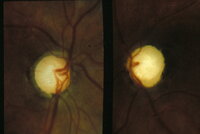
Non-glaucomatous Optic Disc Cupping (Bilateral Compressive Optic Neuropathy)
Image
600
Non-glaucomatous Optic Disc Cupping (Bilateral Compressive Optic Neuropathy)
Image
576
-
600
of
785
<
19
20
21
22
23
24
25
26
27
28
>