The Health Education Assets Library (HEAL) is a collection of over 22,000 freely available digital materials for health sciences education. The collection is now housed at the University of Utah J. Willard Marriott Digital Library.
Filters: Collection: "ehsl_heal" Subject: "PAS"
1 - 25 of 6
| Title | Description | Subject | Collection | ||
|---|---|---|---|---|---|
| 1 |
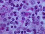 |
Thymus - Epithelial Reticular Cells | This image from the medulla shows many epithelial reticular cells, which have a pale cytoplasm, large nucleus, and prominent nucleolus. These cells form a meshwork which contains developing lymphocytes. One macrophage or PAS cell is visible. PAS stains its phagocytic inclusions bright pink. Note the... | epithelial reticular; Hassall's corpuscle; PAS | UCLA Histology |
| 2 |
 |
Thymus - Medulla | In this image of the medulla, identify lymphocytes, PAS cells, and an epithelial reticular cell. UCLA Histology Collection. | medulla; PAS | UCLA Histology |
| 3 |
 |
Thyroid | A low magnification of the thyroid gland stained with PAS. The fact that the colloid is PAS+ indicates that the colloid (thyroglobulin) is a glycoprotein. UCLA Histology Collection. | PAS; Thyroid | UCLA Histology |
| 4 |
|
Thymus - Epithelial Reticular Cells | This image from the medulla shows many epithelial reticular cells, which have a pale cytoplasm, large nucleus, and prominent nucleolus. These cells form a meshwork which contains developing lymphocytes. One macrophage or PAS cell is visible. PAS stains its phagocytic inclusions bright pink. Note the... | epithelial reticular; Hassall's corpuscle; PAS | UCLA Histology |
| 5 |
 |
Cells / Organelles - Hepatocytes | PAS stains cytoplasmic glycogen granules pink. Identify the nucleus and the nucleolus of a hepatocyte. UCLA Histology Collection. | PAS | UCLA Histology |
| 6 |
 |
Thyroid | A higher magnification of the PAS stained thyroid gland. Again, the follicles are filled with PAS positive colloid or thyroglobulin, a glycoprotein which is the stored form of thyroid hormone. Shrinkage artifacts are present within the colloid but not within the epithelial cells. The arrow indicates... | PAS; Thyroid | UCLA Histology |
1 - 25 of 6