The Health Education Assets Library (HEAL) is a collection of over 22,000 freely available digital materials for health sciences education. The collection is now housed at the University of Utah J. Willard Marriott Digital Library.
TO
Filters: Collection: "ehsl_heal" Subject: "Liver"
1 - 25 of 16
| Title | Description | Subject | Collection | ||
|---|---|---|---|---|---|
| 1 |
 |
Gallbladder, normal, unfixed, with liver | Gallbladder, normal, unfixed, with liver. Photograph. Multimedia. | Gallbladder; Liver; Gastrointestinal system; Anatomy | Slice of Life |
| 2 |
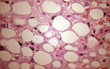 |
Fatty liver | Fatty liver | Gastrointestinal System; Liver | Slice of Life |
| 3 |
 |
Liver, normal, unfixed, overview of organ, serosal surface | Liver, normal, unfixed, overview of organ, serosal surface. Photograph. Multimedia. | Liver; Gastrointestinal system; Anatomy | Slice of Life |
| 4 |
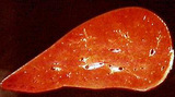 |
Liver unfixed, normal, cut surface | Liver unfixed, normal, cut surface. Photograph. Multimedia. | Liver; Gastrointestinal System; Anatomy | Slice of Life |
| 5 |
 |
Liver unfixed, normal, cut surface | Liver unfixed, normal, cut surface. Photograph. Multimedia. | Liver; Gastrointestinal System; Anatomy | Slice of Life |
| 6 |
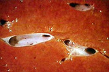 |
Liver unfixed, normal, cut surface | Liver unfixed, normal. Closeup cut surface with veins. Photograph. Multimedia. | Liver; Veins; Gastrointestinal System; Anatomy | Slice of Life |
| 7 |
 |
Glands of GI Tract | Higher magnification image of liver stained with PAS showing different levels of glycogen storage in hepatocytes. Some hepatocytes have numerous glycogen granules, whereas there are other hepatocytes without glycogen granules, indicating depletion of stored glycogen. UCLA Histology Collection. | glycogen; Liver; PAS stain; gastrointestinal tract | UCLA Histology |
| 8 |
 |
Glands of GI Tract | This images illustrates a section of the liver stained with PAS to visualize the glycogen storage in hepatocytes. Hepatocytes have granules in their cytoplasm due to the presence of stored glycogen. Shown here are also sinusoids and a central vein. UCLA Histology Collection. | gastrointestinal tract; glycogen; Liver; pas stain | UCLA Histology |
| 9 |
 |
Glands of GI Tract | Higher magnification image showing the portal triad with a biliary duct, a branch of hepatic artery and a branch of portal vein embedded within connective tissue, and the hepatocytes. UCLA Histology Collection. | Gastrointestinal tract; Liver; portal triad | UCLA Histology |
| 10 |
 |
Glands of GI Tract | This image shows Kupffer cells, which are macrophages that are distinguished by their phagocytation of a particulate dye. Note the hepatocyte cords and the sinusoids which separate them. UCLA Histology Collection. | Kupffer cells; Liver | UCLA Histology |
| 11 |
 |
Glands of GI Tract | In this image you can see a portal triad containing a branch of the portal vein, a small hepatic artery, and a biliary duct embedded in collagenous connective tissue (stained blue by trichrome). Also shown are dark red hepatocytes and pinkish sinusoids. UCLA Histology Collection. | gastrointestinal tract; Liver; trichrome | UCLA Histology |
| 12 |
 |
Glands of GI Tract | Another area of the liver showing a central vein surrounded by hepatocytes organized into cords which are separated by sinusoids. Nutrient-rich blood from the hepatic portal vein percolates through the sinusoids, where the hepatocytes regulate the nutrient content. The sinusoids drain into central v... | gastrointestinal tract; Hepatocyte; Liver | UCLA Histology |
| 13 |
 |
Connective Tissue | The lining of the gallbladder is a tall simple columnar epithelium. The underlying loose connective tissue is characterized by a large number of cells relative to collagen fibers. Look for plasma cells here. UCLA Histology Collection. | Connective Tissue; Gall bladder; Liver | UCLA Histology |
| 14 |
 |
Circulatory System | The lumen is on the far right, with no evident endothelium. The tunica intima and tunica media seem to blend together; the two are relatively small compared with more extensive tunica adventitia. Note the vasa vasorum and blue stained collagen fibers that can be identified within the tunica adventit... | Circulatory System; Hepatic cords; hepatic vein; Liver; tunica adventitia; tunica intima; tunica media; vasa vasorum | UCLA Histology |
| 15 |
 |
Glands of GI Tract | This low power view of the liver shows the mass of hepatocytes, a central vein and a portal triad composed of a biliary duct, a branch of hepatic artery and a branch of portal vein embedded within connective tissue. UCLA Histology Collection. | gastrointestinal tract; hepatic triad; Liver | UCLA Histology |
| 16 |
 |
Glands of GI Tract | The staining used in this preparation reveals reticular fibers supporting the hepatocytes which line the sinusoids. Bile canaliculi also bind the dye used here, so they appear black. UCLA Histology Collection. | Gastrointestinal tract; Hepatocyte; Liver; reticular fibers | UCLA Histology |
1 - 25 of 16