Home
Browse
Ask Us
Chat
Harmful Language Statement
Log in
NOVEL - Neil R. Miller Collection
Advanced Search
About
The Neil R. Miller Collection covers a broad range of neuro-ophthalmologic conditions and syndromes in videos, images, and lecture presentations.
Year
2024
TO
2024
Type
Image
87
Format
image/jpeg
86
application/pdf
1
Collection
NOVEL - Neil R. Miller Collection
87
Filters:
Collection:
"ehsl_novel_nrm"
Subject:
"Brain"
1
-
25
of
87
<
1
2
3
4
>
Gallery view
Number of results to display per page
10
25
50
100
200
Sort by Relevance
Sort by Title A-Z
Sort by Title Z-A
Sort by Date Ascending
Sort by Date Descending
Sort by Last Modified Ascending
Sort by Last Modified Descending
Title
Date
Type
1
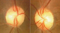
Bilateral Band Atrophy of Optic Nerves from Optic Chiasmal Compression
2024-07
Image
2
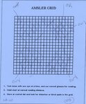
Bilateral Homonymous Hemianopia with Bilateral Macular Sparing in a Patient with Bilateral Cortical Infarction (Amsler Grid)
2024-07
Image
3
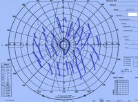
Bilateral Macular Sparing in a Patient with Bilateral Homonymous Hemianopias Due to Bilateral Occipital Lobe Infarcts (Kinetic Perimetry)
2024-07
Image
4
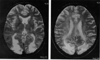
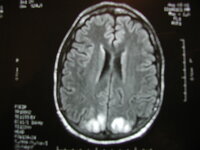
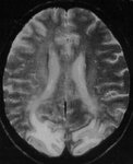
Bilateral Occipital Infarcts Causing Cortical Blindness
2024-07
Image
5
Bilateral Occipital Infarcts Causing Cortical Blindness
2024-07
Image
6
Bilateral Occipital Infarcts Causing Cortical Blindness
2024-07
Image
7
Bilateral Occipital Infarcts Causing Cortical Blindness
2024-07
Image
8
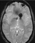
Cavernoma in Region of Chiasm (SWI MRI)
2024-07
Image
9
Cavernoma of the Optic Chiasm Causing Chiasmal Apoplexy
2024-07
Image
10
Cavernoma of the Optic Chiasm Causing Chiasmal Apoplexy
2024-07
Image
11
Cavernoma of the Optic Chiasm Causing Chiasmal Apoplexy
2024-07
Image
12
Cavernoma of the Optic Chiasm Causing Chiasmal Apoplexy
2024-07
Image
13
Cavernoma of the Optic Chiasm Causing Chiasmal Apoplexy
2024-07
Image
14
Cavernoma of the Optic Chiasm Causing Chiasmal Apoplexy
2024-07
Image
15
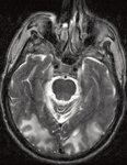
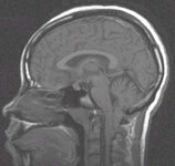
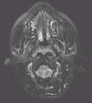
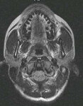
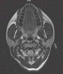
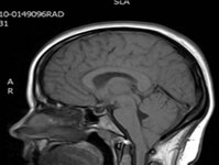
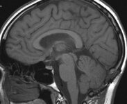
Chiari Malformation - 01
2024-07
Image
16
Chiari Malformation - 02
2024-07
Image
17
Chiari Malformation - 03
2024-07
Image
18
Chiari Malformation - 04
2024-07
Image
19
Chiari Malformation - 05
2024-07
Image
20
Chiari Malformation - 06
2024-07
Image
21
Chiari Malformation - a
2024-07
Image
22
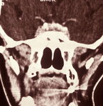
Chiasm Anatomy - 001
2024-07
Image
23
Chiasm Anatomy - Birdbeak
2024-07
Image
24
Chiasm Anatomy - Chiasmal Adenohypophysis - 1
2024-07
Image
25
Chiasm Anatomy - Chiasmal Adenohypophysis - 2
2024-07
Image
1
-
25
of
87
<
1
2
3
4
>