The Health Education Assets Library (HEAL) is a collection of over 22,000 freely available digital materials for health sciences education. The collection is now housed at the University of Utah J. Willard Marriott Digital Library.
TO
Filters: Collection: "ehsl_heal" Subject: "Lung"
1 - 25 of 12
| Title | Description | Subject | Collection | ||
|---|---|---|---|---|---|
| 1 |
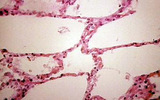 |
Lung, normal | Lung, normal | Respiratory System; Lung | Slice of Life |
| 2 |
 |
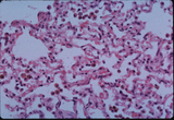
Hyaline membrane formation, lung | Hyaline membrane formation, lung | Respiratory System; Lung | Slice of Life |
| 3 |
 |
Emboli, lima bean, lungs | Emboli, lima bean, lungs | Respiratory System; Lung; Embolism | Slice of Life |
| 4 |
 |
Respiratory System | Bronchus-associated lymphoid tissue BALT consists of submucosal lymphoid tissue in intermediate and small airways. They are part of the diffuse mucosa-associated lymphoid tissue (MALT) also found in many other tissues. These structures are normal, but similar collections in lung alveolar tissue are ... | Lung; lymphoid tissue; Respiratory System | UCLA Histology |
| 5 |
 |
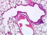
Respiratory System | In this image, a respiratory bronchiole (with intermittent alveoli) branches into alveolar ducts (walled by continuous alveoli). Alveolar sacs and alveoli can also be seen. UCLA Histology Collection. | Lung; respiratory bronchiole; Respiratory System | UCLA Histology |
| 6 |
 |
Respiratory System | In this small bronchus, the respiratory epithelium is less tall, with a thick basal lamina. The submucosa contains connective tissue, smooth muscle, and the mixed glands made up of serous and mucous components. UCLA Histology Collection. | epithelium; Lung; respiratory epithelium; Respiratory System | UCLA Histology |
| 7 |
 |
Respiratory System | Observe the narrowing of this small bronchus (with cartilage plates) into a bronchiole, terminal bronchiole, then respiratory bronchiole (with intermittent alveoli). A branch of the pulmonary artery is also visible. UCLA Histology Collection. | Lung; Respiratory System | UCLA Histology |
| 8 |
 |
Respiratory System | A macrophage rests on the alveolar wall. It is difficult to differentiate between type I pneumocytes and endothelial cells lining the alveolar capillaries. The type II pneumocytes, which produce surfactant, have large round nuclei with prominent nucleoli. UCLA Histology Collection. | Lung; pneumocytes; Respiratory System | UCLA Histology |
| 9 |
 |
Respiratory System | Pulmonary arteries are unusually thin-walled for an artery and usually travel with a bronchus. Here these conduits can be seen surrounded by pulmonary alveolar components. UCLA Histology Collection. | Lung; Pulmonary artery; respiratory system | UCLA Histology |
| 10 |
 |
Respiratory System | This high power view shows the short ciliated cells and goblet cells of a small bronchus. Remember that the epithelium and basal lamina together constitute the mucosa. In the submucosa, find fibroblasts and the cigar-shaped nuclei of smooth muscle cells. UCLA Histology Collection. | bronchus; Lung | UCLA Histology |
| 11 |
 |
Respiratory System | A small number of alveolar macrophages is found in alveolar spaces of normal healthy lungs. They are part of the lung's defense mechanism. UCLA Histology Collection. | alveolar spaces; Lung; macrophage; Respiratory System | UCLA Histology |
| 12 |
 |
Respiratory System | Identify an alveolus and a small blood vessel. Macrophages are usually found resting on the thin alveolar wall. UCLA Histology Collection. | Lung; Respiratory System | UCLA Histology |
1 - 25 of 12