The Health Education Assets Library (HEAL) is a collection of over 22,000 freely available digital materials for health sciences education. The collection is now housed at the University of Utah J. Willard Marriott Digital Library.
Filters: Collection: "ehsl_heal" Subject: "Skin"
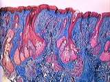
1 - 25 of 19
| Title | Description | Subject | Collection | ||
|---|---|---|---|---|---|
| 1 |
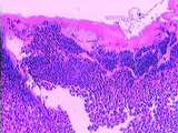 |
Skin | Healing skin (rat). See lymphocyte infiltration. UCLA Histology Collection. | Skin | UCLA Histology |
| 2 |
 |
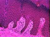
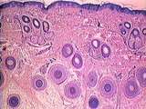
Skin | This high power view illustrates the relationship of epidermal pegs, which invaginate into the dermis, with the dermal papillae which extend into the epidermis. This sample has a highly pigmented basal layer, composed of keratinocytes with melanosomes. Melanocytes produce the pigment melanin and are... | epidermis; Skin | UCLA Histology |
| 3 |
 |
Skin | Moderate power view of the base of a hair follicle in skin. Good view of the dermal papilla and other coverings of the hair follicle bulb. UCLA Histology Collection. | Skin | UCLA Histology |
| 4 |
 |
Skin | This section of abdominal skin is a good example of thin skin. Note the thin layer of epidermis. The dermis is composed of two layers: the superficial papillary layer, and the deeper reticular layer. Deep in the dermis, hair follicles and associated sebaceous glands are present. Again note the inter... | epidermis; Meissner's corpuscles; Skin | UCLA Histology |
| 5 |
 |
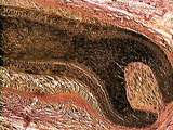
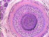
Skin | This Pacinian corpuscle was found within the hypodermis (a layer beneath the dermis). Pacinian corpuscles are sensitive to pressure and vibration. The corpuscle resembles an onion cut in x-section. The lamellar appearance is due to concentric layers of flattened endoneuronal cells. Identify the bloo... | Pacinian corpuscle; Skin | UCLA Histology |
| 6 |
 |
Skin | Longitudinal section of two hair follicles. The hair bulb is composed of the hair root and the invaginating dermal papilla. The cells in the hair root are the matrix. As the hair grows the matrix cells become differentiated into the inner medulla and outer cortex. External to the cortex is the inter... | hair follicles; Skin | UCLA Histology |
| 7 |
 |
Connective Tissue | In the dermis of the skin, adipocytes (fat cells) are also present. These cells appear empty because the organic solvents used in tissue preparation dissolve away fat. Sweat glands are also found deep within the dermis. UCLA Histology Collection. | connective tissue; Skin; Sweat glands | UCLA Histology |
| 8 |
 |
Skin | A nail is a keratinized plate located on the dorsal aspect of the distal phalanges. Above and below, the nail is continuous with the cornified layer of the epidermis. The highly keratinized edge of epidermis above the nail is called the epon or eponychium. Under the nail toward the bone is the epith... | epidermis; Nail; Skin | UCLA Histology |
| 9 |
 |
Skin | Eccrine sweat glands are not associated with hair follicles. Their ducts open independently on the surface and are composed of stratified cuboidal cells (usually 2 layers). These sweat glands are simple coiled tubular glands. The secretory portion is composed of low columnar to cuboidal cells deep i... | Eccrine sweat glands; Skin | UCLA Histology |
| 10 |
 |
Skin | Observe the basal layer of the epidermis. In addition to the keratinocytes one can observe melanocytes. Note the difference between the cells of the basal layer and the spiny layer. Note the collagen and fibroblasts of the dermis. UCLA Histology Collection. | epidermis; keratinocytes; melanocytes; Skin | UCLA Histology |
| 11 |
 |
Skin | Trichrome stained skin showing a sebaceous gland and an arrector pili muscle. UCLA Histology Collection. | Skin | UCLA Histology |
| 12 |
 |
Skin | This section of the fingertip illustrates the layers of relatively thick skin. We can easily distinguish the basal layer and spiny layer (both proliferative), the granular layer, and the layer of enucleated, dead cells, the cornified layer. Within the dermal papillae numerous capillaries can be obse... | Skin | UCLA Histology |
| 13 |
 |
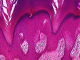
Skin | Relatively low power view of thin skin. Many cross sections of hair follicles are seen as well as some small sebaceous glands. UCLA Histology Collection. | Skin | UCLA Histology |
| 14 |
 |
Skin | The skin of the finger tip is characterized by a thick keratinized epithelium, and an abundance of Meissner's corpuscles in the dermal papillae. Meissner's corpuscle is an oval, encapsulated mechanoreceptor lying immediately under the basal layer. The nuclei in the corpuscle belong to flattened Schw... | Meissner's corpuscles; Skin | UCLA Histology |
| 15 |
 |
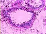
Skin | This cross section of a hair shaft is useful to delineate the various layers. The most interior cells form the medulla, then cortex, and finally a layer of flatter cells, the cuticle. The internal root sheath is composed of three layers, and is covered by the external root sheath. The basal lamina i... | hair; Skin | UCLA Histology |
| 16 |
 |
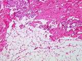
healing Skin | Healing skin. The wound has disrupted the epidermis and dermis. At the base of the wound a lymphocytic infiltration has formed. Note the hair follicles and sebaceous gland. UCLA Histology Collection. | epidermis; Skin | UCLA Histology |
| 17 |
 |
Skin | In selected areas of the body, such as the axilla, specialized sweat glands release an apocrine secretion via conventional exocytosis not via the method in which part of the apical cytoplasm is lost.Their ducts open into hair shafts. The secretory cells of these sweat glands are simple cuboidal to l... | Myoepithelial cells; Skin; sweat glands | UCLA Histology |
| 18 |
 |
Skin | This section illustrates the appearance of cells in the granular layer. They are filled with granules of basophilic keratohyaline. The cells of the overlying cornified layer lack nuclei. In thick skin, a clear layer of cells is present over the granular layer. These cells also lack nuclei and organe... | Skin | UCLA Histology |
| 19 |
 |
Skin | Another view of the association between sebaceous glands and hair follicles. Note that the collagenous connective tissue of the dermis is stained blue. Identify the epidermis and sweat glands. UCLA Histology Collection. | epidermis; hair follicles; Skin | UCLA Histology |
1 - 25 of 19