The Health Education Assets Library (HEAL) is a collection of over 22,000 freely available digital materials for health sciences education. The collection is now housed at the University of Utah J. Willard Marriott Digital Library.
1 - 25 of 10
| Title | Description | Subject | Collection | ||
|---|---|---|---|---|---|
| 1 |
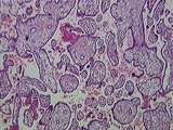 |
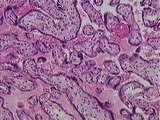
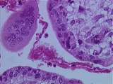
Placenta | This image presents the basic organization of the placenta. Embedded within the maternal blood space are numerous fetal villi, some of them clearly tertiary villi , all lined by the syncytiotrophoblast . The only things missing in this image are maternal decidual cells and fetal/maternal fibrinoid. ... | Placenta | UCLA Histology |
| 2 |
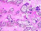 |
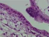
Placenta | This image presents all that you have to know about placental histology for this course. Fetal villi, some clearly tertiary villi , are completely surrounded by the maternal blood space. Lining everything in this image is the syncytiotrophoblast . The only other maternal component in this image is a... | Placenta | UCLA Histology |
| 3 |
 |
Placenta | This image is very useful for differentiating the fetal and maternal components of the placenta . One can readily identify the maternal blood space , fetal villi including one obvious tertiary villus . Fibrinoid is present in the area of fetal villi as well as in a region dominated by maternal decid... | Placenta | UCLA Histology |
| 4 |
 |
Placenta | The fetus is located in the amniotic cavity which is lined by the epithelium of the amnion and its mesenchyme . Below the amnion is the beginning of the chorion, starting first with chorionic mesenchyme which contains fetal blood vessels . Within the chorion one will find the maternal blood space an... | Placenta | UCLA Histology |
| 5 |
 |
Placenta | This image illustrates numerous fetal villi surrounded by syncytiotrophoblast and the maternal blood space part of which is prominently filled with maternal red blood cells . There also appears to be a small region of fibrinoid. UCLA Histology Collection. | Placenta | UCLA Histology |
| 6 |
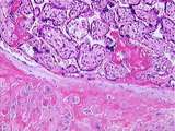 |
Placenta | The base of the placenta lies adjacent to the maternal endometrium which has decidual cells embedded in fibrinoid . Fibrinoid is also seen among the fetal villi . In this image the maternal blood space is evident. UCLA Histology Collection. | Placenta | UCLA Histology |
| 7 |
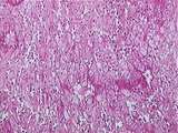 |
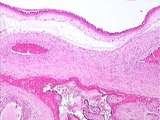
Placenta | Uterine Decidual Cells. This image of the postpartum uterus illustrates the marked change in the endometrial stromal cells as they become decidual cells. UCLA Histology Collection. | Decidual Cells; Placenta; uterus | UCLA Histology |
| 8 |
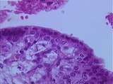 |
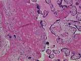
Placenta | Most of this image consists of a tertiary fetal villus. Note the fetal blood vessels and the fetal mesenchyme. The fetal villus is lined by the syncytiotrophoblast but cytotrophoblast cells are still present. The maternal blood space is obvious. UCLA Histology Collection. | Placenta | UCLA Histology |
| 9 |
 |
Placenta | This is an unusual image. Although it shows the maternal blood space and typical fetal villi , there is one villus that is composed only of syncytiotrophoblast and is therefore a possible primary villus . However, this putative primary villus could also be a tangential or grazing section of a second... | Placenta | UCLA Histology |
| 10 |
 |
Placenta | In this image recognize fetal villi lined by the syncytiotrophoblast and filled by fetal mesenchyme . At 3 months of age, cytotrophoblasts are still present as is the critical maternal blood space. UCLA Histology Collection. | Placenta | UCLA Histology |
1 - 25 of 10