The Walsh Society Annual Meeting Archives: Proceedings of the annual meeting of the Walsh Society, which is now part of the North American Neuro-Ophthalmology Association (NANOS) Annual Meeting. Contains records from the first meeting in 1969, through the present.
NOVEL: https://novel.utah.edu/
| Title | Creator | History | ||
|---|---|---|---|---|
| 1 |
 |
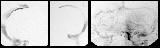
Star Light, Star Bright (9 Gaze Image) | Shannon C. Lynch, MD, University of Nebraska Medical Center | An 83-year female with ptosis and binocular oblique diplopia. |
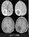
| 2 |
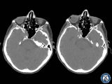 |
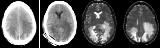
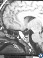
Star Light, Star Bright (Axial CT without Gadolinium) | Shannon C. Lynch, MD, University of Nebraska Medical Center | An 83-year female with ptosis and binocular oblique diplopia. |
| 3 |
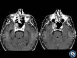 |
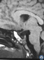
Star Light, Star Bright (Axial MRI T1 Post) | Shannon C. Lynch, MD, University of Nebraska Medical Center | An 83-year female with ptosis and binocular oblique diplopia. |
| 4 |
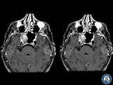 |
Star Light, Star Bright: (Axial MRI T1 Post) | Shannon C. Lynch, MD, University of Nebraska Medical Center | An 83-year female with ptosis and binocular oblique diplopia. |
| 5 |
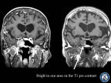 |
Star Light, Star Bright: Axial MRI T1 Pre and Post | Shannon C. Lynch, MD, University of Nebraska Medical Center | An 83-year female with ptosis and binocular oblique diplopia. |
| 6 |
 |
Star Light, Star Bright (Coronal MRI T1 Post) | Shannon C. Lynch, MD, University of Nebraska Medical Center | An 83-year female with ptosis and binocular oblique diplopia. |
| 7 |
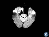 |
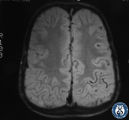
Star Light, Star Bright (Diffusion MRI) | Shannon C. Lynch, MD, University of Nebraska Medical Center | An 83-year female with ptosis and binocular oblique diplopia. |
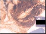
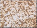
| 8 |
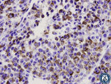 |
Star Light, Star Bright (Pathology HMB 45 600) | Shannon C. Lynch, MD, University of Nebraska Medical Center | An 83-year female with ptosis and binocular oblique diplopia. |
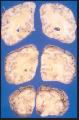
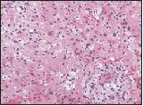
| 9 |
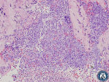 |
Star Light, Star Bright (Pathology) | Shannon C. Lynch, MD, University of Nebraska Medical Center | An 83-year female with ptosis and binocular oblique diplopia. |
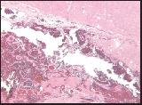
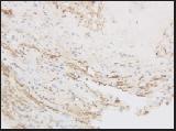
| 10 |
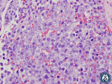 |
Star Light, Star Bright (Pathology) | Shannon C. Lynch, MD, University of Nebraska Medical Center | An 83-year female with ptosis and binocular oblique diplopia. |
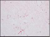
| 11 |
 |
Brain Drain Strain (Pathology) | John Kerrison, MD, Retina Consultants of Charleston | A 55-year old female with complaints of bilateral blurred vision, headaches and general malaise of a 1-year duration. Previous history significant for left occipital dural ateriovenous malformation, cerebral hemorrhage and left hemiplegia. |
| 12 |
 |
Brain Drain Strain (Pathology) | John Kerrison, MD, Retina Consultants of Charleston | A 55-year old female with complaints of bilateral blurred vision, headaches and general malaise of a 1-year duration. Previous history significant for left occipital dural arteriovenous malformation, cerebral hemorrhage and left hemiplegia. |
| 13 |
 |
Brain Drain Strain (Pathology) | John Kerrison, MD, Retina Consultants of Charleston | A 55-year old female with complaints of bilateral blurred vision, headaches and general malaise of a 1-year duration. Previous history significant for left occipital dural arteriovenous malformation, cerebral hemorrhage and left hemiplegia. |
| 14 |
 |
Brain Drain Strain (Pathology) | John Kerrison, MD, Retina Consultants of Charleston | A 55-year old female with complaints of bilateral blurred vision, headaches and general malaise of a 1-year duration. Previous history significant for left occipital dural ateriovenous malformation, cerebral hemorrhage and left hemiplegia. |
| 15 |
 |
Brain Drain Strain (Angiogram) | John Kerrison, MD, Retina Consultants of Charleston | A 55-year old female with complaints of bilateral blurred vision, headache and general malaise of a 1-year duration. Previous history significant for left occipital dural ateriovenous malformation, cerebral hemorrhage and left hemiplegia. |
| 16 |
 |
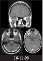
Brain Drain Strain (MRI) | John Kerrison, MD, Retina Consultants of Charleston | A 55-year old female with complaints of bilateral blurred vision, headaches and general malaise of a 1-year duration. Previoous history significant for left occipital dural arteriovenous malformation, cerebral hemorrhage and left hemiplegia. |
| 17 |
 |
Parieto-occipital Mass 1 Year After Painful Permanent Acute Optical Neuropathy (MRI) | Jeffrey G. Odel, MD, Columbia University Medical Center | A 46-year old male with painful loss of vision OS. Previous history significant for ambylopia OS. |
| 18 |
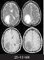 |
Parietooccipital Mass 1 Year After Painful Permanent Acute Optical Neuropathy (MRI) | Jeffrey G. Odel, MD, Columbia University Medical Center | A 46-year old male with painful loss of vision OS. Previous history significant for ambylopia OS. |
| 19 |
 |
Parietooccipital Mass 1 Year After Painful Permanent Acute Optical Neuropathy (MRI) | Jeffrey G. Odel, MD, Columbia University Medical Center | A 46-year old male with painful loss of vision OS. Previous history significant for ambylopia OS. |
| 20 |
 |
Parieto-occipital Mass 1 Year After Painful Permanent Acute Optical Neuropathy (Pathology) | Jeffrey G. Odel, MD, Columbia University Medical Center | A 46-year old male with loss of vision OS. Previous history significant for ambylopia OS. |
| 21 |
 |
Parieto-occipital Mass 1 Year After Painful Permanent Acute Optical Neuropathy (Pathology) | Jeffrey G. Odel, MD, Columbia University Medical Center | A 46-year-old with painful loss of vision OS. Previous history significant for ambylopia OS. |
| 22 |
 |
Parieto-occipital Mass 1 Year After Painful Permanent Acute Optical Neuropathy (Pathology) | Jeffrey G. Odel, MD, Columbia University Medical Center | A 46-year old male with painful loss of vision OS. Previous history significant for ambylopia OS. |
| 23 |
 |
Highly Impossible (MRI) | William A. Fletcher, MD, Departments of Clinical Neurosciences & Surgery, University of Calgary | A 4-year old male with a 1 1/2-year history of visual loss OU. Previous history significant for megacephaly. |
| 24 |
 |
Highly Impossible (MRI) | William A. Fletcher, MD, Departments of Clinical Neurosciences & Surgery, University of Calgary | A 4-year old male with a 1 1/2-year history of visual loss OU. Previous history significant for megacephaly. |
| 25 |
 |
Highly Impossible (MRI) | William A. Fletcher, MD, Departments of Clinical Neurosciences & Surgery, University of Calgary | A 4-year old male with a 1 1/2-year history of visual loss OU. Previous history significant for macrocephaly. |