John A. Moran Eye Center Neuro-Ophthalmology Collection: A variety of lectures, videos and images relating to topics in Neuro-Ophthalmology created by faculty at the Moran Eye Center, University of Utah, in Salt Lake City.
NOVEL: https://novel.utah.edu/
TO
| Title | Description | Type | ||
|---|---|---|---|---|
| 101 |
 |
Spasm of the Near Reflex | Example of patient with spasm of the near reflex and voluntary nystagmus. Discussion of similar-looking conditions (e.g. six nerve palsy, limitation of abduction, lateral rectus muscle problems) and how to tell them apart from spasm of the near reflex by observing the myosis evoked by the near respo... | Image/MovingImage |
| 102 |
 |
Spasmus Nutans | Example of patient with spasmus nutans. Discussion of characteristics of this disorder, such as dissociated or monocular nystagmus, abnormal head position, and to-and-fro head oscillation. Sometimes an eccentric gaze is seen as well (as in patient). Patient has a monocular horizontal nystagmus in th... | Image/MovingImage |
| 103 |
 |
Spasmus Nutans | Example of patient with spasmus nutans. | Image/MovingImage |
| 104 |
 |
Spontaneous Venous Pulsations | This clips shows a spontaneous venous pulsation viewed during an ocular examination. | Image/MovingImage |
| 105 |
 |
Square Wave Jerks | Example of patient with square wave jerks. Discussion of difference between square wave jerks (saccadic oscillations) and horizontal nystagmus. | Image/MovingImage |
| 106 |
 |
Stereoacuity Testing | Demonstration of examination for stereoacuity. | Text |
| 107 |
 |
Superior Oblique Myokymia | Close-up video of a patient with superior oblique myokymia (no audio.) | Image/MovingImage |
| 108 |
 |
Superior Oblique Myokymia | Example of patients with superior oblique myokymia, a saccadic intrusion. First patient is seen to have intermittent, intorting movements with superimposed slight vertical deviations in right eye. Discussion of disorder as benign, but frequently disabling, as patients experience episodes of diplopia... | Image/MovingImage |
| 109 |
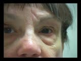 |
Test Duane | Image/MovingImage | |
| 110 |
 |
Testing the Visual Fields | Demonstration of various methods of testing visual fields, including counting fingers, motion, and color of several objects. | Image/MovingImage |
| 111 |
 |
Third Nerve Palsy | Patient with third nerve palsy (no audio) | Image/MovingImage |
| 112 |
 |
Third Nerve Palsy, Pupil Involving | Example of patient with third nerve palsy. Left eye shows pupilary involvement. Left eye doesn't immediately duct, but abducts well, with impaired superduction. Secondary and primary deviations are demonstrated. Anisocoria is more prominent when light is on, showing a parasympathetic defect to the p... | Image/MovingImage |
| 113 |
 |
Tour of the Direct Ophthalmoscope | This clip describes the parts and operation of the ophthalmoscope as an ocular examination tool. Includes adjustment of aperture size and adjustment of lenses. | Image/MovingImage |
| 114 |
 |
Tour of the Fundus | This clip demonstrates the funduscopic examination technique. | Image/MovingImage |
| 115 |
 |
Transillumination - Ciliary Body Neurofibromas | Example of transillumination on a patient with neurofibromatosis, but without Lisch nodules. Shows suspected neurofibromas in the ciliary body. | Image/MovingImage |
| 116 |
 |
Transillumination - Lisch Nodules | Demonstration of transillumination of the Lisch nodules on a patient with neurofibromatosis. Shows how Lisch nodules that were not very visible in slit-lamp examination are better seen with transillumination, which may therefore be useful in detecting Lisch nodules earlier in children where they are... | Image/MovingImage |
| 117 |
 |
Transillumination Ocular Melanoma | Video describing condition. | Image/MovingImage |
| 118 |
 |
Trigeminal Nerve Exam | Explanation of a trigeminal nerve exam. | Image/MovingImage |
| 119 |
 |
Ultrasonography Techniques | This video describes and demonstrates the various techniques for examination of the eye using ultrasonography, including A-scan, B-scan and immersion. | Image/MovingImage |
| 120 |
 |
Ultrasonography: Immersion Technique | This video describes and demonstrates the immersion technique for examination of the eye using ultrasonography. | Image/MovingImage |
| 121 |
 |
Unilateral Blepharospasm | Example of patient with unilateral blepharospasm. | Image/MovingImage |
| 122 |
 |
Upbeat Nystagmus | Example of a patient with upbeat nystagmus. Shows vertical jerk nystagmus with fast phases in the up direction. Localizes to brain stem, and occurs with strokes, demyelination, and tumors. | Image/MovingImage |
| 123 |
 |
Vestibular Nystagmus | Example of patient with vestibular nystagmus. Patient is led through instructions for direction of gaze. Shown also with Frenzel goggles. | Image/MovingImage |
| 124 |
 |
Vestibular Nystagmus | Discussion of vestibular nystagmus. Seen with peripheral disorders and central disorders, and in two varieties: spontaneous and positional. Horizontal jerk with small amplitude. | Image/MovingImage |
| 125 |
 |
Voluntary Nystagmus | Example of patient with voluntary nystagmus. Discussion of how a lack of uniform, patterned movement of the eyes along with associated lid movements suggests that activity is voluntary. | Image/MovingImage |
