| OCR Text |
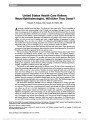
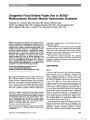
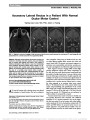
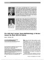
Show Assessment of Optic Nerve Head Drusen Using Enhanced Depth Imaging and Swept Source Optical Coherence Tomography Anna L. Silverman, BA, Andrew J. Tatham, FRCOphth, Felipe A. Medeiros, MD, PhD, Robert N. Weinreb, MD Background: Optic nerve head drusen (ONHD) are calcific deposits buried or at the surface of the optic disc. Although ONHD may be associated with progressive visual field defects, the mechanism of drusen-related field loss is poorly understood. Methods for detecting and imaging disc drusen include B-scan ultrasonography, fundus auto-fluorescence, and optical coherence tomography (OCT). These modalities are useful for drusen detection but are limited by low resolution or poor penetration of deep struc-tures. This review was designed to assess the potential role of new OCT technologies in imaging ONHD. Evidence Acquisition: Critical appraisal of published litera-ture and comparison of new imaging devices to established technology. Results: The new imaging modalities of enhanced depth imaging optical coherence tomography (EDI-OCT) and swept source optical coherence tomography (SS-OCT) are able to provide unprecedented in vivo detail of ONHD. Using these devices it is now possible to quantify optic disc drusen dimensions and assess integrity of neighboring retinal structures, including the retinal nerve fiber layer. Conclusions: EDI-OCT and SS-OCT have the potential to allow better detection of longitudinal changes in drusen and neural retina and improve our understanding of drusen-related visual field loss. Journal of Neuro-Ophthalmology 2014;34:198-205 doi: 10.1097/WNO.0000000000000115 © 2014 by North American Neuro-Ophthalmology Society Optic nerve head drusen (ONHD) are acellular deposits of calcium, amino and nucleic acids, and mucopoly-saccharides, buried or at the surface of the optic disc (1-3). When located near the surface, drusen can be directly visu-alized by ophthalmoscopy. Superficial drusen typically con-fer an irregular lumpy appearance to the optic disc (4). It is hypothesized that some superficial drusen become visible with age as a result of drusen growth or loss of the neural tissue that obscures the drusen. In contrast, when disc dru-sen are located closer to the lamina cribrosa, they can be difficult to detect and may require imaging for confirmatory diagnosis (5,6). Although ONHD are normally asymptom-atic, they are associated with visual field defects in 24%- 87% of affected adults (4,5,7). Wilkens et al (8) found that superficial drusen were more commonly associated with visual field defects than deep drusen. The mechanism of drusen-related visual field loss is poorly understood. ONHD typically enlarge slowly throughout life and a slow progres-sion of visual field loss is common (4,5,7). In rare cases, acute decreases in vision can occur due to vascular occlusion (9). Recent advances in ocular imaging have improved our ability to image ONHD and have provided a means to obtain objective, quantitative measurements of ONHD and neighboring structures, including the retinal nerve fiber layer (RNFL) (5,10). Better in vivo imaging has the poten-tial to improve our understanding of the pathogenesis of drusen-related visual field damage. The purpose of this review was to describe the use of 2 new optical coherence tomography (OCT) methods, enhanced depth imaging optical coherence tomography (EDI-OCT) and swept Department of Ophthalmology, Hamilton Glaucoma Center, Uni-versity of California, San Diego, California. Supported in part by the National Institutes of Health/National Eye Institute grant EY021818 (F.A.M.) and Core grant P30EY022589; unrestricted grant from Research to Prevent Blindness. F. A. Medeiros receives research support from Carl-Zeiss Meditec, Topcon, and Reichert; R. N. Weinreb receives research support from Carl-Zeiss Meditec, Optovue, Kowa, Nidek, and Topcon; A. J. Tatham, F. A. Medeiros, and R. N. Weinreb receive research support from Heidelberg Engineering; F. A. Medeiros and R. N. Weinreb are consultants to Carl-Zeiss Meditec, Inc; and R. N. Weinreb is a con-sultant to Topcon. Address correspondence to Anna L. Silverman, BA, Department of Ophthalmology, Hamilton Glaucoma Center, University of California, San Diego, 9500 Gilman Drive, La Jolla, CA 92093-0946; E-mail: anna.l.silverman@gmail.com 198 Silverman et al: J Neuro-Ophthalmol 2014; 34: 198-205 State-of-the-Art Review Section Editors: Val erie Biousse, MD Steven Galetta, MD Copyright © North American Neuro-Ophthalmology Society. Unauthorized reproduction of this article is prohibited. source optical coherence tomography (SS-OCT), and to evaluate their application in the assessment of ONHD. CURRENT UNDERSTANDING Prevalence of ONHD Clinically recognized ONHD are estimated to occur in 0.3% of the population, with both genders affected equally. However, an autopsy series found a higher prevalence of 2.4% (11,12). The discrepancy between the clinical and autopsy findings is likely due to a high prevalence of undi-agnosed drusen (4,5). ONHD are usually asymptomatic and therefore tend to present incidentally, either following routine ophthalmoscopy or following detection of an abnor-mality on visual field testing (13,14). In approximately 75% of individuals, drusen are bilateral, with a higher prepon-derance in the nasal rather than temporal optic disc sectors (4,15). ONHD appear to vary in prevalence among those of different racial backgrounds, with ONHD less common in those of African and Asian descent compared to other ethnic backgrounds (16,17). ONHD are more common in con-junction with systemic and ocular diseases, such as retinitis pigmentosa, pseudoxanthoma elasticum, and Alagille syn-drome (5); yet the majority of patients have no predisposing ocular or systemic conditions (13,14). Diagnosis When superficial ONHD are present, they are often detected on ophthalmoscopy. If drusen are located deep in the optic nerve head (ONH), they may not be directly visible or may be confused with disc swelling due to papilledema, ischemic optic neuropathy, or other neurolog-ical conditions. Care must be taken to avoid overlooking genuine neurologic conditions, but in most cases, careful examination and supplementary imaging can readily differ-entiate these disorders and avoid unnecessary neurological investigations (4). The use of imaging devices to differenti-ate ONHD and papilledema is specifically discussed later in this review. Etiology Even though the first histopathological account of ONHD was more than 150 years ago, the mechanism underlying drusen formation is yet to be fully elucidated (4,18,19). It has been proposed that disc drusen might arise as a conse-quence of abnormal axonal metabolism leading to the depo-sition of calcium crystals in mitochondria, disruption of axons, and extrusion of mitochondria into the extracellular space with further accumulation of calcified cellular con-tents (20). There is some support for this concept from histological findings. For example, following surgical exci-sion of a druse, Kapur et al (21) found it to be composed of calcium phosphate [Ca3(PO4)2], which has been observed to be a trigger for cell death. Congenital anomalies of the ONH have been sug-gested as possible contributory factors. It has been proposed that the presence of a small scleral canal could lead to interruption of axoplasmic transport and ischemic changes (22), with resultant phosphate-dependent calci-fication of intracellular neural mitochondria and accom-panying extracellular calcium accumulation (21). Nerve fiber degeneration and accumulation of calcified intracel-lular contents may also occur due to reduced axoplasmic flow secondary to a congenital anomaly of the ONH (23). An increased prevalence of ONHD has been observed in those with a family history, suggesting that ONHD, or an anatomical predisposition to drusen, might be inherited (22). ONHD-Induced Visual Field Defects Although disc drusen are usually asymptomatic, they frequently are associated with visual field defects (4,5,7). It is hypothesized that drusen-related visual field loss may occur as a result of mechanical stress on delicate structures within the prelaminar scleral canal (3). In addition, drusen may compress neighboring retinal ganglion cell axons, resulting in retrograde axonal degradation and further gan-glion cell death (7). The visual field defects seen in ONHD range from an enlarged blind spot to defects sim-ilar to that seen with glaucomatous optic neuropathy (24,25). Vascular Complications in ONHD Congestion of the optic disc secondary to ONHD may lead to impaired blood flow and predispose to acute vascular events, such as retinal vein occlusion, retinal artery occlusion, and anterior ischemic optic neuropathy (3). In rare cases, dramatic visual field loss can occur due to vascu-lar complications. In addition, chronic ischemia in parapa-pillary tissues can result in subretinal neovascularization, even in younger patients (3). Severe visual field loss has also been reported in eyes without evidence of an acute vascular event (4). ONHD Progression The size and relative location of ONHD may change over time, as evident from the change from deep buried drusen typical of childhood to the more visible superficial drusen of older age. Visual field defects may also progress, with an age-related increase in both frequency and severity of drusen-related field loss (26,27). Lee and Zimmerman (9) reported a 1.6% per year increase in severity of drusen-related field loss during a 36-month period. Both the location and size of drusen within the optic disc may impact the risk of visual field defect (2,4). It is notable that the relationship between disc drusen and defects of the RNFL and visual field has not been well documented, perhaps due to the limitations of previous imaging technology. Silverman et al: J Neuro-Ophthalmol 2014; 34: 198-205 199 State-of-the-Art Review Copyright © North American Neuro-Ophthalmology Society. Unauthorized reproduction of this article is prohibited. ESTABLISHED IMAGING TECHNIQUES In addition to ophthalmoscopy, established imaging tests that have been useful for the detection of ONHD include B-scan ultrasonography, fundus autofluorescence (FAF), and spectral domain optical coherence tomography (SD-OCT). Fundus fluorescein angiography has also been used with drusen demonstrating irregular hyperfluorescence during the late frames. B-scan ultrasonography and FAF rely respectively on the hyperechoic and autofluorescent properties of the calcific drusen. We briefly discuss each of these technologies. B-Scan Ultrasonography Using B-scan ultrasonography, disc drusen appear as highly reflective round structures that can also be identified by their acoustic shadowing (Figs. 1, 2). B-scan imaging also may reveal additional calcium deposits invisible on ophthalmos-copy (28) and has been found to have good ability to differ-entiate ONHD and optic disc adema, with superior accuracy compared to modalities, such as FAF and computed tomog-raphy (CT) (29). B-scan is fast, relatively inexpensive, and practical enough for use even in children who are unable to sit still for long periods. Ultrasonography also provides some detail regarding the posterior limit of drusen and drusen dimensions; however, it has a relatively poor resolution and provides little information regarding the structural integrity of the neural retina. Fundus Autofluorescence FAF makes use of the inherent autofluorescent properties of ONHD. Autofluorescence occurs in macular degeneration due to the natural fluorophores, particularly lipofuscin, within the retinal pigment epithelium. The specific auto-fluorescent component(s) of disc drusen are not known (12). FAF can be performed using a standard fundus camera with appropriate filters or using a confocal scanning laser ophthalmoscope (cSLO) (Figs. 1, 2). FAF is useful for dif-ferentiating ONHD from optic disc adema (30). However, a major disadvantage of FAF is that it performs poorly in detection of deeper, buried drusen (4,31-33). Fluorescein Angiography Fluorescein angiography (FA) uses a fluorescent dye and camera to capture information regarding retinal and FIG. 1. Fundus photograph (A), B-scan ultrasound (B), fundus autofluorescence images (C, E) and enhanced depth imaging optical coherence tomography (EDI-OCT) images (D, F) of left eye of subject with optic nerve head drusen. The green lines shown in the red-free images indicate the direction of the respective EDI-OCT line scans. 200 Silverman et al: J Neuro-Ophthalmol 2014; 34: 198-205 State-of-the-Art Review Copyright © North American Neuro-Ophthalmology Society. Unauthorized reproduction of this article is prohibited. choroidal circulation. Pineles and Arnold (34) reported that FA can be used to reliably differentiate between ONHD and optic disc edema, even in cases where drusen were buried. A key characteristic of optic disc edema was the presence of diffuse, early fluorescein leakage. Buried optic disc drusen were evident by late peripapillary staining, which could be circumferential (80%) or nodular (20%). In addition, FA was useful in diagnosing coexisting disc FIG. 2. Fundus photograph (A), B-scan ultrasound (B), fundus autofluorescence images (C) and enhanced depth imaging optical coherence tomography (EDI-OCT) (D) of the left eye of subject with optic nerve head drusen. The green line in the red-free image indicates the direction of the EDI-OCT line scan. Superficial (small arrows) and buried drusen (large arrows) are shown. TABLE 1. Comparison of strengths and weaknesses of imaging modalities for the detection of optic nerve head drusen Imaging Modality Strengths Weaknesses B-scan ultrasonography Able to image deep drusen Poor resolution Noninvasive No information regarding retinal nerve fiber integrity Fundus autofluorescence Requires only a standard fundus camera with filters Limited ability to detect deeper buried drusen Noninvasive No 3-dimensional images Fluorescein angiography Able to differentiate between ONHD and optic disc adema Invasive Small risk of serious allergic reaction SD-OCT Relatively easy to operate Resolution decreases as depth increases High resolution Unable to visualize posterior limits of drusen Able to differentiate between ONHD and optic disc adema Quantitative assessment of retinal nerve fiber layer EDI-OCT and SS-OCT Able to image the posterior limits and shape of optic disc drusen SS-OCT is not yet widely available, whereas EDI-OCT can be performed using modified SD-OCT Relatively easy to operate High resolution Quantitative assessment of retinal nerve fiber layer EDI-OCT, enhanced depth imaging optical coherence tomography; ONHD, optic nerve head drusen; SD-OCT, spectral domain optical coherence tomography; SS-OCT, swept source optical coherence tomography. Silverman et al: J Neuro-Ophthalmol 2014; 34: 198-205 201 State-of-the-Art Review Copyright © North American Neuro-Ophthalmology Society. Unauthorized reproduction of this article is prohibited. edema and ONHD. But FA is an invasive procedure, and therefore, in cases of diagnostic uncertainty, effective non-invasive alternatives would be preferable. Optical Coherence Tomography SD-OCT may be used to image ONHD (35), providing high-resolution images compared to techniques such as B-scan ultrasonography and allows measurement of retinal layers, including the RNFL (4,36,37). SD-OCT may be useful for distinguishing between buried ONHD and optic disc edema (29,38). Using SD-OCT, both ONHD and disc edema typically result in elevation of the ONH; the internal optic nerve contour is smooth in cases of disc edema but irregular in cases of ONHD (29). In a study of 92 patients, SD-OCT was able distinguish 45 patients with ONHD from 15 with disc edema and 35 controls (8). It was also noted in this study that RNFL thinning in patients with ONHD was particularly prevalent in the in-feronasal and nasal areas (8). Savini et al (39) suggested that a structure known as the subretinal hyporeflective space (SHYPS), which is located between the retinal pigment epithelium and the choriocapillaris may be useful for differ-entiating ONHD and disc edema. Using OCT, it has been reported that SHYPS thickness is greater in eyes with disc edema compared to those with ONHD (36). SHYPS thick-ness greater than 464 mm had 85% sensitivity and 60% specificity in distinguishing between the 2 pathologies (36). Although SD-OCT has shown promise as a tool for detection and diagnosis of ONHD, a disadvantage of conventional OCT technology is that as depth increases, the resolution of SD-OCT decreases, meaning deeper disc drusen are often poorly demarcated (10,35) (Table 1). Imaging the posterior limits of drusen is also difficult due to the hyperreflective anterior surface causing shadowing (5). NEW IMAGING TECHNIQUES Enhanced Depth OCT EDI-OCT was first reported in 2008 by Spaide et al (40) to address the limitations of conventional SD-OCT for imag-ing deep ocular structures. The method initially described involved placing the OCT apparatus close enough to the eye to create an inverted view of the fundus (40,41). This places the coherence gate at a deeper plane than its usual position in the vitreous and moves the position of peak sensitivity from near the posterior vitreous in conventional OCT to the inner sclera for EDI-OCT (41). In EDI-OCT, the deeper layers are closer to the zero delay, with the result that these structures have a smaller frequency and lower shift. Using EDI-OCT it is possible to visualize structures 500-800 mm deeper than with conventional OCT. EDI-OCT has been used to examine the choroid (40), but recently, its application for imaging ONHD has been explored (5,10). Sato et al (5) demonstrated that EDI-OCT had a high ability to detect ONHD, obtaining images of the posterior limits of disc drusen and measuring drusen area. Figure 3 shows an EDI-OCT image of a large optic disc druse. The circumference of the druse is clearly visible, which allows the cross-sectional area of the druse to be calculated. Druse volume could also be calculated from the complete volume scan. EDI-OCT provides more information regarding the extent of disc drusen than FFA or B-scan ultrasonography. In a prospective comparative cross-sectional study, Merchant et al (10) found that EDI-OCT was able to detect ONHD more frequently than B-scan ultrasonography.When disc dru-sen were visible on dilated optic disc photographs or stereo-photographs, both EDI-OCT and B-scan ultrasonography identified the ONHD. However, in 25 eyes with suspected ONHD, EDI-OCT detected drusen in 17 eyes compared to B-scan which detected drusen in only 7 eyes. Drusen were evident either as signal poor regions surrounded by short hy-perreflective bands or as isolated or clustered hyperreflective bands without a signal poor core. A further advantage of EDI-OCT is that it provides greater ability to assess the shape and structure of the drusen, which may have implications for visual function. EDI-OCT has proved useful for investigating the relationship between drusen and RNFL. Using EDI-OCT, Sato et al (5) found significant negative correlation between the diameter of disc drusen and the global RNFL thickness (r = 20.61, P = 0.001). In addition, there was a significant positive correlation FIG. 3. Enhanced depth imaging optical coherence tomogra-phy images of left eye of a subject with optic nerve head drusen (A), with the borders of the drusen outlined in yellow (B). 202 Silverman et al: J Neuro-Ophthalmol 2014; 34: 198-205 State-of-the-Art Review Copyright © North American Neuro-Ophthalmology Society. Unauthorized reproduction of this article is prohibited. between the disc drusen diameter and the number of sectors of thinned RNFL. Increased presence of drusen within the optic canal was also associated with thinner RNFL (5). It has been proposed that EDI-OCT might also be able to detect early drusen formation, which may be indicated by the pres-ence of deep hyperreflective bands within the ONH (10). EDI-OCT may also be used in conjunction with cSLO FAF to allow precise localization of the EDI-OCT scan to the region of interest (Figs. 1, 3). Swept Source OCT SS-OCT such as the deep range imaging OCT (Topcon, Tokyo, Japan) has been introduced recently with several modifications compared to conventional SD-OCT. SS-OCT uses a laser that sweeps across a range of wavelengths to produce an image in almost real-time with a scanning speed of 100,000 Hz at the 1 mm wavelength region (41,42). Despite differences in light source and detection methods, SS-OCT acquisition times are fast since the detector is much simpler (41). Deeper penetration of ocular tissue is achieved by using light of a longer wavelength, which is less affected by light scattering by the photoreceptors and retinal pigment epithe-lium. The SS-OCT light source has a center wavelength of 1,050 nm, yielding approximately 8 mm axial resolution (42). SS-OCT has been shown to significantly improve visualization of the posterior ocular structures compared FIG. 4. Swept source optical coherence tomography of the left eye of a subject with optic nerve head drusen (A, B). The yellow lines on the red-free images indicate the direction of the enhanced depth imaging optical coherence tomography. The corresponding retinal nerve fiber layer thickness "heat-map" is shown (lower image) (C). Silverman et al: J Neuro-Ophthalmol 2014; 34: 198-205 203 State-of-the-Art Review Copyright © North American Neuro-Ophthalmology Society. Unauthorized reproduction of this article is prohibited. to conventional OCT (43). Similar to EDI-OCT, the advantage of SS-OCT in ONHD imaging is its ability to image the complete cross-sectional area of the druse. SS-OCT provides improved resolution compared to previ-ous imaging methodologies, such as B-scan ultrasonogra-phy. SS-OCT also provides a wide 12.0 · 9.0 mm RNFL thickness map that allows evaluation of drusen-associated RNFL thinning. Being a new technology, there are few stud-ies evaluating the use of SS-OCT for ONHD. Sato et al (5) used SS-OCT to image 4 eyes with ONHD and demon-strated that drusen were visible as ovoid regions of low re-flectivity with hyperreflective curvilinear borders (Fig. 4). CONCLUSIONS New imaging technologies, such as EDI-OCT and SS-OCT, provide a means to quantify optic disc drusen dimensions and examine the integrity of neighboring structures in the retina and optic disc. These devices therefore provide the potential to develop better under-standing of the relationships between disc drusen, RNFL loss, and visual field defects. They also provide a means to allow longitudinal assessment of drusen and may help explain disease mechanisms. Further research using EDI-OCT and SS-OCT may identify risk factors associated with drusen-related visual field loss and help provide prognostic information. Although there are presently no known treat-ments for drusen-related field loss, improved understanding of the mechanism of neuronal damage through enhanced imaging may lead to developments in this area (44). REFERENCES 1. Lam BL, Morais CG Jr, Pasol J. Drusen of the optic disc. Curr Neurol Neurosci Rep. 2008;8:404-408. 2. Choi SS, Zawadzki RJ, Greiner MA, Werner JS, Keltner JL. Fourier-domain optical coherence tomography and adaptive optics reveal nerve fiber layer loss and photoreceptor changes in a patient with optic nerve drusen. J Neuroophthalmol. 2008;28:120-125. 3. Grippo TA. Optic disc drusen. Glaucoma Today. 2012;10:19- 23. 4. Auw-Haedrich C, Staubach F, Witschel H. Optic disk drusen. Surv Ophthalmol. 2002;47:515-532. 5. Sato T, Mrejen S, Spaide RF. Multimodal imaging of optic disc drusen. Am J Ophthalmol. 2013;156:275-282 e1. 6. Spencer WH. XXXIV Edward Jackson memorial lecture: drusen of the optic disc and aberrant axoplasmic transport. Ophthalmology. 1978;85:21-38. 7. Katz BJ, Pomeranz HD. Visual field defects and retinal nerve fiber layer defects in eyes with buried optic nerve drusen. Am J Ophthalmol. 2006;141:248-253. 8. Wilkens JM, Pomeranz HD. Visual manifestations of visible and buried optic disc drusen. J Neuroophthalmol. 2004:24: 125-129. 9. Lee AG, Zimmerman MB. The rate of visual field loss in optic nerve head drusen. Am J Ophthalmol. 2005;139:1062-1066. 10. Merchant KY, Su D, Park SC, Qayum S, Banik R, Liebmann JM, Ritch R. Enhanced depth imaging optical coherence tomography of optic nerve head drusen. Ophthalmology. 2013;120:1409-1414. 11. Friedman AH, Gartner S, Modi SS. Drusen of the optic disc. A retrospective study in cadaver eyes. Br J Ophthalmol. 1975;59:413-421. 12. Dinc AU, Tatlipinar S, Gorgun E, Yenerel M. Fundus autofluorescence in optic disc drusen: comparison of confocal scanning laser ophthalmoscope and standard fundus camera. J Neuroophthalmol. 2009;33:318-321. 13. Aumiller MS. Optic disc drusen: complications and management. Optometry. 2007;78:10-16. 14. Davis PL, Jay WM. Optic nerve head drusen. Semin Ophthalmol. 2003;18:222-242. 15. Friedman AH, Beckerman B, Gold DH, Walsh JB, Gartner S. Drusen of the optic disc. Surv Ophthalmol. 1977;21:373-390. 16. Thurtell MJ, Biousse V, Bruce BB, Newman NJ. Optic nerve head drusen in black patients. J Neuroophthalmol. 2012;32:13-16. 17. You QS, Xu L, Wang YX, Jonas JB. Prevalence of optic disc drusen in an adult Chinese population: the Beijing eye study. Acta Ophthalmol. 2009;87:227-228. 18. Muller H. Anatomische beitrage zur ophthalmologie. Arch F Ophthalmol. 1858;4:363-388. 19. Liebrich R. In discussion of Iwanoff A. Ueber neuritis optica. Klin Monatsbl Augenheilkd. 1868;6:426-427. 20. Tso MO. Pathology and pathogenesis of drusen of the optic nerve head. Ophthalmology. 1981;88:1066-1080. 21. Kapur R, Pulido JS, Abraham JL, Sharma M, Buerk B, Edward DP. Histologic finding after surgical excision of optic nerve head drusen. Retina. 2008;28:143-146. 22. Antcliff RJ, Spalton DJ. Are optic disc drusen inherited? Ophthalmology. 1999;106:1278-1281. 23. Seitz R. The intraocular drusen. Klin Monbl Augenheilkd. 1968;152:203-211. 24. Pasol J. Neuro-ophthalmic disease and optical coherence tomography: glaucoma look-alikes. Curr Opin Ophthalmol. 2011;22:124-132. 25. Roh S, Noecker RJ, Schuman JS. Evaluation of coexisting optic nerve head drusen and glaucoma with optical coherence tomography. Ophthalmology. 1997;104:1138-1144. 26. Noval S, Contreras I, Rebolleda G, Muñoz-Negrete FJ. Optical coherence tomography versus automated perimetry for follow-up of optic neuritis. Acta Ophthalmol Scand. 2006;84:790- 794. 27. Spencer TS, Katz BJ, Weber SW, Digre KB. Progression from anomalous optic discs to visible optic disc drusen. J Neuroophthalmol. 2004;24:297-298. 28. Pollack P. Hyaline bodies (drusen) of the optic nerve. Am J Ophthalmol. 1962;54:651-654. 29. Sarac O, Tasci YY, Gurdal C, Can I. Differentiation of optic disc edema from optic nerve head drusen with spectral-domain optical coherence tomography. J Neuroophthalmol. 2012;32:207-211. 30. Gili P, Flores-Rodríguez P, Yangüela J, Orduña-Azcona J, Martín- Ríos MD. Sensitivity and specificity of monochromatic photography of the ocular fundus in differentiating optic nerve head drusen and optic disc oedema: optic disc drusen and oedema. Graefes Arch Clin Exp Ophthalmol. 2013;251: 923-928. 31. Kurz-Levin MM, Landau K. A comparison of imaging techniques for diagnosing drusen of the optic nerve head. Arch Ophthalmol. 1999;117:1045-1049. 32. Lee KM, Woo SJ, Hwang JM. Morphologic characteristics of optic nerve head drusen on spectral-domain optical coherence tomography. Am J Ophthalmol. 2013;155:1139-1147. 33. Morris RW, Ellerbrock JM, Hamp AM, Joy JT, Roels P, Davis CN. Advanced visual field loss secondary to optic nerve head drusen: case report and literature review. Optometry. 2009;80:83-100. 34. Pineles SL, Arnold AC. Fluorescein angiographic identification of optic disc drusen with and without optic disc edema. J Neuroophthalmol. 2012;32:17-22. 35. Slotnick S, Sherman J. Disc drusen. Ophthalmology. 2012;119:704-708. 36. Johnson LN, Diehl ML, Hamm CW, Sommerville DN, Petroski GF. Differentiating optic disc edema from optic nerve head drusen on optical coherence tomography. Arch Ophthalmol. 2009;127:45-49. 204 Silverman et al: J Neuro-Ophthalmol 2014; 34: 198-205 State-of-the-Art Review Copyright © North American Neuro-Ophthalmology Society. Unauthorized reproduction of this article is prohibited. 37. Murthy RK, Storm L, Grover S, Brar VS, Chalam KV. In-vivo high resolution imaging of optic nerve head drusen using spectral-domain optical coherence tomography. BMC Med Imaging. 2010;10:11. 38. Flores-Rodríguez P, Gili P, Martín-Ríos MD. Sensitivity and specificity of time-domain and spectral-domain optical coherence tomography in differentiating optic nerve head drusen and optic disc oedema. Ophthalmic Physiol Opt. 2012;32:213-221. 39. Savini G, Bellusci C, Carbonelli M, Zanini M, Carelli V, Sadun AA, Barboni P. Detection and quantification of retinal nerve fiber layer thickness in optic disc edema using stratus OCT. Arch Ophthalmol. 2006;124:1111- 1117. 40. Spaide RF, Koizumi H, Pozonni MC. Enhanced depth imaging spectral-domain optical coherence tomography. Am J Ophthalmol. 2008;146:496-500. 41. Mrejen S, Spaide RF. Optical coherence tomography: imaging of the choroid and beyond. Surv Ophthalmol. 2013;58:387-429. 42. Nuyen B, Mansouri K, Weinreb RN. Imaging of the lamina cribrosa using swept-source optical coherence tomography. JOCGP. 2012;6:113-119. 43. Ikuno Y, Kawaguchi K, Nouchi T, Yasuno Y. Choroidal thickness in healthy Japanese subjects. Invest Ophthalmol Vis Sci. 2010;51:2173-2176. 44. Nentwich MM, Remy M, Haritoglou C, Kampik A. Radial optic neurotomy to treat patients with visual field defects associated with optic nerve drusen. Retina. 2011;31:612-615. Silverman et al: J Neuro-Ophthalmol 2014; 34: 198-205 205 State-of-the-Art Review Copyright © North American Neuro-Ophthalmology Society. Unauthorized reproduction of this article is prohibited. |