| OCR Text |
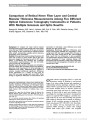
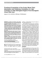
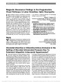
Show Functional Constriction of the Ocular Motor Field: Description and Preliminary Evaluation of a New Technique to Help Distinguish Organic From Nonorganic Visual Field Loss Nadeem Ali, MA, MB BChir, MRCOphth, FRCSEd(Ophth) Background: To describe and evaluate a novel clinical method to assess patients with constricted fields using the Goldmann perimeter, with the aim of distinguishing nonorganic from organic field constriction. Methods: Ten patients with constricted visual fields who were undergoing kinetic perimetry as part of their routine workup were included. Five of them had suspected functional visual loss (FVL), and 5 had organic field loss. Patients were assessed on the Goldmann perimeter using a test that combines kinetic perimetry (visual field) with a modified uniocular field of fixation (motor field). The main outcome measure was the size of the visual and motor fields. Results: In all patients with organic visual loss, the motor field was expanded relative to the visual field, as would be expected (P = 0.02). In all patients with suspected FVL, the motor field was markedly constricted and was not significantly different from the visual field (P = 0.27). The motor fields of the 2 groups were significantly different sizes (P = 0.001). Conclusion: Patients with FVL may exhibit functional behavior on a motor task, believing that it is a test of vision. Functional constriction of the ocular motor field may help distinguish organic from nonorganic visual field loss, but further evaluation is required. Journal of Neuro-Ophthalmology 2011;31:131-134 doi: 10.1097/WNO.0b013e31820a45b2 2011 by North American Neuro-Ophthalmology Society Subjectively impaired vision without a detectable organic basis is termed functional visual loss (FVL). Patients with FVL pose significant diagnostic and therapeutic challenges to ophthalmologists and may also present to other medical specialties (1-4). FVL can present as loss of visual acuity, visual field, or both (5). In all cases, an underlying organic cause must first be excluded. A common pattern of functional field loss is marked visual field constriction. The field area is often the same at different testing distances (a tubular field), a feature that helps distinguish it from a physiological conical field. In some patients, however, it can be difficult to be certain that marked field constriction is functional rather than organic. The Goldmann perimeter is an ideal tool to demonstrate functional features in suspected nonorganic field loss. Overlapping of isopters, spiraling, stellate patterns, and centrifugal expansion are evidence of functional behavior (6,7). However, the Goldmann perimeter has also been used as a way of measuring ocular ductions in conditions such as thyroid eye disease (8-10) and chronic progressive external ophthalmoplegia (11). The quantitative record of the ocular motility obtained, which is plotted on standard kinetic perimetry paper, is called the Uniocular Field of Fixation (UFOF). The Goldmann perimeter provides the opportu-nity to switch from a visual task (perimetry) to a motor task (UFOF) without changing the testing environment. In our study, we aimed to determine 1) if functional be-havior observed on perimetry would be translated into func-tional motor behavior when the task was switched and 2) if the responses obtained might be helpful in distinguishing a func-tional cause from an organic cause of field constriction. METHODS Ten patients undergoing kinetic perimetry as part of their standard ophthalmic workup were studied. All had pre-viously demonstrated constricted visual fields with preserved central vision and no limitation of ocular motility. The first group (‘‘organic'') comprised 5 patients with known disease thought to be wholly responsible for their visual field loss. Moorfields Eye Hospital, London, United Kingdom. The author reports no conflicts of interest or sources of funding to acknowledge. Address correspondence to Nadeem Ali, MA, MB BChir, MRCOphth, FRCSEd(Ophth), Moorfields Eye Hospital, 162 City Road, London EC1V 2PD, United Kingdom; E-mail: nadeem.ali@nhs.net Ali: J Neuro-Ophthalmol 2011; 31: 131-134 131 Original Contribution Copyright © North American Neuro-Ophthalmology Society. Unauthorized reproduction of this article is prohibited. Their characteristics are shown in Table 1. The second group (‘‘functional'') comprised 5 patients with suspected FVL, based on typical functional features as shown in Table 2. Three of these functional patients (#6, 7, 8) had no concurrent organic ophthalmic or neurological disease and were considered to have pure FVL. In the remaining 2 patients, the diagnosis of FVL on a background of organic disease (functional overlay) had been made. Case 9 had long-standing hydrocephalus, and Case 10 had cranio-pharyngioma resection earlier in life. Immediately following conventional perimetry, the right eye of each patient was retested using the procedure de-scribed below. Testing was performed in all cases by an experienced perimetrist (N.A.). The testing method was designed to be quick, simple to understand, repeatable, and easily incorporated into a rou-tine kinetic perimetry session. It consisted of 2 steps: the first visual and the second motor. The first step was a simplified visual field plot using a single stimulus target (V4e). The patient was instructed to ‘‘look at the central spot all the time and press the buzzer when the light first appears.'' The target was moved toward the center along each of the 8 cardinal meridians in random order, and the responses were plotted in the standard way. This was termed the ‘‘visual field.'' The second step was a modified UFOF and followed immediately. The patient was instructed, ‘‘Now the target is going to start in the centre and move outwards, but this time you should follow it with your eyes as it moves. Press the button as soon as it disappears.'' The V4e target was then moved centrifugally from the center along each of the 8 cardinal meridians, and the responses were plotted. This was termed the ‘‘motor field.'' For quantitative analysis of the visual and motor fields, the mean radial distance from the center for each of the plotted responses was measured in millimeters, and the 8 distances summed to give a total field score. T tests (5% level, 2 tailed), paired for within group and independent for between group, were performed on SPSS (ver 17). RESULTS The visual and motor field plots for each patient are shown in Figure 1. Table 3 shows the visual field and motor field scores. All patients had markedly constricted visual fields, the size of which was not significantly different between the 2 groups (mean field scores: organic, 139 6 44; functional, 111 6 30; P = 0.61). For the organic group, the motor fields were significantly larger than the visual fields (mean field scores: visual, 139 6 44; motor, 446 6 48; P = 0.02). For the functional group, the motor fields were markedly constricted and were, on average, the same size as the visual fields (mean field scores: visual, 111 6 30; ocular motor, 142 6 30; P = 0.27). Comparing the motor fields between the 2 groups, the difference was highly significant (mean field scores: organic, 446 6 48; functional, 142 6 30; P = 0.001). DISCUSSION Most of the techniques for demonstrating functional fea-tures in patients with suspected nonorganic visual loss are tricks (7). They rely on the patient's inadequate un-derstanding of visual physiology. In the technique described here, the patient is led to believe that step 2 is another visual field task when in reality, because the target is foveated as it moves, it is a test of ocular motility. We found that patients with suspected FVL demonstrated a functional gaze paresis, which mapped quantitatively in visual space to their perceived field of vision, a feature that can distinguish them from patients with organic field loss. Other techniques have been described to expose func-tional field loss. A commonly used method is to tell the TABLE 1. Patients with organic visual field loss No. Age/Gender Diagnosis 1 46/Male Idiopathic intracranial hypertension with chronic papilledema 2 61/Male Advanced retinitis pigmentosa 3 21/Male Chronic relapsing idiopathic optic neuropathy 4 84/Male Advanced primary open angle glaucoma 5 38/Female Advanced pigmentary retinopathy TABLE 2. Patients with functional visual field loss No. Age/Gender Functional Features 6 19/Female Crossing isopters, factitious hemianopia on automated field (not present on kinetic field) 7 15/Female Spiraling isopters on kinetic perimetry 8 40/Male History of pseudohemiparesis and pseudoseizures; triplopia on 4 prism-diopter base up test 9 16/Female Improvement in visual acuity with minimal prescription; Ishihara numbers traced accurately but not read 10 60/Female Tubular fields; normal visuomotor behavior incompatible with claimed fields 132 Ali: J Neuro-Ophthalmol 2011; 31: 131-134 Original Contribution Copyright © North American Neuro-Ophthalmology Society. Unauthorized reproduction of this article is prohibited. patient to make saccades to targets in the supposed blind field, disguising the test as an assessment of eye movements (7). This has recently been examined quantitatively for its value in discriminating organic from nonorganic visual loss (12). More objective tests, which demonstrate intact visual pathways and are less reliant on patient cooperation, include FIG. 1. Visual (blue) and motor (red) fields plotted with the V4e stimulus for all patients. Ali: J Neuro-Ophthalmol 2011; 31: 131-134 133 Original Contribution Copyright © North American Neuro-Ophthalmology Society. Unauthorized reproduction of this article is prohibited. pupil perimetry (13,14) and measuring multifocal visual evoked potentials (15). The technique described in this study differs from these in that no attempt is made by the examiner to prove sight in the blind field. Rather the goal is to demonstrate additional unequivocally functional behavior, in this case restriction of ocular ductions where none exists. This is a limitation of the technique since other tests would still be needed to prove a scotoma is factitious. The advantages of our technique are that it is simple to perform and can be used within the setting of the Gold-mann perimeter as soon as functional perimetry features are suspected. In addition, in this preliminary series of patients, there were no false-positive results. This technique may prove to be a useful way of distinguishing organic from nonorganic field loss. However, larger patient groups are required to validate sensitivity and specificity and to see whether patients with pure FVL differ from patients with functional overlay. We are planning additional studies to address these issues. REFERENCES 1. Griffiths PG, Eddyshaw D. Medically unexplained visual loss in adult patients. Eye. 2004;18:917-922. 2. Scott JA, Egan RA. Prevalence of organic neuro-ophthalmologic disease in patients with functional visual loss. Am J Ophthalmol. 2003;135:670-675. 3. Miller NR. Neuro-ophthalmologic manifestations of psychogenic disease. Semin Neurol. 2006;26:310-320. 4. Griffiths PG, Ali N. Medically unexplained visual loss in adult patients Curr Opin Neurol. 2009;22:41-45. 5. Lim SA, Siatkowski RM, Farris BK. Functional visual loss in adults and children patient characteristics, management, and outcomes. Ophthalmology. 2005;112:1821- 1828. 6. Scott GI. Traquair's Clinical Perimetry. 7th edition. London, United Kingdom: Henry Kimpton, 1957. 7. Chen CS, Lee AW, Karagiannis A, Crompton JL, Selva D. Practical clinical approaches to functional visual loss. J Clin Neurosci. 2007;14:1-7. 8. Mourits MP, Prummel MF, Wiersinga WM, Koornneef L. Measuring eye movements in Graves' ophthalmopathy. Ophthalmology. 1994;101:1341-1346. 9. Steel DH, Hoh HB, Potts MJ, Harrad RA. Uniocular fields of fixation in thyroid eye disease. Eye. 1995;9:348-351. 10. Haggerty H, Richardson S, Mitchell KW, Dickinson, AJ. A modified method for measuring uniocular fields of fixation: reliability in healthy subjects and in patients with Graves orbitopathy. Arch Ophthalmol. 2005;123:356-362. 11. Richardson C, Smith T, Schafer A, Turnbull D, Griffiths P. Ocular motility findings in chronic progressive external ophthalmoplegia. Eye. 2005; 19:258-263. 12. Zinkernagel MS, Pellanda N, Kunz A, Mojon DS. Saccade testing to distinguish between non-organic and organic visual-field restriction. Br J Ophthalmol. 2009;93: 1247-1250. 13. Rajan MS, Bremner FD, Riordan-Eva P. Pupil perimetry in the diagnosis of functional visual field loss. J R Soc Med. 2002;95:498-500. 14. Skorkovska´ K, Lu¨dtke H, Wilhelm H, Wilhelm B. Pupil campimetry in patients with retinitis pigmentosa and functional visual field loss. Graefes Arch Clin Exp Ophthalmol. 2009;247:847-853. 15. Massicotte EC, Semela, L, Hedges TR III. Multifocal visual evoked potential in nonorganic visual field loss. Arch Ophthalmol. 2005;123:364-367. TABLE 3. Individual field score (in millimeters) Organic Group Functional Group Patient Visual Motor Patient Visual Motor 1 172 377 6 37 98 2 40 509 7 194 239 3 271 595 8 91 109 4 40 329 9 167 186 5 172 419 10 65 79 Mean 139 448 111 142 SE 44 48 30 30 SE, standard error. 134 Ali: J Neuro-Ophthalmol 2011; 31: 131-134 Original Contribution Copyright © North American Neuro-Ophthalmology Society. Unauthorized reproduction of this article is prohibited. |