| OCR Text |
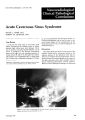
Show J, C1in. Npuro-ophth,dmol. I: 269-211, IQ81. Abnormal Visual-Evoked Responses and Opsodonus BAHMAN JABBARI. M.D. EDWARD URBAN. M.D. Abstuct A n-yu.r-old wom.n presented wilh cLlssic opsoclonus two wtc'ks following .n .cutt febrile iIlntss. Four wtc'k!> 1.ltr. complett diniul recovery wu tvidtnl. Si.. wetks .fter onStt of the probltm, or 2 wtc'ks .fttr rnolulion of tht ..bnorm.al tye movements• .a p..«tm revuwl visu.al tyoktd r"ponst showtd .a dtl.ayN rtsponst in both eyts .and h..d.an unusu.aUy high .amplitudt. For tM nut 2 months tht p.atient compl..intd of occuian..1 blu"ing of vision_ A rtpt.at visu.al evoktd responst test ..fter " months tfvnltd norm..1Lalency.and ..mplitude In both eytsThe possible mtdu.nisms f(ll" the visu..1evoktd mponse ..bnorlrWllty.lff discussed. Opsoclonus is d~hned u .... blurre ocul.ar oscilI.. tlon consisting of upid, Iftvolunt.ary, chaotic. conjug.Jte eye movements 1ft .. II directions persistIng during sl«p.'·' II occurs 1ft ..ssoci..hon with a v.llnety of pathologiul conditions, including infbmm. lltory and postinft'Ctious encephalitides, neuroblastomas in children,u .nd intr.cranial4 and eXlr.acranial neoplasms in adults.s-7 A benign course with complete r~covery after sevual w~ek.s is not uncommon." Whelher prolong~d opsoclonic movements damage optic fibers and thert'fore account for intermittt'nt visual complaints of some patients during recovery rl'mains a mallt'r of spt'("ulation. The postmortem examin.lliion of pdtients with opsoclonus has not yel related data concerning the optic nerves."" MorMver, on those who rKovered• .lI reliable qU.llntlt.allve method WdS not .av.ailable to me.llsure possible del.ll)'S of optic nerve conduction. This is .lI report of • p..tlent in whom .lI tr.ansient bib.teral OptiC neurop.lthy W.llS detected by p.lttern revers.aJ visU<lI.evoked rrspanse test 2 weeks .lifter recovery from opsoclonus. Several mKh.llnisms were postul.llled to expl.ain this finding. including trauma to lhe optic nerves from agjt.lltory movements of the globes. From 1M N",roIosY ~rvKe g,f W... tltl Rttd Army Mrdiul Cel'lter. W.sh,ngton. D. C...nd 1M Dtp.artmtnl of Nturolop;y. Uniform~ SorrvlCn U",~r5'ty of 1M Ht...hh SntRCcs. Stlhnd. a. M.ryl...nd_ December 1981 Cue Report A 73-year-old female experienced dIffuse weak.ness, .1R0rexia, moderatl' dbdominat pain, diarrhea, vomiting, and fever 2 wl'eks prior to admission. The ddy befort' old mission shl' became lethargic, complained of dizziness, and was noted to have abnorm.llJ eye movements. On admission she hold a blood prrssure of 144/75, pulse rate of 68. and temper.alur~ of 100.00 and weighed 124 tbs. She was .lIgilaled, disoriented, .lind diffusely tremulous. The tremor W.llS most prominent in the right upper 11mb and increased markedly dunng volitional movements. She tud continuous SlcudlC, chaotic. r.llpid. lnvolunt.llry rye movements In .lI1I dirrctions of g.Jze. These movements incrt'.llsed during excilement and persisted during sl«p. Th~re W.lS moder. ale we.akness of upward gue. Motor .lind sensory eumin.lltions reveal...d no .lIbnorm.ality. Her dt't'p tendon reflexes were brisk lhroughout. She had bll.lller.lli B.1binski rl!'Sponses. The pertinent laboutory findings consist...d of CBC 10,600 with a differenti.lll of 53Cl1l polys and 33Cl1l lymphocyles, hemalocrit 40.5, sed.mentation rat... 35, serum sodium 130, and Sl'rum triglycerides 161 mg%. Urinalysis revedled 5-15 RBC's per highpower field and 1+ dtbumin. Thyroid functions tests, rheumatoid fdclor, and urine drug screening tesls r('vedled no dbnonnJlity. A chesl rOt'ntgt'nogram showed a righllowt'r lobE' infiltrJte. An t'tt'c o Irocardiogrdm rl~v('alE'd old ischemic chJngE's in the precordi.lll le.llds. On computed tomographic sun .a sm.all rddiolucent drea was preSE'nt .It the region of Ihe lefl lenticular nucleus. An electroencephdlogr.. m showed mild diffuSE' slowIRg. A lumbdr punc1ur(' reve.llied dedr and colorlrss spm,)1 nuid with .lin opening prpssurf' of 170 mm. ('> Iymphocytrs. protein 18 mg'lr• .lind glucose 04 m~. B.lIcleri. 1, fung.J1. ... nd vir.lll cultures were- neg.atlve. four weeks .lifter admission the p.lhent enjoyed complPle recovery from h('r symptoms. A repe.llt chest roentgenogr.llm showt'd ~Iullon of the righl Iowt'r lobe infiltrate whIch wu .lIllribuled to .lI vir.al infection. Six weeks .lifter .lid mission. an ophth.llimoiogic.lli examindtion ft'Ve.llled normdl s.JIccadic .lind pursuit functions .lind no involuntary movemenlS. Her pupils were 3 mm in diameter '69 I R L Figurf I. High_~mplitudfvisu~I-~vohd rfspon5~<; showi"g I~tfndes of 128 ..nd 132 milhsf<onds .n the rIght ~nd the I.," fyfS. rfSpe<:I,vfly (Forourl..boutory. mun ,5101 m.lhSf'Conds. 1st 50 _ 4 milhs.«onds.t Thf "n"lysls t,mf,S ~SO m,lhSf<onds ..nd thf ",libr"I,on m.. rk represenh 5 microvolts. dnd redcted briskly to light. She hdd d normdl near reflelt. Her visudl dcuity WdS 20/30 in the right eye dnd 20/25 in the left eye. A tdng£>nt screen t£>st showed intact central visu<ll fields in both eyes. A ViSUdl evoked response tesl W<lS performed using .I TV p<lttem medsuring 21 cm verticdlly and 28 cm horizont<llly, pldced .It .I dist,mce of ISO cm. This pdttem consisted of a 16 x 10 m.ltrix of rect<lnguldr checks, edch ch£>ck thus subtending dpproltimdtely 30 minutes verticdlly .md 40 minutes horizont<llly. The reversal r<lte WdS 2.1 Hertz. The response W.lS deldyed in both eyes and hdd dn unusudlly high dmplitude (Fig. I). For 2 months dfter ct'ssation of opsodonus, th£> pdtient compl,lln('d of occdsiondl blurring of vision. A repe<lt visual-('vokt'd response test afler 4 months revedled normJI IJtency Jnd amplitude in both eyes (Fig. 2). Discussion In IQf'.~, (,,~.ln df'~cribed ,m .1(ute self-limited ·,hl', '1 ,1",r"1 INtf-l'd by 0rsI,dl1nus dnd Figurf 2. Four months I"IN. v,su"l-evo[..ed responses show norm~1 l..tfnc" (0<> Jnd 100 m,I1'sf<ondsl Jnd Jmplltude s"me c.lhbr.llion .Ind In.lySlS um.. .IS FIg I diffuse body Iremulousness.~He beli£>ved thai this disorder was caused by a benign encephalitis beCduse of the pdtients prodromdl symptoms, the remitting course of the illness. and presence of cen~brospinal fluid pleocytosis. OUf patient obviously suffered from a simil<IT condition. There <lTe threE' possible explanations for her abnormal visudl-£>voked responses: I) encephalitis. 2) demyelindtion. Jnd .3) dbnormal £>ye movements. Considering these possibilities, if the patient suffered .In encephJ1itis, the optic nerves or chiasm mJy hdv£> been directly involved by the inflammdlory process or by ischemic changes as the result of d diffuse vdsculopilthy. On the other hilnd, l"('ntrd[ nervous system demyelindtion should be dlwdyS considered dS one of the first Cduses of a bil<lterdlly del<lyed visual evoked r£>sponse. IO If this WdS the cause of OUf patient's VER abnonnality, then the VER dbnormdlity itself served as an importdnt clue to the pdthophysiology of the syndrome of "benign opsoclonus dnd diffuse body tremulousness" for which no postmortem data Journal of Clinicdl Neuro-ophthdlmology have been available. Delayed pattern rl'verS.:l1 vis_ ual-evoked responses in demyelinating, opti..: n('uropathies, however, rarely returned to nornl.ll v.:llues after recovery from the ,lcute illness. Moreov('r. the central nervous system demyclin.ltion itsl'lf could have been secondary to .:I prim,lry dise.lsl' which has involved the br.:lin diffusdy. Th(' third possibility was a transient trJumJtic opti\" n('uropathy caused by violent movements of both optic globes. In our opinion, closeness of thl' latencies of pattern reversal visu.l1 eVl)j..ed n.'spI.)nses in both "delayed" .md "returned to norm.ll" stJh.' Jnd return of these latencies t\) normal values Jfter recovery from acute illness bvored this l.lt!er mechanism. In optic neurop.lthies, del.:lyed visual·evoked re+ sponses often demonstrate decreased amplitude. ll - 1J The increased amplitude of our patient's visual-evoked response therefore demanded an el(planation. BrooksH reported increased amplitude and raised threshold of visual~evoked responses to flash in normal individuals during saccadic eye movements. On the other hand, brief ocular flutter and bursts of opsodonic movements have been noted persisting for some time after cessation of opsoclonus. 1 Our patient did not have such eye movements during visual-evoked response testing, and her saccadic and pursuit functions revealed no abnormality. Moreover, one cannot apply findings with flash to pattern reversal method since these two techniques test the functional integrity of two different visual pathways. In our opinion, the high amplitude of our patient's abnormal visual-evoked response resulted from a remaining state of hyper+ el(citability in a large number of macular receptors, causing the phenomenon of temporal summation. We hope that this report will evoke enthusiasm for further el(ploration of the cause and mechanism of pattern reversal visual-evoked response abnormalities in relation to opsodonus. References 1. D.Hoff, R.B., Troost, T.B., .md Dell"oss, l.F.: Nystagmus and related ocular oscillations. In Neurooph. thd/mo/ogy, IS Glaser, Ed. Harper &. Row, New York, 1978. p. 238. December 1981 [JbbJri, UrbJn 2. Solomon, G.E., Jnd Chutori.lll, A.M.: Opsoclonus .lnd neurobl.lstoma. N. [nxl. /. Med. 279: 475+477, 1%8 J. Dyken. P.. .lnd Kol.lr, 0.: Dancing eyl.'~. d.lncing f("{'!: InfJntiJ(" polymyoclonus. Brain 91: 305-320, ,q~. 4. K("Jnl'. I.R., .lnd Dt'vereau>;, M.W.: Opsoclonus .lSs< lci.l!("d with .In intr.l(r,mial tumor. Arch. Ophrh.lJmol. 92: 443~445, 1974. 5. Ross, A.T., Jnd Z("m"n, W.: OpsocJonus, occult C.lrcinom.l Jnd chl'mic"J poltholo~y in d("nt.lte nuclei. Arch. Nf'urol. 17: 545-551, 1967. o. Bellur. S.N., Opsoclonus: Its clinical v"lue. Nt'uroJ". l:y 25: 502,~507, 1975. 7. EJI("nber~er, c., Camp", J.F.. .lnd W("tsky. M.G., Opsoclonus and parenchymatous degener"tion of the c("rebelJum. Neurology (Minn,) 18: 1041-1046, 1968. 8. Smith,l.L., .lnd W.llsh, r.B.: Opsoclonus-ata>;ic con· jug.lte movements of the I.'y(>s. Arch. OphlhollmoJ. 64: 244~250, 1960. 9. Cog"n, D.G.: Opsoclonus, body tremulousness and benign encl.'phalitis. Arch. Ophlh.lJmol. 79: 545-551, 1%8. 10. Halliday, A.M.: Clinical applications of evoked potentials. In Recent Adv4nces in ClinicolJ Neurology, Vol. .2, M.lUhews .lnd jS GI.lser, Eds Churchilllivingston, London, 1978. p. 53. II. Chiappa, K.H.: Pattl.'rn shift visual. brainstem audi. tory and short-I"tency somatosensory evoked potenti. lls in multiple sclerosis. Neurology 30/21: 110-123. 1980. 12. Sokol, S.: Visual evoked potentials in eleetrodiagnosis. In Clinical Neurology, M.r. Aminoff, Churchill-livingston, London, 1980, pp. 348+369. 13. Stockard, n, Hughl.'s, I.F., and Sarbrough, FW.: Visual evoked response to electronic pattern revers. ll: 1...ltency v"ri.l!ions with gender. agl.'. .lnd tech. nical factors. Am. j, E.E.G Tt'ch. 19: 171+204, 1979. 14. Brooks, B.A.: Vision and visual evoked potentials during saccadic eye movements. In ViSUoll E"oked Potenti.lJs in M,m: New Devt'lopments, F.E. Dt'smedt. Ed. C1.lrendon PrI.'SS, O>;ford, i977, pp. 301315. 15 Sokol. S.: Visual ("voked potenti.lls: Theory, techniques .Jnd c1inic.l] .Jpplic.ltions. 5ur\'. Ophth.l/mt'1. 2'1: 18-44. 1970. Writl.' for rt'prints to; Bahm.ln IJbbJn. MD.. Nl'uf<)loI:Y Service, Bo>; 310, Walter Reed Army M("dicJI (ent('f, Washington, D.C. 20012. 271 |