| OCR Text |
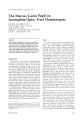
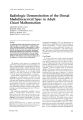
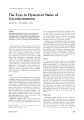
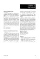
Show f. Clin. Neuro·<>phth.l/l1l<l/. 2: 271-272, 101\2. Neurinoma of the Oculomotor Nerve Case Report TULAY KAN5U, MD. OSMAN E. OZCAN, M.D. EMIRE OZOIRIM, M.D. BEH5AN l1NOL, M.D. 6ZDEMIR CURCAY, M.D. Abstract ,.\ tS-year-old boy with d recurrent third nerve pdlsy who was found to have a neurinoma of the third nerve is described. Unusual features of his presentation and review of the literature relative to this rare tumor are discussed. Tumors derived from Schwann cells, which wrap around the axons of peripheral nerves, are called neurinomas, neurofibromas, plexiform neurofibromas, neurilemomas, and schwannomas. Neurinomas are usually localized in the peripheral nerves.· The most frequent site of occurrence of an intracranial schwannoma is the acoustic nerve; however, the tumor may occur along the second, fifth, seventh, 11th, and 12th cranial nerves! Neurinomas of the oculomotor nerves are extremely rare. In the literature, there are only four reported cases clinically manifested and pathologically verified as neurinomas of the third nerve.3 .' Case Report A lS-year-old boy was admitted in May 1980, with a 3-day history of periorbital pain and complete ptosis on the right side. Past history revealed that in 1969, when he was 4 years old, he had the sudden onset of headache, and external deviation of the right eye was noted by his family. All symptoms cleared in 2 weeks without any specific treatment. He was then well until 1974, when he had the sudden onset of headache, ptosis, and mydriasis on the right side. He was given antibiotics with corticosteroids, and the sym ptoms resolved in 3 months. He was seen in our institution for the first time in 1976, when he presented with a 10-day history of an enlarged right pupil. On examination, he was found to have a dilated right pupil with no light reaction. There was a 150 external devi'ltion of the right eye in primary position, .1l1d eye movements From the DepJrtml'nt' "f Nl'un>l,,~y. N('"n"LJr~l'ry, ,'nd 1'.,thology. HJccttl'pc Univer,ity H,,,pit,,I,. Ank.H.l, T"rk('y. December 1982 were normal. Visual acuity was 20/70 in the right eye and 20/20 in the left. Neurological examination was within normal limits. Laboratory investigations, including skull x-rays, lumbar puncture, right carotid angiography, and computerized tomography (CT) did not reveal any abnormalities. A marked right-sided ptosis was observed after the angiography. The diagnosis of painful ophthalmoplegia was made, and the patient was started on corticosteroids. On follow-up examinations at 6-month intervals, a persistent right mydriasis was recorded with varying degrees of ptosis. Progressive limitation of eye movements was noted on adduction, depression, and elevation. He received steroids intermittently. A repeat CT scan in June 1977 failed to reveal any abnormality. Examination on his last admission in May 1980, when he presented with a 3-day history of periorbital pain and ptosis, and revealed a complete right third nerve palsy. Visual acuity was unchanged from previous examinations, and neurological examination was normal. Repeat right carotid angiography was also normal. Vertebral angiography was performed to visualize the posterior circulation, and an aneurysmal dilatation of the right posterior cerebral artery originating from the top of the basil.H artery was suspected. Suspecting d posterior cerebr.ll .Hterv .1l1eurysm, .1 right subtemporal Cf.ll1iotomy W.1S' perfon~1ed. When the posterior cerebr.11 dnd posterior communicdting arteries were explored, .1 tumoral widening of the third nerve W.1S seen in the precavernous portion of the nerve. The tumor was 3 X 4 mm in di.1meter. No .1Ileurysm W.1S found in the posterior cerebr.11 .1rtery. Fl)lIl)wing dissectil)f1 from surrounding tissucs, the tuml)r W.1S resected tot.llly. The histologic di.1gnosis l)f the surgic.1l1v rel1lllVed tissue W.1S d ncurinom.l ch.H.lcterized by fibrous tissue bundlcs .1l1d structures resembling pNiphcral nerve sections (Fig. 1). Discussion Intr,lCf,lIlidl neurinomds most frequently arise from the vestibuldr nerve .1l1d less frequently from 271 t kulllllllltllr Nerve N('Ur;nlllll,1 Fil\ure I. Micn"clIp'c JppeMJnC(' ,.f th" tumt'r M"lng from the third nerve, showing the loos" p.JUern l)f tunHlr c('l1~ .lnd pcnphcr.lJ nerve fiber.. -..urnlundcd by (l..lnnCcdIV(' t ..;~u(' t,b('r~ (H,'mJltl\vlin & 1''''ln, X250.) the trigeminal nerve or ganglion; they rarely involve a cranial motor nerve. Neurinomas of the cranial nerves innervating the extraocular muscles are particularly unusual. There are two reported cases of trochlear nerve neurinomas and four cases of third nerve neurinomas in the Iiterature."-h All previously reported third nerve neurinomas were unsuspected preoperatively. The three cases described by Huber presented with a discrete progressive palsy of the oculomotor nerve, leading in later stages to complete ophthalmoplegiJ. In two of his cases there was a simultaneous or preceding unilateral optic neuropathy, leading finally to amaurosis. All signs were similar to those of the syndrome of the orbit.ll apex, superior orbit.ll fissure, or the .lnterior C,lVernous sinus.:1 The patient described by Shuangshoti presented with right hemip.Hesis Jnd left third nerve paresis, .lnd W.lS found to h.lve .l fusiform .lneurysm of the termin,ll part of the left intern,ll c.ln)tid ,1rtery due tl) ,1thefO!> r1efO!>i!>. which W,lS not compressing the third nerve. At autopsy. the third nerve cont,lined ,1 cy!>tic m,lS!> from its pointl)f ('xit fwm the midbr,lin ,md !>howed the chJr,lcteristic fe,ltures of ,1 neurilemom, 1. I The mo..,t unusu,ll fe.lture of our p,ltient W,lS the recurrent ophth,]lmoplegi,l with complete remission of thl' first tWll epiSl)des. The symptl)I11S h,ld bl'l'n present fur 1110re th,1I1 10 years, They were f1udu,lting in the bl'ginning but bter were progressive. In thi!> p,ltient, spont,1I1eous relief of he,ld,1l1H' ,Inc! l'ye devi,ltion which h,HJ occurred at age -l are not well explained Later fluctuations of the findings probably were rebted to the corticosteroid treatment. and the visual loss in the right eye was likely due to suppression amblyopia. Mydriasis was an early manifestation in this patient, and it can be explained bv eJrlv involvement of parasympathetic fibers with a tumor that originated from the nerve sheath. This patient and four others reported in the literature emphasize the need to consider the diagnosis of neurinoma in patients presenting with third nerve palsies. References I. Rubinstein, LI.: Tuml)rs l't the centroll nervous svstem. In Atl,I" ,,{ Tunlllr PJthl,log\' (tasc 0). A~ed FllrlTs Institute l't Polthl1log\', Washington, D.C, 1072. pp. 205-217 2 Ausmoln. 1.1.. French. LA., olnd Bolker, A.B.: Intracran;,) 1 nel1pl,lsms. In ClinicJI Neuro/og~', Vol. 1. A.B. B.lker, .lnJ LH. B,lker, EJs. H.lrper and Row, Marv- I.\I1J, 10 7(" Ch,lpter 0, pp. 52-53. . 3. Huber, A.. D,lSl)kull'motoriusneurinom. Klin. Mbl. Aug<'nheIiI-. 172: o27-t>35, 1978. 4. Shu,1I1gshl1ti, S.: Neurilemom.l of the oculomotor ner\,('. 8r. I. l )phth.l/mol. 59: o4-ot>, 1975. 5. Bllgg.ln, I.E., Rosenb,lum, M.L.. Wilson, C.B.: Neurilemnlllnhl of the tl)urth cranial nerve. f. Neurosurg. 50: 51°-521, 10 70. t>. King, 1.5.: TrlKhlear nerve sheath tumor. f. Neurosurg. 44: 245-247, 1976. ,",Vrite for reprint" to: Tulay Kansu, M.D., Department of Neumlogy. Neuro-ophthalmology Unit, Ankara, Turkey. Journal of Clinical Neuro-ophthalmology |