| OCR Text |
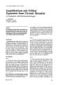
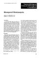
Show J. elin. NeuTo-ophthalmol. 4: 167-172, 1984. Enophthalmos and Orbital Expansion from Chronic Sinusitis CT Evaluation with Reformatted Images A MARTELLI WILLIAM F. HOYT T. HANS NEWTON Abstracl Five patients were found to have an unusual variety of enophthalmos fhat results from chronic constricting sinusitis with expansion of the orbit. We illustrate the usefulness of computed tomogr.J.phy in the diagnosis of this process, emphasizing the role of oblique sagittal reformatted images. Introduclion Enophthalmos may result from any process in which the walls of the orbit bend or collapse into Ihe adjacent maxillary or ethmoid sinuses. Although trauma causing -blow-out- fractures of either the floor or the medial wall of the orbit is the most common cause of this disorder,l-5 there are cases in which it may result from the effects of chronic maxillary and ethmoid sinusitis.6 We report five patients in whom computed tomography (CT) with oblique sagittal reformatted images was instrumental in making the diagnosis of this unusual variety of enophthalmos. Case Reports Case 1 A 52-year-old woman had a sunken appearance of her left eye and a slight drooping of the left upper lid for 2 years. She did not have double vision, nor did she have an orbital injury or known sinus infections. Plain x-ray films of the skull demonstrated opacification of the left maxillary antrum. Axial CT scans showed that the left maxillary sinus was smaller than the right, and the region of density indicated that it was filled almost completely with soft tissue (Fig. la). On From the Dep.:utm£,nt of Neuroradiology (AM), Neurological Clinic. University of Pavia. Pavia. Italy; Department of Neuroophthalmology (WfH) and Neuroradiology (THN), University of California. San francisco, San Francisco. California. September 1984 coronal (Figs. Ib and Ie) and oblique sagittal (Fig. Id) reformations, the floor of the left orbit appeared thickened and depressed. The extraocular muscles and optic nerves were normal. Evidence of enophthalmos was not clearly defined. The diagnosis was chronic sinusitis of the left maxillary sinus resulting in enophthalmos. No treatment was advised. Case 2 A 29~year-old woman had noticed that her right eye was gradually receding into the orbil. She had no pain or double vision. At the age of 8 years, she had acute maxillary sinusitis. The examination confirmed signs of enophthalmos of the right eye, deepening of the superior palpebral fold, and slight depression of the right globe. Ocular movement and vision were not impaired. Plain x-ray films and CT in axial and direct coronal projections showed opacification and decreased volume of the right maxillary sinus (Fig. 2). After surgical removal of gelatinous material from the right antrum, the enophthalmos regressed, but did nol resolve completely. Case 3 A 62-year-old man had vertical double vision when he looked upward; displacement of the vertical images gradually increased in upward fields of gaze. Simultaneously, he had become aware of recession of the soft tissue beneath his right brow which gave the appearance of insinking of his right eyeball. He also noticed a slight indentation of the bone of his right cheek overlying the right maxillary sinus. He had no pain and did not recall haVing previous attacks of sinusitis. The examination revealed 2-mm enophthalmos of the right eye, a deep superior palp.ebral fold, a small indentation in the right maxdlary bone located 5 mm beneath the inferior o.rbital rim, ~nd a 5-diopter hypotropia of the nght eye dunng upward gaze. Axial (Fig. 3a) and 167 Enophthalmos from Chronic Sinusitis •,j direct coronal (Fig. 3b) CT scans showed thickening of the wall of the right maxillary sinus, which was also opacified and was much smaller than the well-aerated left maxillary sinus. Similar findings were shown on tomograms of the paranasal sinuses. Chronic inflammatory tissue was excised from the right maxillary sinus cavity; on palpation, the superior and medial walls of the antrum were softened. After the operation, the patient's enophthalmos was unchanged, but he reported that the diplopia was less severe. Case 4 A 49-year-old man developed diplopia associated with right-sided periorbital edema, pain in the right maxillary region, and low-grade fever. His posterior cervical nodes were enlarged and there was a slight numbness of the right cheek and upper lip. Upward motion of the right eye was mechanically restricted. A tomogram revealed soft tissue opacity of the right maxillary sinus, the right nasal cavity, and anterior ethmoid cells. The diagnosis was acute maxillary sinusitis. 168 .,j Figures la-It. Case 1. (a) AXIal CT scan. Opacification indicating that the left maxillary sinus. which is smaller than the right is filled with soft tissue. (b) Oblique coronal reformation. left orbit The floor of the orbit (aITOws) is thickened and markedly depressed. The maxillary sinus is opacified. Extraocular muscles and optic nerve are normal. (e) Oblique coronal n.-formation, right orbit. The floor of the orbit (alTows) is relatively thin and has an upward convexity. (d) Oblique sagittal reformation, left orbit. The floor of the orbit is thickened and depressed. (t) Oblique reformation, right orbit. The floor of the orbit is thin and shows tht normal slight upward convex.ity. The maxillary sinus is well aerated. 'ournal of Clinical Neuro-ophthalmology Martelli, Hoyt, Newlon I') I') The patient was treated with aqueous penicillin. Within I week, the signs of infection resolved. and double vision diminished. Six weeks later, he had mild (2 mm) enophthalmos and restriction of upward gaze on the right. Axial CT scans with coronal and sagittal reformations of the orbit showed a soft tissue density extending in the right maxillary antrum and distortion of the floor of the right orbit (Fig. 4). The floor of the orbit was thinned, discontinuous, and depressed (Figs. 4b and 4d). Case 5 A 65-year-old man reported a mild swelling of his left upper lid. The examination revealed. normal vision and apparent proptosis of the left eye. Exophthalmometry readings were 18 mm on the right and 22 mm on the left. Eye movements were full in all directions. The lids were not swollen. The patient denied any history of sinusitis. CT scans of the orbits and sinuses showed soft tissue densities in the ethmoid sinuses bilaterally (Fig. 5). The right ethmoid sinuses appeared to be reduced in size and the medial wall of the right orbit was markedly concave. In this patient, the right-sided enophthalmos resulting from chronic ethmoid sinusitis was asymptomatic and ~ptember 1984 was not recognized until he was studied by CT for the apparent left-sided proptosis. Discussion Although enophthalmos most commonly is caused by traumatic ~blow-out~ fractures of the orbital floor, any process that results in diplacement of the orbital walls into the adjacent paranasal sinus may cause enophthalmos. Chronic sinusitis may cause thickening of the orbital wall from osteoblastic activity. Remodeling of the walls, especially downward displacement of the floor of the orbit, may cause the globe to be displaced posteriorly and inferiorly. Ophthalmic signs other than enophthalmos include extraocular muscle imbalance, malar swelling or indentation, blepha~oftosis, eye~id r~traction, epiphora, and proptosIS. Metastatic sarrhous adenocarcinoma and congenital absence of the sphenoid wing are less common causes of enophthalmos. The principal symptom in our five patients was chronic, painless enophthalmos. in some cases accompanied by diplopia (cases 3 and 4). One patient noted. slight maxillary indentation. ThE'Se symptoms have been reported in patients with ·congenital hypoplasia~ of the maxillary sinus'·! '6' Enophthalmos from Chronic Sinusitis and maxillary mucocele."·9 In our patients, the symptoms were related to chronic sinusitis, as shown by radiologic evaluations; in two of them, these findings were confirmed during surgery. A previous acute episode of sinusitis was reported by two of our five patients. Radiologically, the Figure 2. Case 2. Plain frontal roentgenogram of the paranasal sinuses. The right maxillary sinus is small and opacified by soft tiS&ue density. affected sinuses were opacified with thickened walls and were smaller than the contralateral sinus. In one patient, the maxillary roof was thinned and irregular, probably because of a recent episode of sinusitis (case 4). The loss of the normal upward convexity of the maxillary roof was a characteristic feature observed on CT scans. Outward convexity may occur in the medial orbital wall when the ethmoid sinus is affected. These findings, which are most clearly shown on coronal and parasagittal CT scans, explain the orbital expansion and consequent enophthalmos. Enophthalmos also was clearly evident on CT in most of the cases, depending on the correct positioning of the patient's head in the midline. Opacification and constriction of the sinuses and thickening of the sinus walls can be recognized on plain x-ray films and tomograms, as well. CT, however, provides superior information regarding soft tissues, displacement of the globe, and bone structureeasily discriminating chronic sinusitis from other pathologic processes causing enophthalmos. While there is general agreement·· 10-15 that both axial and coronal CT scans should be performed in the evaluation of orbital and paranasal sin~s pathology, these. cases illustrate that oblique sagittal and parasagtttal reformations provide other useful imag~ to evaluate the effect of sinus diseases on the orbit. 'hi Figures J.:r anci -h . .36..Case J. (0) Axial.a.nd (tt) dirE"Ct (o. ronal CT suns. There is mark.-..".. "ymm~ •.-,of 'hemaJ(-JIIarysm- uses. The ng I maXillary smus IS smaller, opaaf,e<t and has shgh'l, thickened walls. Th~-I'-- ', maJ(J-'Iary S-inUS -IS nonnaIIy aerat...."... 170 Journal of Clinical Neuro-ophthalmology St>ptem~r 1984 Martelli, Hoyt. Newton ,., ,b) '" 1'1 Figures 41l-4d. Case 4. (a) Axial CTscan. A soft tissue density is noted in the right maxillary sinus, which is smaller than the left. (b) Coronal reformdtion. The floor of the right orbit is irreguldrly thickened with a soft tissue density extending in the maxillary antrum. (e) Oblique sagittal reformation of the right orbit. The floor of the right orbit is discontinuous and depressed. (d) Oblique sagittal reformation of the Ii'ft orbit. Thi' floor of the orbit and adjacent maxillary sinus are normal. 171 Enophthalmos from Chronic Sinusitis Figure S. A"ial CT scan. The ethmoid air cells are partially opacified. The medial wall of the right orbit has a marked medial concavity. Note a slight enophthalmos on the right. References 1. Berkowitz, R.A, Putterman, AM., and Patel. D.8.: Prolapse of the globe into the maxillary sinus after orbital floor fracture. Am. f. Ophtlzalmol. 91: 253257,1981. 2. Crove, A.S.: Computed tomography in the management of orbital trauma. Ophthalm%gy 89: 433440, 1982. 3. Hammt'rschlag. S.B., Hughes,S., O'Reilly, G.V., et al.: Blow-out fractures of the orbit: A comparison of computed tomography and convt'ntional radiography with anatomical correlation. Radiology 143: 487-492,1982. 4. Ht'Sselink, 1.R., Nt'w, P.F., Davis, K.R., et al.: Computed tomography of the paranasal sinuses and face: Part Il. Pathological anatomy. J. Compul. Assist. Tomogr. 2: 568-576,1978. 5. Miller, C.R., and GlaSt"r, J.5.: The retraction syndrome and trauma. Arch. Ophtha/mol. 76: 662-663. 1966. 6. Montgomery, W.W.: Mucocele of the maxillary sinus causing enophthalmos. Eyl' Ear Nosl' Throat Monthly 42: 41-44, 1964. 7. Bierny, J.P., and Dryden, R.: Orbital t'nlargement secondary to paranasal sinus hypoplasia. Am. f. ROf'/ltgl'l1ol. 128: 850-852,1977. 8. ludisch, G.F., Jacoby, CG., and Ossonig. K.C: Developmental orbital t'nlargement and acquired 172 vertical diplopia. AmI. OphthalmoJ. 12: 1282-1286, 1980. 9. Traustason, 0.1., and Feldon, S.E.: CauSt" of enophthalmos secondary to maxillary sinus muce«le. Am·f. Ophtha/mol. 95: 838-840,1983. 10. Bilaniuk, L.T., and Zimmerman, R.A: Computerassisted tomography: Sinus lesions with orbital involvement. Hl'ad Nuk Surg. 2: 293-304, 1979. 11. Forbes, G.: Computed tomography of the orbit. Radial. Gin. North Am. 20: 37-49, 1982. 12. HesSt"link, J.R., New, P.F.. Davis, KR., et al.: Computed tomography of the paranasal sinuses and face: Part I. Normal anatomy. f. Comput. Assist. Tomogr. 2: 559-567,1978. 13. Nicolle, D.A., Ethier, R., and Peters, T.M.: Clinical application of multiplanar computerized tomography of the orbit. Can. f. Ophtha/moJ. 16: 124-131, 1981. 14. Perugini, 5" Pasquini, U., Menichelli, F., Salvolini, U., t't al.: Mucocelt' in tht' paranasal sinuses involving the orbit: CT signs in 43 cases. Neuroradi% gy 23: 133-139, 1982. 15. Zilkha, A.: Multiplanar reconstruction in computed tomography of the orbit. Comput. Radiol. 6: 57-62 1982. ' Write for reprints to: William F. Hoyt M.D.. c/o Tht' Editorial Office, Department of Neurological Surgery, 350 Parnassus Avenut', Suite 807, San Francisco, Califomia94117. Journal of Clinical Neuro-ophthalmology |