| OCR Text |
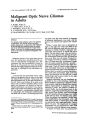
Show ,. Clill. Nl.'lIro-Clphtlwlllol. 5:258-262, 1985 © 1985 Raven Press, New York Cavernous Hemangioma of the Retina and Agenesis of Internal Carotid Artery with Bilateral Oculomotor Palsies MAY-YUNG YEN, M.D. CHIH-CHIOU WU/ M.D. Abstract Cavernous hemangioma of the retina is an unusual vascular hamartoma. It is recognized as a neurooculo- cutaneous syndrome. Association with systemic abnormalities is extremely rare. This report describes an 18-year-old man who presented with relinal cavernous hemangioma of the left eye and bilateral oculomotor palsies since early childhood. Cerebral angiography showed agenesis of the right internal carotid artery. Because of this unusual combination, we feel justified to report this case. Cavernous hemangioma of the retina is excular hamartoma comprised of sacular aneurysms containing venous blood and located on the surface of the retina or at the optic nerve head. It is usually unilateral. Fluorescein angiography demonstrates delayed perfusion and a characteristic appearance of plasma erythrocytic layering within the aneurysm.) Leakage of dye does not occur. Hemorrhages from it are rare and exudations have never been reported. The long-term course is believed to be nonprogressive. 2 Cavernous hemangioma of the retina is extremely rare. Only 70 cases have been reported in the Iiterature. 3 The first clinical case was reported in 1934 by Niccol and Moore.4 The first report of cavernous hemangioma of the retina associated with similar lesions of the skin and brain was published in 1940 by Weskamp and Cotlier.5 The histopathology of this tumor was described by Hogan and Zimmerman6 and by Kogan and Boniuk7 in 1962. In 1971/ Gass) recognized the lesion as a distinct entity and postulated the existence of a neuro-oculo-cuta- From the Department of Ophthalmology, Veterans General Hospital, Yang-Ming College, Taipei, Taiwan, Republic of China. Write for reprints to: M.-V. Yen, M.D., Department of Ophthalmology, Veterans General Hospital, Taipei, Taiwan. R.O.C. 112. 258 neous syndrome that consists of cavernous angioma of the central nervous system, cavernous hemangioma of the retina or optic nerve head, and cutaneous angiomas. Cavernous hemangioma of the retina in association with systemic abnormalities is rarer. 8,9 The patient described here has a cavernous hemangioma of the retina and bilateral oculomotor palsies. Agenesis of the right internal carotid artery was found by cerebral angiography. Case Report This 18-year-old male came to Veterans General Hospital on June 14/ 1983. asking for the correction of a squint that had been present since his childhood. According to his parents, the patient had had a squint and fair vision since sometime in early childhood. He was delivered spontaneously, and transient neonatal jaundice was noted after birth. His growth and development were normal. A picture taken at 5 years of age showed abnormal deviation of both eyes (Fig. 1). He was brought to see a few local eye doctors/ but was told only of amblyopia of the left eye. As he grew up/ an inferiority complex made him come to us for the correction of the squint. His family history was negative. Results of general physical examination were unremarkable. The patient turned his head to the left and used a right-sided gaze for fixation. His best corrected visual acuity was 20/30 in the right eye and finger counting at 3 feet in the left eye. Bilateral ptosis, exotropia, and limitation of extraocular movements were noted in all directions except lateral rectus (Fig. 2). The right eye had 4-mm ptosis with poor levator function. The right pupil was 4.5 mm and lacked direct and indirect light reflex. The left pupil was 3 mm. with sluggish direct light reflex. The conjunctiva/ cornea, lens, and vitreous of both eyes, as well as the fundus of the left eye, showed an Journal of Clinical Neuro-ophthalmology Figure 1. A picture taken at 5 years of age showed abnormal de~tion of both eyes. The head turned to the left side. aneurysmal mass lesion with grape-like clusters of dark cysts covered with grayish fibrous tissue lying over the area between macula and temporaJ lower branches of retinal vessels (Fig. 3). Fluorescein angiographic study showed delayed filling of the saccular lesion till the mid and late venous phases. There was no dye leakage. Perfusion of other retinal vessels were normal. The characteristic finding of plasma erythrocyte separation persisted until the background and retinal fluorescein had faded (Fig. 4). Because of bilateral oculomotor palsies, computed tomography of the brain was done. It turned out to be normal. Four-vessel cerebral angiography was performed by femoral catheterization. The right common carotid artery was relatively smaller than usual (Fig. 5). The right intemaJ carotid artery was not opacified (Figs. Pipue 2. The patient turned his head to the left side and used a right-sided gaze for fixation. December 1985 Yen, Wu Figure 3. The fundus of the left eye showed an aneurysmal mass lesion. with grape-like clusters of dark cysts covered by grayish fibrous tissue lying over the area between macula and temporal lower branches of retinal vessels. 6, 7). The right external carotid artery and the left common carotid artery and their branches were normally demonstrated. The intracranial branches on both sides were well opacified from left-side injection and showed normal caliber and distribution on both sides (Fig. 8). The vertebrobasilar arterial system was normally demonstrated (Fig. 9). There was no mass lesion noted. Because hemorrhages from retinal hemangioma are rare and the long-term course is believed to be nonprogressive, we decided to leave the retinal hemangioma alone and just correct the squint for abnormal head position. Figure 4. Fluorescein angiography showed the characteristic finding of plasma-erythrocyte separation. which persisted until the background and retinal fluorescein had faded. 259 Retinal Cavernous Hemangioma and Systemic Abnormalities Figure 5. The right common carotid artery was relatively smaller than usual. He received 8-mm lateral rectus muscle recession and 8-mm medial rectus muscle resection of the right eye on July 29, 1983. The head position improved moderately. On November 18, Figure 6. The PA view of right carotid angiography did not show the internal carotid artery. Only the external carotid artery and its branches were opacified. 260 Figure 1. The lateral view of right carotid angiography did not show the internal carotid artery and its branches. 1983, we tried to do superior oblique transposition, but we failed. We followed the patient regularly and no significant change has been noted since then. Discussion The patient showed a typical retinal cavernous hemangioma in his left fundus. The tumor was composed of dusters of saccular aneurysms filled with venous blood and presented the appearance of grapes projecting from the Figun 8. The intracranial branches of both sides were opacified. as seen on left carotid angiography, and showed normal caliber and distribution. Journal of Clinical Neuro-ophthalmology Yen, Wu dition, the middle and anterior cerebral arteries on the side of the absent vessels are most frequently supplied through the circle of Willis by the basilar artery and by the opposite internal carotid artery, 13 as in our case. Although the absence of one or both internal carotid arteries may be entirely asymptomatic under usual conditions, this type of anomaly has often been associated with intracranial aneurysm. 14 Symptoms such as epilepsy, transient weakness of limbs, and intracranial hemorrhage have been reported. IS Our case had cavernous hemangioma of the left fundus, bilateral oculomotor palsies, and agenesis of right internal carotid artery. The oculomotor palsies are the nuclear type. It may be due to small cavernous hemangioma in the rostral midbrain, which cannot be demonstrated by angiography. Another possibility is vascular insufficiency of the rostral midbrain because of agenesis of right internal carotid artery. Finally, these three rare things may be combined in our case just by figure 9. The vertebrobasilar arterial system was normally chance. demonstrated. References inner retinal surface. Separation of erythrocytes and plasma had occurred in some of the aneurysms. Fibrosis of some of the aneurysms has developed. There was no evidence of intraretinal exudation. Angiographic study demonstrated the vascular malformation isolated from the retinal circulation. Perfusion of the tumor was delayed and incomplete. Plasma--erythrocyte layering presented during the late phase of angiography. Gassl suggested that in some cases retinal cavernous hemangioma may occur in association with intracranial cavernous hemangioma as well as with angiomatous hamartoma of the skin. Cavernous hemangioma of the central nervous system is difficult to demonstrate by angiography or computed tomography, however. Proof of cerebral cavernous hemangioma has been difficult to obtain before death. The neurooculo- cutaneous syndrome has been documented in only a few cases.I,S,IO The association of systemic abnormalities (as congenital malformations of the heart and great vessels8) with the blue rubber bleb nevus syndrome9 seem to be rare and may occur by chance. Our case demonstrated agenesis of right internal carotid artery, which is extremely rare. Since Todell first described it in 1787, 60 cases of congenital absence of the internal carotid artery have been reported. It is a developmental anomaly that occurs before the 3-mm stage of human embryogenesis. 12 In this con- December 1985 1. Gass, J. M. D.: Cavernous hemangioma of the retina, a neuro-oculo-cutaneous syndrome. Am. ]. Ophthalmol. 71: 799-814, 1971. 2. Lewis, R. A., Cohen, M. H., and Wise, G. N.: Cavernous hemangioma of the retina and optic disc. A report of three cases and a review of the literature. Br. J. Ophtlralmo/. 59: 422-434, 1975. 3. Messer, E., Laqua, H., and Wessing, A.: Nine cases of cavernous hemangioma of the retina. Am. /. Opltlhalmol. 95: 383-390, 1983. 4. Niccol, W., and Moore, R. F.: A case of angiomatosis retinae. Br. /. Ophthalmo/. 18: 454-457, 1934. 5. Weskamp, c., and Cotlier, I.: Angioma cerebro y de la retina con malformaciones cupi lares de la pei!. Arch. Oplltha/mol. 15: 1-10, 1940. 6. Hogan, M. J., and Zimmerman, L. E.: Ophthalmic Pathology (ed. 2) Philadelphia: W. B. Saunders, 1962:533. 7. Kogan, L., and Boniuk. M.: Causes for enucleation in childhood with special reference to pseudogliomas and unsuspected retinoblastoma. Int. Ophtha/mol. Clin. 2: 507-524, 1962. 8. Colvard, D. M., Robertson, D. M., and Trautmann, J. c.: Cavernous hemangioma of the retina. Arch. Oplltl.almo/. 96: 2042-2044, 1978. 9. Crompton, J. L., and Taylor, D.: Ocular lesions in the blue rubber bleb naevus syndrome. Br. J. Ophtha/mol. 65: 133-137, 1981. 10. Schwartz, A. c., Weaver, R. G., Bloomfield, R., and Tyler. M. E.: Cavernous hemangioma of the retina, cutaneous angiomas and intracranial vascular lesions by computed tomography and nuclear magnetic resonance imaging. Am. J. Ophthalmol. 98: 483-487, 1984. 261 Retinal Cavernous Hemangioma and Systemic Abnormalities 11. Lie, T. A.: COl/gel/itaI anomalies of the carotid arteries. Amsterdam: Excerpta Medica, 1968, pp. 44-49. 12. Padget, D. H.: The development of the cranial arteries in the human embryo. Con/rib. Embryol. 32: 205-261, 1948. 13. Turnbull, I.: Agenesis of the internal carotid artery. N,'urology 12: 588-590, 1962. 262 14. Handa, J., Matsuda, I., Nakasu, S., and Nakano, Y.: Agenesis of internal carotid artery: Angiographic, tomographic and computed tomographic correlation. Neuroradiology 19: 207-211, 1980. 15. Huber, G.: Intracranial carotid anastamosis and partial aplasia of an internal carotid artery. Neuroradiology 20: 207-212, 1980. Journal of Clinical Neuro-ophthaImology |