| OCR Text |
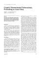
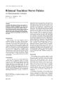
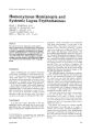
Show /. Gill. Nt'lIn1-tll,htlltl//l/11/. 5: 57-58, 1985. Bilateral Trochlear Nerve Palsies A Clinicoanatomic Correlate RONALD S. MURRAY, M.D. E. T. AIAX, t\t.D. Abstract A patient with bilateral trochlear nerve palsies is presented. Computed tomographic (Cn brain scan localized an anterior cerebellar vermis lesion compressing the area caudal to the inferior colliculi where the fourth nerves decussate and exit the dorsal brain stem. This lesion was probably responsible for the bilateral trochlear nerve dysfunction. Pertinent anatomy and pathologic involvement are discussed. Introduction Head trauma is the most frequent cause of acquired trochlear nerve pals\' followed, respecti\' el\·. by ischemic neuropath'y and neoplasm. l .' Pathologic im'ol"ement of the fourth nerve may occur at any point from its nuclear origin to the superior oblique muscle.~ Bilateral trochlear nerve palsies are extremely rare and ,usually produced b\' sen're frontal head trauma. Neoplasms have been known to compress the trochlear nerves simultaneousl\' as the\' decussate in and exit from the dorsal brain stem below the inferior colliculi. 4 In this case report, a single mass lesion, as demonstrated by CT scan, produced bilateral trochlear nerve palsies. Case Report A 74-year-old male was in good health until 5 months prior to admission when he became aware of vertical diplopia in positions of neutral and down gaze. This caused difficulty both with reading and when descending stairs. He could correct the diplopia by closing either eye. Soon thereafter, he noticed drooping of the left side of his mouth and progressive difficulty with left eye closure. General examination was unremarkable. Neurologic examination revealed a complete pe- From the Departments of Neurologv. Univl'rsitv of Utah Medical Center; and Veterans Administration Ml'dica' Cl'ntl'r. Salt Lake CltV. Utah March 1985 ripheralleft facial nerve paralysis, left hemIataxia, hyperactive lower extremity stretch reflexes, and a left Babinski sign. The patient reported, with a red glass over his right eye, visualizing the red light above the white light in primary and right lateral gaze. On red glass testing, maximal vertical separation of the images occurred upon looking down and to the right or left. As he followed the light from right to left, he reported an instantaneous reversal of red and white light positions such that the red light was below the white light as the midline was crossed. Observing the patient move his eyes in down gaze to the right and left revealed the emergence of a left or right hypertropia, respectively. Admission chest radiograph showed a right upper lobe mass. A head CT scan (Fig. 1.) showed an enhancing lesion in the anterior cerebellar vermis compressing the region beneath the inferior colliculi which was responsible only for the trochlear nerve palsies. The CT scan did not detect any lesion(s) to account for the patient's left Bell's palsy, left hemiataxia, and left Babinski sign. Biopsy of the pulmonary lesion was performed and pathology was consistent with an adenocarcinoma. Radiation therapy was instituted. Discussion The trochlear nerve's nuclear origin is at the level of the inferior colliculus and ventral to the cerebral aqueduct. The nerve crosses the midline dorsal to the cerebral aqueduct, then e'\its the dorsal brain stem caudal tll the inferillr colliculus and lateral to the frenulum of the anterillr med· ullary velum, then courses laterall\' in the cisterna ambiens passing between the supl'rillr cerebellar and posterior cerebral arteries. then around the cerebral peduncle. Prior to inl1l'T\'ating the contrala! l'ral superior oblique muscle, it traverses the caVl'rnous sinus and superior llrbital fissure, respectively. Pathologic involvement of the fourth nerve may occur at any point along its course. The most common etiologies are head trauma, ischemic neuropathy, neoplasms (primary or metastatic), and in rare instances demyelination, aneurysms, 57 Truchk,)f Nerve l'.llsll'S Figure 1. Brain CT scan demonstrates an enhanCIng mass (arTllws) in the antenor cerebellar venms wmpressmg the inferior colhcular region. The serial 4-mm sections begin in ascendmg urder WIth the nght upper cumer. then left upper curner, right luwer curner, and left lower curner, respectivelY In thiS regIl>n. the truchlear nerves are bemg cumpressed as they decussate and exit the dursal brain stem, aCClluntmg fllr the panent s bIlateral twchlear nerve palSIes. herpes zoster, mastoiditis, ethmoiditis, meningitis, and neurosurgical complications. ~ Severe head trauma is the usual cause of bilateral trochlear nerve palsies with the lesion proposed to be at anterior medullary velum, the site of the trochlear decussation.:' In our case, a presumed metastatic lesion in the anterior cerebellar vermis appeared to be compressing the dorsal brain stem in the area of the trochlear nerve decussation. In this location, a single lesion may produce bilateral trochlear nerve palsies. Prognosis for recovery of function in these cases is poor unless secondary to ischemic neuropathy. 1 ; , In summary, this case demonstrates by CT brain scan the unique anatomic location where a single lesion may produce bilateral fourth nerve dysfunction. 58 References Younge, BR, and Sutula, F: Analvsis of trochlear nerve pableS: Dlagnl)51s. etwlL)gv' and treatment. Maw ellll. Prl'" 52: 11-18, 1977. , I(hil\vam, L SCl)tt. AB., and Jampolsky, A.: Acquired superil)r ,)bhque palsy Arch. Ophtlltl/mol. 77: 7bl-7b8, 19b7 J. Burger, LJ I(ah'in, :\H. and Smith, J.L: Acquired leswns l)f the fl)urth cranial nef\·e. Bralll 93: 56757- l,1970 -l. LeIgh, IR, and Zee, OS: The NeUr,)/ogy of Eye Mll('elllellt. The Ltmtemporanl Nelir%~'Y Series, F.A. Dans, Philadelphia 1983, PI'. 169-171. 5 Rush, lA, and Younge, B.R.: Paralysis of cranial ner\'t's III. IV, and VI: Cause and' prognosis in 1.000 Cases. Ardl. Ophtha/mo/. 99: 76-79,1981. Journal of Clinical Neuro-ophthalmology |