| OCR Text |
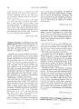
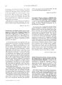
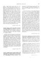
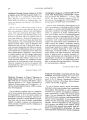
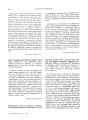
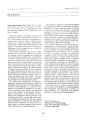
Show Jour/wi of Cli"iCIII Nl'IIn>-"I'''tlW[",olo,~1/ 6(41: 205-208, lY8b. Resolution of Optociliary Shunt Vessels Sydney L. Tyson, B.A., M.P.H., and Simmons Lessell, M.D. IY86 Raven Press, New York A -lO-year-old woman developed profound bilateral loss of vision irom chronic atrophic papilledema. There were striking optociliarv shunt vl'ssels on thl' right optic disc Within 1 ml1l1th ,liter remov,ll of a massive intracranial meningilmla, the shunt vessels had regressed markedly. This regression was evident on tluorescein angiograms. We attribute their resolution to the normalization oi intracranial pressure. Key Words: Brain tumor-Optic nerve-Optociliary veins- Papilledema. From the Neuro-ophthalmology Unit, Massachusl'tts EyL' and Ear Infirmary, Boston, Massachusl'lts. Address correspondence and reprint rl'LJul'sts III Dr. Simmons Lessell, 243 Charles St., Boston, MA lJ2114, USA 205 Acquired optociliary shunt veins are thought to develop as a result of progressive interference with outflow through the central retinal vein. This interference fosters the development of shunts between retinal and choroidal veins. The evolution of these bypass channels has been well documented by fluorescein angiography (1). However, little has been written on their resolution. We present the case of a patient whose optociliary shunt vessels had resolved completely 1 month after surgical decompression of a large sphenoid ridge meningioma. CASE REPORT A 40-year-old woman was well until June 1985, when she began to complain of transient obscuration of vision in her left eye. This was followed several months later by transient obscurations in her right eye and progressive loss of vision in both eyes. By December 1985, she was blind. The patient did not consult a physician until April 1986. His examination revealed bilateral visual loss with optic atrophy. A large contrast-enhancing mass, extending from the greater wing of the sphenoid on the right with edema and subfaIcial and uncal herniation, was imaged on a computerized tomographic (CT) scan (Fig. 1). Neuro-ophthalmologic examination was performed 1 week later. Visual acuity was no light perception in the right eye and light perception in the left. Exophthalmometry measured 23 mm on both sides, but the family reported that the patient's eyes had always been prominent. Motility examination was abnormal. The right eye tended to drift slowly during fixation in the primary position, and the left eye could not fully adduct, abduct, or depress. Slit-lamp examination showed normal anterior segments. Her pupils were equal, with no direct response on the right and a minimal response on the left. Ophthalmoscopy revealed bilateral secondary optic atrophy with prominent s /./rs( W liNn s. I.LSSLLL FIG. 1. (a) Axial CT scan showing large right hemispheric mass with surrounding edema and subfalclan herniatIOn (b) Coronal CT scan showing right hemispheriC mass with bilateral distention of OptiC nerve sheaths. llptociliarv shunt vessels lln the right llptic di:,c (Fig. 2). No shunt vesseb were seen lHl the left. Phvsical and neurllll)gical t':--aminatillns were nllrmaL In May 1980, a meningil)ma lli the right sphenoid ridge WdS ren1l1\'ed tlltally. Four week:, later, visual acuity was h,lnd motions in bllth eves, and the :,hunt \'essels had disappeared irllm tilt' right disc (Fig. 3). Minimal shunt vessel rl'sidu,) FrG. 2. Fundus of right eye showing chronic atrophic papilledema. Note shunt vessels. could be identitied l)n a ilul)rescein angiogram (Fig . .f). Tht' remainder l)i the e\.amination was unchanged. DISCUSSION Acquired l)ptl)ciliclrv \eins h,l\·e been described mllst irequenth· in cl)nditil)ns assl1Ciated with cllmpressil1n l)t the central rdinal \'ein: chronic FIG. 3. Postoperative appearance of right fundus, Shunt vessels have disappeared, RESOLUTION or OPTOc/UARY SHUNT VESSELS 207 FIG. 4. Fluorescein angiogram (arteriovenous phase) shows filling of small. residual shunt vessels. glaucoma (2--1). chronic papilledema (5). occlusion of the central retinal vein (6), and in tumors such as glioma (7) and optic nerve sheath meningioma (8-1-1). They also have been reported with drusen of the optic disc (15), arachnoid cysts of the optic nerve sheath (16), and phacomatosis (17). Four publications describe reduction in caliber of shunt vessels (18-21). One describes their regression after radiation therapy for optic nerve sheath meningioma (18). Two other reports noted partial regression after surgical decompression of the optic nerve sheath in patients with chronic papilledema (19,20). There is only one reported case of total resolution of shunt vessels (21). This occurred 11 days after surgical removal of a subfrontal meningioma. Postresolution angiography, if done, was not mentioned. No mention was made of the extent to which visual acuity was restored. Our case is quite similar, with nea; total resolution I month after surgical decompression. What is the pathogenesis of these abnormal shunt vessels? Two mechanisms can be postulated. Cogan (11) postulated that in optic nerve sheath meningiomas, shunt vessels may represent drainage from the tumor itself. In cases associated with venous obstruction, the shunts presumably represent enlargement of pre-existing capillary vessels bridging the retinal and choroidal circulations (10-12,14). Boschetti, Smith, Osher, Gass, and Norton (22) have reported that the fluorescein angiographic appearance of the optociliary shunt vessels in these two groups are characteristically different. In the optic nerve sheath meningioma group, it was noted that the shunt vessels filled earlier (in the arteriovenous phase), the flow drained to central venous tributaries, and the late staining was hyperfluorescent to other veins. However, in the central vein occlusion group, the shunts filled later (in the venous phase), showed a flow draining to the outer disc margin, and late staining was eufluorescent to other veins. In our patient, the fluoroangiographic appearance of the vessels was more typical of changes noted in patients with optic nerve sheath meningiomas. Fluoroangiography revealed early filling of shunts in the arteriovenous phase and drainage to central venous tributaries. We were not able to characterize the staining pattern in the late stages of angiography. Despite these analogous changes, there was no evidence of meningiomatous infiltration of the optic nerve in our patient. This discrepancy calls into question the value of fluoroangiography in differentiating their pathogenesis. What accounts for the decrease in the caliber of these shunt vessels? We speculate that in normal individuals, these channels are invisible, because pressure in the retinal and choroidal circulations is nearly equal. However, when pressure in the central retinal vein becomes greater than that in the uveal venous circulation, blood flow is diverted through these channels, and they become ophthalmoscopically and angiographically visible. When the obstruction of the central retinal vein is relieved, the differential pressure which initiated the development of these shunts falls, allowing regression. In our patient and in the reported cases with chronically elevated intracranial pressure (18-21), normalization of the intracranial pressure presumably relieved the obstruction of the central retinal vein in the subarachnoid space and restored the balance of pressure between retinal and choroidal circulations. The angiographically documented regression of shunt vessels after decompression reinforces the hypotheses that these shunts are formed from pre-existing vascular channels that connect retinal and choroidal venous systems, and that their appearance and disappearance are, in great part, dependent upon the balance of central retinal and choroidal Vl'11llUS pressures. REFERENCES 1. 1n1<'s RK, SCh,ltz H, Hllvt WF, Mllntiero MLR, Narahara M. EVlllllti,)Il "' llptllcili,ln; Vl'ins in llptiC nerve sheath meningillm, l. :11<// l )l'iJIIlIlllIl<l/ 14HS;1ll3:59-60. , Stellwag C Vlln C. LciJrlJ//ciJ der I'raktisd/ell AIIgCllI/ei/kliIlde. 2nd I'd. Vil'nn,l: Bralillmliller, 11'64:283. I ell1l NCIIW-lIp1lt/1Il1I11tl!. Vol. b, No.4, 19Sti 208 S. L. TYSON AND S. LESSELL J. Kraupa E. Oil' An,lstonlllsl'n an Papilll'n- und Nl'lzhautvenl'n. ;1,'['/1 /IIIXl'llll('//~ 1915;7H: IH2-207. -to Hill'hings RA, Sp,ll'lh Cl.. Chronic rdindl vein occlusion in gl,llICllm,l. HI I 01"111/111111,,/ I97h;hO:h')-t _l). 5. Hedges rl'. l'.lpilll'dl'lll,l. lis rl'l,llion 10 incrl'dsl'd inlr,]cr, 1I1i,]I pn'ssun'. SlIn' 01,111111//11I,,1 197'i; 19:201. h. II.l\'rl'h SS. l kclusion 01 till' n'nlrdl rdindl vl'ssl'ls. Hr I l1/'IIIIII/I/lI,,1 19h5;-t9:h2h. 7 • h'~'t WF, Bl'l'Stlll1 D. rlre (lelllllr fuudlls ill flt'ur%xic di ../,t1s/'. St. Louis: CV Mosbv. 19hhA2. H. Z,lkkd KA, SUlllllll'rl'r RW, Yl'l' RD, Foos [{Y, Kim J. Opticocili. JrY veins in ,] primary optic lll'rVl' slu'dlh Illl'ningioma. ;1/11 I 01,111//11/11I,,1 1979;H7:91. 9. M.Jrtin VAl', Schofield Pl'. Ml'ningioma invading Ihl' optic nerve. HI I 01'111111/1/1,,,1 1957;41:1hl. 10 Frisl'n L, Hoyt WI', Tengrolh BM. Oplociliary veins, disc pallor and visual loss. Ada 0l'lIIlIalm,,/ 197J;51 :241. II. Hollenhorst RW, Hollenhorst RW )r, McCarthy CS. Visual prognosis of the optic nerve sheath ml'ningiomas producing shunt vessels on the optic disc. Mall" ellll Pr"c 1971\;5J:1\-t. 12. Rodrigues MM, Savino 1'), Schatz N). Spheno-orbital meningioma with optociliary shunt veins. Am I 0l'lIll1alm,,/ 1976;1\1:666. 13. Ellenberger C. Perioptic meningiomas. Ardl Nell,,,l 1976;33:671. I Cli/l Ncuro-ophthnllllol. Vol. 6. No.4, 198b 14. Spencer WHo Primary neoplasms of the optic nerve and its sheaths. Clinical features and current concepts of pathogenetic mechanisms. Trans Am Ophlha/mol Soc 1972;70:490. 15. Karel I, Otradovec J, Peleska M. Fluorescein angiography in circulatory disturbances in drusen of the optic disc. 0l'h/llalllloloxica 1972;164:449. 10. Miller N, Green R. Arachnoid cysts involving a portion of the intraorbital optic nerve. Arch OphthalmoI1985;93:1117. 17. Zarel CR, Chromokos EA, Meisler OM. Cilio-optic vein associated with phacomatosis. Ophthalmology 1980;87(4): JJ(J-o. 1H. Smith)L, Vuksanovic MM, Yates BM, Bienfang DC. Radiation therapy for primary optic nerve meningiomas. JClin Nellro 0l'ht/w/lllo/19HI;1:85-99. 19. Galbraith )EK, Sulivan )H. Decompression of the perioptic meninges for relief of papilledema. Am J Ophthalmol 1973;70:01\7-92. 20. Perlmutter )c. Klingele TG, Hart WM, Burde RM. Disappearing optociliary shunt vessels and pseudotumor cerebri. Alii I OphlhalllloI1980;89:703-7. 21 Egger" HM, Sanders MD. Acquired optociliary shunt vessels in papilloedema. Br f Ophtha/moI1980;64:267-71. 22. Boschetti NV, Smith )L, Osher RH, Gass OM, Norton EWO. Fluorescein angiography of optociliary shunt vessels. f Clln Nell,o·ophlhalmoI1981;1:9-30. |