| OCR Text |
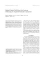
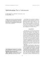
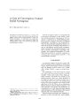
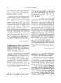
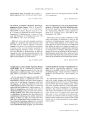
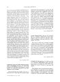
Show loumal of Cti/liml N"/Iro-ol'"t/lIIllIJology 6(3): 137-/43. 1986. Treatment of Optic Neuritis with Megadose Corticosteroids Thomas C. Spoor, M.D., F.A.C.S. ,\) 1986 Raven Press. New York Difficulty in distinguishing bt"twet"n monosymptomatic optic neuritis due to autoimmune disease or to intrinsic demyelination in young adults prompted a trial administration of megadose corticosteroids in a series of patients. Treatment with 1 g daily of intravenous methylprednisolone (Solu-Medrol) for 2 to 5 days led to the rapid resolution and rt"storation of visual function in five patients. Two untreated patients sufiert"d irreversible loss of vision. One patient with known autoimmune disease required a higher dose. The author suggests a trial of megadose. intravenous corticosteroids in patients between the ages of 21 and 45 years with optic neuropathies of uncertain or suspected autoimmune etiology after appropriate neuro-ophthalmologic evaluation. Address correspondence and reprint requests to Thom,lS C. Spoor. M.D.• F.A.C.S., Kresge Eye Institute. 3'1'14 John R.. Detroit, MI 48201 /37 Optic neuritis may result from inflammation of the optic nerve secondary to viral, demyelinating, or inflammatory disease. Demyelinating disease may cause an acute neuritis (1-4), and spontaneous recovery without treatment is the rule (1,2,5). Corticosteroids are thought to be of little benefit in altering the eventual visual outcome (6), but may shorten the clinical course (6-8). Dutton et al. (9) have described an autoimmune optic neuropathy and suggest screening patients with optic neuritis for an underlying autoimmune disease to discover those who may benefit from corticosteroid or immune suppressive therapy. Their three patients responded well to intensive pulse therapy with high-dose, intravenous methylprednisolone (Solu-Medrol) (9). Difficulty in distinguishing clinically demyelinating and autoimmune inflammatory optic neuritis, as well as poor visual results in two consecutive patients without treatment, prompted treatment of a series of patients presenting with optic neuritis of unknown etiology with megadose corticosteroids to determine which patients would benefit from this treatment. The results of treating optic neuropathies of unknown or uncertain etiologies in young adults with intravenous megadose corticosteroids are described herein. MATERIALS AND METHODS Patients between the ages of 21 and 45 presenting with acute visual loss due to an optic neuritis of unknown etiology were offered treatment. All patients were admitted to the hospital and underwent high-resolution computed tomography of the brain and orbits, neurologic consultation with lumbar puncture, and medical consultation with serologic tests for autoimmune diseases and syphilis. After the possibility of either an occult sinus infection or a compressive lesion had /38 r. C SPOOR been ruled out, patients were started on a 1 to 5 day regimen of pUISl' tlwrapy with intr,wenous nwthylpl"L'dnisololw. f.ive hundrl'd milligrams were ,1dministered initi,llly, followed by 25() mg l'\'l'rv 6 h. Visu,11 ,lcuities Wl'rL' followed daily, while visu,11 fil'lds Wl'rL' followl'd periodically. AtlL'r J to .5 d,1ys, corticoslL'wids werl' discontinued ,lbruptlv or a rapidly tapered oral dose was ,1dminislL'red. Tlw dosage of corticos!l'roids was diminished much more slowly in patients with documented autoimmune disease. No patients sultert'd significant complications from this regimen. Case 1 A 35-year-old woman was referred with an optic neuropathy in the left eye. Visual acuity was 20/20 in the right eye, and 20/50 - in the left eye. Visual helds demonstrated a central scotoma and inferonasal defect on the left, and a full field for the right eye. An afferent pupillary defect was present in the left eye. Funduscopy revealed a mildly swollen disc. An intlammatory optic neuropathY was presumed, and she was observed. One week later, visual acuity in the left eye had deteriorated to counting fingers and her visual field defect had progressed (Fig. 1). The afferent pupillary defect was more obvious and the disc was more swollen. Computed tomography with appropriate views and contrast was normal. Sedimentation rate was 10, antinuclear antibody (ANA) 1:20. A rheumatologist failed to find any clinical stigmata of systemic lupus erythematosus and advised against treatment with systemic steroids. After 3 months, visual acuity remained at FIG. 1. Case 1: deterioration of visual field after 1 week of observation. Visual acuity: counting fingers. I ellll ,.\J('Itrl l -I'J'liIlwlllWI, Vol. 6, No, 3, 1986 counting fingers and the central scotoma persisted. The disc swelling had resolved partially. ANA was 1:80, C3 complement decreased, sedimentation rate was 10 mm/h, and serum complement was normal. The patient declined systemic corticosteroids. When last examined, visual acuity remained at counting fingers with a dense central scotoma and secondary optic atrophy was present. Case 2 A 32-year-old woman was referred with a 1month history of decreased vision in the right eye, accompanied by periorbital pain. Examination revealed a visual acuity in the right eye of counting fingers at 2 feet. Visual acuity in the left eye was 20/20. An afferent pupillary defect was present in the right eye. Versions and ductions were full; the right disc was swollen with peripapillary hemorrhages. The left disc was normal. An inferior altitudinal defect involving fixation was present in the right eye (Fig. 2); the visual field of the left eye was normal. There were no other localizing neuro-ophthalmologic signs. Computed tomography was normal. There was no serologic evidence of autoimmune disease or syphilis. No treatment was offered. Visual acuity stabilized at 10/400 with a relative altitudinal defect and absolute central scotoma (Fig. 3). The disc was pale and atrophic. Case 3 A 48-~lear-old woman with known systemic lupus erythematosus, maintained on 40 ~g of in- FIG. 2. Case 2: inferior altitudinal defect involving~fixation. Visual aCUity: counting fingers. TREATMENT or OPTIC NEURITIS /39 FIG. 3. Case 2: visual field 1 year later with relative altitudinal defect and absolute central scotoma. Visual acuity: 10/400. tramuscular Depo-Medrol weekly, was hospitalized with a presumed lupus cerebritis presenting as slurred speech and dysarthria. Neuro-ophthalmologic examination documented visual acuity of 20120 in the right eye and 20/50 in the left eye with an afferent pupillary defect. A temporal field defect was present in the left eye. The visual field in the right eye was full. Computed tomography was normal. Thirty-six hours after admission, she developed a right sixth nerve palsy and no light perception acuity with an amaurotic pupil in the left eye. Fundus examination was normal. A diagnosis of posterior ischemic optic neuropathy secondary to an autoimmune vasculitis was made. She was treated with a 1.5 g bolus of intravenous methylprednisolone every 6 h. Eight hours later, visual acuity had improved to hand motion. The following morning, acuity was at counting fingers and the sixth nerve palsy had resolved 50(7". Methylprednisolone was decreased to 250 mg every 6 h. Twenty-four hours later, acuity had improved to 20/800. Two days later, the sixth nerve palsy had resolved, but acuity had decreased to counting fingers. Solu-Medrol was increased to 1 g every 6 h for 48 h, and acuity improved to 20/200. Administration of Cytoxan 100 mg and oral prednisone 100 mg daily was started and the dosage of intravenous methylprednisolone was rapidly tapered. One week later, the patient was discharged on prednisone and Cytoxan. Visual acuity was 20170. Two months later, visual acuity was 20/20 and visual fields were full. Very mild optic atrophy, nerve fiber layer dropout, and a 1+ afferent pupillary defect persisted. She presently maintains 20/20 acuity, and a full visual field. Case 4 A 45-year-old woman was referred for evaluation of a painful papillitis of 1 week's duration. Visual acuity was 20/25 in the right eye and 20/20 in the left eye. An inferonasal field defect sparing fixation was present in the right eye. The right disc was swollen, and peripapillary hemorrhages and exudates were present. The remainder of the examination was normal. Computed tomography demonstrated mild enlargement of the right optic nerve, compatible with an inflammatory optic neuropathy, but was otherwise normal. Serologic studies for syphilis and autoimmune diseases were negative. Sedimentation rate was 10. Spinal fluid revealed three lymphocytes but was otherwise normal. The patient followed without treatment. Two weeks later, visual acuity in the right eye had decreased to counting fingers. A 4 + afferent pupillary defect and right exotropia were present. A large, inferonasal defect involving fixation was present (Fig. 4). The left field remained normal. The right disc remained swollen with peripapillary hemorrhages; the left disc was normal. The patient was admitted and treated with a 500 mg bolus of intravenous methylprednisolone, followed by 250 mg every 6 h. Twelve hours later, visual acuity had improved to 201100. Two days later, acuity had improved to 20/40. After as-day course of intravenous methylprednisolone, treatment was discontinued abruptly and the patient was discharged. Visual acuity was 20/30. The following week, visual acuity had improved to 20/20. She maintained this acuity without medication for 20 months. FIG. 4. Case 4: visual field demonstrating an inferonasal defect involving fixation. 1Cl;" NCllro-ol,hthalmol, Vol. 6, No.3. 1986 140 r. c. SPOOR Case 5 A 28-year-old woman presented with retrobulbar n-euritis in the right eye. Visual acuity was 20/100 in the right l'yl', <1I1d 20/20 in the left ~y:. Compull'd tomography was normal. Spinal tlUld reveall'd a Iym phocytic pleocytosis, normal protein, and IgC with oligoclonal bands in the gamma zone. She was treated with a 500 mg bolus ot Intravenous methylprednisolone, followed by 250 mg every b h. T-he following day, acuity had improved to 20/50. A day later, 20/20 acuIty was attained. Steroids were discontinued abruptly due to abdominal pain. Extensive evaluation revealed an ileus which responded to medical management. Sedimentation rate was 6. Serologic studies revealed no evidence of autoimmune disease. Serum FTA-ABS was positive x 2. The patient was discharged on 2.4 million units of Bicillin weekly for 3 weeks. Acuity has remained 20/20. Case 6 A 41-year-old man was referred for evaluation of decreased vision in the left eye. In November 1984, visual acuity was documented as 20/40 in the left eye. Upon referral in April 1985, acuity in the left eye had deteriorated to counting fingers. Examination revealed visual acuity of 20/20 in the right eye and counting fingers in the left eye with an afferent pupillary defect. Visual fields were normal in the right eye; a superior altitudinal defect involving fixation was present in the left eye (Fig. 5). The right fundus was normal, but the left disc was obviously atrophic (Fig. 6). Com- FIG. 5. Case 6: visual field demonstrating a superior altitudinal defect involving fixation. Visual acuity: counting fingers O.S. , elm Nt'uro-ophtllllill/ol, Vol. b, No.1, 1986 puted tomography was normal.. Sedimen~ation rate and serologic testing for autOImmu.ne dIsease and syphilis were negative. S~inal flUid demonstrated a mildly elevated protem, but was otherwise normal. The patient was treated with a 500 mg bolus of intravenous methylprednisolone, followed by 250 mg every 6 h. The following morning, ac~ity had improved to 20/70. After a 4-da~ course of mtravenous methylprednisolone, aCUity ~as 20/25 a.nd the field was improved markedly (FIg. 7). SterOids were discontinued abruptly. The patient has maintained 20/25 acuity, an almost full field, and a pale disc for 16 months. Case 7 A 24-year-old woman was referred fo.r e~aluation of a retrobulbar neuritis. Visual acUity m the right eye was counting fingers and in the left eye was 20/20, and a 3 + afferent pupil was present. An absolute central scotoma was present in the right visual field; the left visual field was normal. The remainder of the neuro-ophthalmologic examination was normal. Both discs appeared normal. The patient was treated with a short course of oral prednisone. Over the following 3 weeks, acuity deteriorated to no light perception in the right eye. She was admitted for further evaluation. Examination revealed poor light perception in the right eye, a '* + afferent pupillary defect, and a mild swelling of the right disc. Computed tomography was normal. Sedimentation rate was 1, ANA was positive 1:80 with a speckled pattern. Additional serologic tests for autoimmune disease were negative. Spinal fluid revealed three lymphocytes but was otherwise normal. Rheumatology evaluation failed to reveal any systemic signs supporting the diagnosis of an autoimmune disease. An intravenous bolus of 500 mg methylprednisolone was given, followed by 250 mg every 6 h. After 3 days, visual acuity had improved to counting fingers. Visual fields were full peripherally, but an absolute central scotoma remained. The patient was discharged on 100 mg predisone a day, which was tapered over a month. Visual acuity improved to 20/200 and a central scotoma persisted in an otherwise full field. Biopsy of a facial lesion was considered consistent with the diagnosis of lupus erythematosus. Case 8 A 29-year-old man was referred for evaluation of rapid visual loss in his only functional eye. Ex- TREATMENT or OPTIC NEURITIS 141 FIG. 6. Case 6: normal disc 0.0. and obviously atrophic disc O.S. amination revealed hand motion acuity in the right eye and 20/60 acuity in the left eye, secondary to anisometropic amblyopia. A 4 + afferent pupillary defect was present in the right eye. The right visual field revealed a large nasal defect involving fixation; the left field was normal. The ANA was 1:20 with a speckled pattern (normal 1:20). Other serologic tests for autoimmune disease and syphilis were normal. A 500 mg intravenous bolus of methylprednisolone was given, followed by 250 mg every 6 h for 3 days. Acuity improved dramatically and the patient was discharged on 80 mg prednisone daily, which WilS tapered rapidly over 3 weeks. Two weeks after admission, visual acuity had improved to 20/15 in the right eye, and visual fields were full. DISCUSSION There are optic neuropathies that are exquisitely responsive to pulse therapy with high doses of intravenous methylprednisolone and patients whose vision deteriorates without such treatment. Some patients (cases 1, 3, 7, and 8) have equivocal serologic evidence of an autoimmune disease. However, only case 3 had unequivocal systemic 10111 Nl'lIro'0I"lti/Il'l/Io'. Vol. 6. No.3, 1986 l.Jl r. c. SPOOR FIG. 7. Case 6: markedly improved visual field O.S. atter 5-day course of intravenous corticosteroids. Visual acuity: 20/25. lupus erythematosus. Patients with optic neuropathies and unequivocal antoimmune disease may require a much higher dosage of methylprednisolone to improve their vision. One patient (case 5) had a reactive FTA-ABS test, nonreactive VORL, and no history of syphilis. This patient responded promptly to systemic methylprednisolone prior to receiving treatment for her reactive serology. False positive FTA-ABS tests have been described with autoimmune vasculitis. Cases 2 and 4 presented with papillitis and no serologic abnormalities. Case 2 developed permanent visual loss and optic atrophy. Case 4 had progressive deterioration of both visual acuity and field while being observed without treatment. She responded dramatically and completely to a short course of intravenous methylprednisolone. These patients may have had an autoimmune optic neuropathy and may in time develop serologic and clinical stigmata of an autoimmune disease. Case 6 had documented deterioration of vision and an atrophic disc. No radiologic, serologic, or cerebrospinal fluid abnormalities were evident. There was no evidence of systemic malignancv. He was treated reluctantly, only because he insisted on trying anything rather than lose his vision. There is no explanation for his rapid and gratifying response to treatment, unless an acute inflammatory process was superimposed upon a previously atrophic optic nerve. Cases 7 and 8 are similar to those described by Dutton et al. (9), presenting with retrobulb~r optic neuropathies and minimal serologic abnormalities. They both responded to a 3-day course of intravenous methylprednisolone, followed by sev- , elm Nt'llrn-(I11/,t/llllltlnl, Vol. b, No . .1, 1980 eral weeks of tapered prednisone. They may have had autoimmune optic neuropathies, but few rheumatologists will concur with the diagnosis based upon minimal serologic data. . Conversely, these patients may represent Instances of optic neuropathies due to intrinsic demyelination and demonstrate a rapid clinical response of the acute demyelinating process. to pulse therapy with intravenous methylprednISOlone. Similar beneficial responses have been reported in patients with relapsing multiple sclerosis, decreasing the duration of the acute demyelination, but producing no long-term benefit (11). Optic neuropathies secondary to intrinsic demyelination (multiple sclerosis) result in recovery of good vision in 75 to over 90(k of patients (1-3,8). Visual recovery usually starts after 7 days, and full recovery may take weeks to months. Ninety-six percent of patients younger than 45 years have been reported to regain an acuity of 20/30 or better, (1). Bradley and Whitney (2) reported that 5OCl< of patients with acute optic neuritis regained an acuitv of 20;30 or better within 1 month, and that ove'r 75'1< achieved this level of acuity after 6 months. Perkins and Rose (3) found that 87o/c of patients regained better than 20;-10 acuity at 6 months, while only 8';;, of patients had an acuity of 20/200 or worse at 6 months. These studies indicate that the majority of patients with optic neuritis secondary to intrinsic demvelination recover , , good functional visual acuity. However, 50 to 809, have some degree of optic atrophy (3,-1), and all patients reportedly develop a detectable nerve fiber layer defect after an episode of acute optic neuritis (-1). Dutton et al. (9) described a subgroup of patients with optic neuritis secondary to an occult autoimmune disease. Unlike patients with intrinsic demyelinating optic neuritis, these patients are more likely to suffer irreversible visual loss. Autoimmune optic neuritis mav be difficult to distinguish from intrinsic demy'elination, and autoimmune disease is known to coexist with multiple sclerosis (11,12), but this relationship may be unconvincing (9). Few patients manifest obvious clinical or serologic evidence of autoimmune disease when they initially present with decreased vision and an optic neuropathy, Those who do, often have very equivocal evidence of autoimmune disease. One patient described by Dutton et al. (9) did not develop a positive antinuclear antibody until 5 years after initial visual symptoms (case 2). In two of the three patients described by Hackett and co-workers (13), serologic evidence for autoimmune disease was not evident for 3 and TREATMENT OF OPTIC NEURITIS 143 2.5 years after developing an optic neuritis (cases 2 and 3, respectively). All of Hackett's (13) patients eventually fulfilled the diagnostic criteria for systemic lupus erythematosus as described by the Amp.rican Rheumatologic Association. The diagnosis of autoimmune optic neuritis must be based upon suspicion and screening of patients with optic neuritis for an occult autoimmune disease (9). The ophthalmologist should interpret serologic data liberally and treat these patients aggressively with a short course of intravenous methylprednisolone. It is apparent that some patients without serologic stigmata of autoimmune optic neuritis (cases 4, 5, and 6) may respond promptly and completely to this treatment. These results demonstrate that a short course of megadose intravenous methylprednisolone may be an efficacious and safe treatment for optic neuropathies of uncertain etiologies. REFERENCES 1. Earl. C. J., and Martin. B.: Prognosis in optic neuritis related to age. LlIJct'f 1: 7·t 1967. 2. Bradley. W. G.. and Whitne\,. C. M.: Acute optic neuritis: clinical features and prognusis for recovery of vision. f. Nelll'Ol. Nell roSIl rg. Psychiatry 30: 531. 1967. 3. Perkin, G. D., and Ruse, F. c.: Optic neuritis alld its differential diagllllSis. Oxfurd: Oxfurd Medical Publishers, 1979. 4. Miller, N. R.: Clinical Ill'lIrlJ-ophthalmology. 4th ed. Balti· more: Williams and Wilkins, 1982. 5. Rucker, C. W.: Sympusium-Diseases of optic nerve: optic neuritis of unknown etiology. Trans. Am Assoc OpltIIIIII/ llot OlolarYl/gol 60: 93, 1956. 6. Cohen, M. M., Lessell, S., and Wolf, P. A.: A prospective study of the risk of developing multiple sclerosis in uncomplicated optic neuritis. Nellrology 29: 208, 1979. 7. Bowden. A. N., Bowden. P. M., Friedman, A. I., Perkins, G. D., and Rose, F. c.: A trial of corticotropin gelatin injt'ction in acute optic neuritis. ,. Nellrol. NellrlJslIrg. Psychiatn! 37: 86Y, 1974. 8. Bi~d, A. c.: Is there a place for corticosteroids in the treatment of optic neuritis? In: Brockhurst. R. M.. Boruchoff, 5. A., Hutchinson. B. T., and Lessell, S., eds. COIltrOl'erSl/ ill ophthalmology. Philadelphia: W. B. Saunders, 1977: 882.' 9. Dutton, j. j., Burde, R. M.. and Klingele, T. G.: Autoimmune retrobulbar neuritis. Am. ,. Ophtlltllmol. 94: 11, 1982. 10. Holmes, K. K.: Syphilis. In: Thorn. G. W., et al., eds. Harrisolz's principles of il/temal medicine. New York: McGrawHill, 1981: 716-26. 11. Cendrowski. W., and Stepien, M.: Clinical variant of lupus erythematosus resembling multiple sclerosis. Ellr. Neurol. 11: 373. 1974. 12. Fulford. D.. Catterall. R. D.. and Delhanty. j. j.: A collagen disurder of the nervous system presenting as multiple sclerosis. Brain 95: 373, 1972. 13. Hackett, E. R.. Martinez. R. D.. Larson. P. F., and Paddison. R. M.: Optic neuritis in systemic lUpus erythematosus. Arch. Nellwl. 31: 9. 1974. I eli" Nl'lIro-ophthallllol. Vol. 6. No.3. 1986 |