| OCR Text |
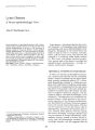
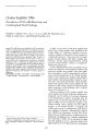
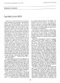
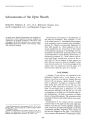
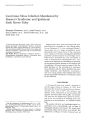
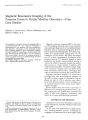
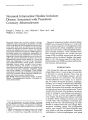
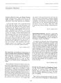
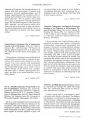
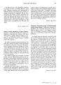
Show ]oumal of Clinical Neuro-ophlhalmology 7(4): 235-240, 1987. 'o 1987 Raven Press, Ltd., New York Magnetic Resonance Imaging of the Posterior Fossa in Ocular Motility Disorders-Four Case Studies Michael A. Lewis, M.D., Steven Goldstein, M.D., and Robert S. Baker, M.D. The superiority of magnetic resonance imaging (MRI) of the posterior fossa over x-ray computed tomography is demonstrated by four patients with neuro-ophthalmologic disorders. Advantages of this technique include lack of ionizing radiation, direct multiplanar imaging, increased sensitivity to pathology, increased tissue contrast, and no known adverse effects. It is concluded that MR, when available, should be the imaging modality of choice in patients with neuro-ophthalmologic disorders of suspected brainstem origin. Key Words: Magnetic resonance imaging-Posterior fossa-Supranuclear motility disorders. From the Departments of Ophthalmology, Neurology, Neurosurgery, and Pediatrics (M.A.L., R.S.B.) and Radiology (S.C), University of Kentucky Medical Center, Lexington, Kentucky. Address correspondence and reprint requests to Dr. R. S. Baker at Department of Ophthalmology, University of Kentucky Medical Center, 800 Rose Street, Lexington, KY 405360084, U.S.A. 235 Magnetic resonance imaging (MRI) is the newest CNS imaging technique used to aid anatomic and pathologic diagnosis of neuro-ophthalmologic disorders. Just as computed tomography (CT) replaced pneumoencephalography and polytomography and supplemented angiography for the evaluation of CNS disease, MR offers advantages over CT in many circumstances. Evaluation of the posterior fossa by CT is seriously limited by beam hardening and partial volume artifacts leaving a void in our ability to precisely localize the pathologic process in a large number of patients with ocular motility disorders of brainstem origin. Magnetic resonance imaging does not suffer from these drawbacks and has other advantages that make it well suited to studying the posterior fossa. Magnetic resonance imaging is a noninvasive technique that uses electromagnetic radiation in the radio frequency range. In contrast to x-ray radiation (as used in CT) no adverse tissue effects have been documented by current techniques or in experimental protocols using much higher magnet strengths. Several studies have shown the superiority of MRI over CT in imaging the posterior fossa (1-6). We present four case studies that exemplify the utility of MRI in the diagnosis, localization, and follow-up of neuro-ophthalmologic disorders. MATERIALS AND METHODS All patients were seen by the authors at the University of Kentucky Chandler Medical Center from 1984 to 1986. All CT scans were performed on a high resolution, fourth-generation scanner. Computed tomography was performed in an axial plane with 5 236 M. A. LEWIS ET AL. mm thick overlapping slices, and a 512 x 512 matrix was used for image reconstruction. Forty grams of iodinated contrast material was administered intravenously by rapid bolus infusion prior to CT. Magnetic resonance was performed on a 0.15 T resistive magnetic scanner operating at 6.25 mHz. Principles of MRI technique have previously been discribed (1,3,7). Briefly, two-dimensional multiecho data were obtained using both Tl- and T2weighted scan sequences. Typical spin-echo scan sequences included a repetition time (TR) of 2,120 ms and echo recovery times (TEs) of 30, 60, 90, and 120 ms. Inversion recovery sequences used a TR of 1,600 ms, a TE of 30 ms, and an inversion time (TI) of 400 ms. No contrast material was administered for MR. CASE REPORTS Case 1 A 70-year-old man awoke with right-sided weakness and a feeling of dizziness. Diplopia soon followed. Neurologic examination revealed slurred speech and right hemiparesis with an upgoing right toe. Sensation to pin prick was decreased on the right. Cortical sensory function was preserved. The right eye was in an abducted position when the patient fixated with the left eye in primary position. On attempted horizontal gaze the left eye failed to move to either side while the right eye had full abduction but decreased adduction. Both eyes abducted on upgaze, and downgaze evoked up beat nystagmus and was accompanied by extreme abduction of the left eye. Convergence could not be elicited. CT exam was normal. MRI revealed a focal lesion involving the left anterior pons which exhibited marked T2 prolongation on the spin-echo images, most likely an infarction (Fig. lA and B). Case 2 Over a period of 2 years, this 19-year-old boy noted increasing difficulty with diplopia on right gaze. At first the double vision was intermittent and noticed only in extreme right gaze, but over the next 2 years it became constant and was present with less deviation of the eyes. By early 1983, he was unable to look into the right field of gaze at all without diplopia. He consulted an ophthalmologist who diagnosed a right sixth nerve palsy. The CT scan, glucose tolerance test, thyroid function studies, and SMA-12 were obtained J n,~ r.~p"rt(-_'d ,)<; I'll '.rn1.11 FIG. 1. Case 1. Axial (A) and coronal (B) MRI with repetition time 2,120 ms and echo time 64 ms. There is an area of increased signal intensity (arrows) suggesting prolongation of T2 in the left anterior pontine tegmentum. Nine months before admission at the University of Kentucky, he developed progressive weakness of the left side, most noticeable during exercise. Immediately preceding admission he also noted mild dysphagia. On admission in October 1983 visual acuity was 20/20 in each eye and single binocular vision could be achieved only with a head turn to the left and chin tilted down. Ocular motility examination revealed a 16 diopter right esotropia in primary position, with limited abduction of the right eye. There was also vertical incomi- MRI OF POSTERIOR FOSSA 237 tance with 18 diopters of esotropia in downgaze and 0 in upgaze. The pupils, visual fields, and fundi were normal. Neurologic examination revealed minimal right facial weakness, a mild left hemiparesis, increased deep tendon reflexes that were brisker on the left, and a left extensor plantar response. Sensory examination was normal. CT scan performed with and without contrast media suggested an area of low density in the right pons with some evidence of mass effect. A metrizamide study was performed to confirm these findings. It demonstrated an enlarged pons, significant narrowing of surrounding cerebrospinal fluid cisterns, and indentation of the floor of the right side of the fourth ventricle. Subsequent angiography did not reveal evidence of abnormal vascularity. MRI revealed a lesion in the ventral aspect of the pons extending posteriorly into the middle cerebellar peduncle on the right. This lesion, which displaced the ventricle to the left, was best demonstrated on a heavily T2-weighted spin-echo sequence (Fig. 2A). On the initial MR study the mass demonstrated prolongation of both the II and T2 relaxation times and was thought to be a pontine glioma. The patient completed a course of radiation therapy and was followed both clinically and with imaging studies. Follow-up MRI performed 1 year later demonstrated resumption of a normal pontine contour, but with a residual region of abnormal signal intensity (Fig. 28). MRI performed 31J2 years after the initial diagnosis was thought to be entirely normal (Fig. 2C). Case 3 A 16-year-old had a 2 month history of diplopia, worse at distance than near, and photophobia. FIG. 2. Case 2. (A) Axial plane spin-echo MRI [repetition time (TR) 2,000 ms, echo time (TE) 90 msj demonstrates a high signal-intensity mass (arrow) involving the belly of the pons and displacing and compressing the fourth ventricle. (8) Saggital plane spin-echo image (TR 2,240 ms, TE 30 ms) performed 1 year later reveals a lesion with T2 prolongation in the ventral pons (arrows). There is little or no associated mass effect. (C) Spin-echo, axial plane MR scan (TR 2,120 ms, TE 32 ms) obtained 3% years follOWing initial diagnosis reveals normal anatomy. The fourth ventricle has resumed its normal position and configuration. I Clin Neuro-ol'lzthalmol, Vol. 7. No.4. 1987 238 M. A. LEWIS ET AL. On examination the visual acuity was 20/20 in each eye. Ocular motility showed limited adduction in each eye and abducting nystagmus on attempted lateral gaze. Quantitative direct current electro-oculography confirmed slow adduction and nystagmus in the abducting eye on lateral gaze to both sides. General neurological examination revealed a mild left facial palsy but was otherwise normal. Head CT with contrast enhancement showed no abnormality. MRI revealed a Chiari Type 1 malformation on sagittal view (Fig. 3). Metrizamide CT of the cervical spine and brainstem showed moderate to marked cervical medullary compression secondary to herniated cerebellar tonsils. The patient underwent a posterior fossa craniectomy with CI and C2 laminectomy, dural decompression, and insertion of dural graft. During the operative procedure, it was noted that the cerebellar tonsils were down to the level of C2. The diplopia gradually resolved and he is symptom-free 1 year after surgery. Case 4 A 13-year-old girl noticed the onset of numbness in her feet 5 weeks prior to admission to this hospital. This was followed by intermittent vom-iting, weakness in her arms and legs, diplopia, and difficulty with speech. On examination the pupils were reactive to light and accommodation. Ocular motility examination revealed bilateral internuclear ophthalmoplegia. Other neurological findings were hyperactive deep tendon reflexes, more on the left than right, bilateral extensor plantar reflex, clonus of left ankle, and left hemiparesis. CT scan with and without contrast showed no abnormality. MRI showed multiple discrete areas of T2 prolongation including right and left pons (Fig. 4), right cerebellum, right and left temporal lobes, right occipital lobe, right thalamus, and right and left periventricular parietal area. A diagnosis of demyelinating disease, probably multiple sclerosis, was made based on the history and MRI findings. After 2 weeks of steroids, neurological function improved markedly and ocular motility returned to normal. DISCUSSION These four cases exemplify the usefulness of MRI in the evaluation, localization, and follow-up of patients with neuro-ophthalmologic disorders originating in the posterior fossa. In three of the FIG. 3. Case 3. Spin echo MRI with slight T1 weighting (repetition time 530 ms. echo time 30 ms). Sagittal vIew shows tonsillar herniation (smaller arrows) through foramen magnum (larger arrow) to C2 level. MRI OF POSTERIOR FOSSA 239 FIG. 4. Case 4. Spin-echo scan (repetition time, 2,120 ms, TE 128) shows multiple areas of T2 prolongation, including bilateral pontine lesions (arrows) as well as right cerebellar and bilateral right temporal lobe lesions. Note areas of low signal surrounding the pontine lesions which represent edema. four cases no abnormality could be detected on CT scan, demonstrating the increased sensitivity of MRI. Other investigators have also found MRI to be equal or superior to CT in posterior fossa disease (1-6). MRI is emerging as the procedure of choice in the initial evaluation of suspected posterior fossa pathology. Case 1 had a unilateral left pontine infarction. The clinical findings included a one-and-a-half syndrome (8), characterized by a lateral gaze palsy and an internuclear ophthalmoplegia, gazeevoked upbeat nystagmus, skew deviation, and absent convergence. All of these findings have been previously reported in association with the one-and-a-half syndrome (9). The syndrome is due to a unilateral lesion in the lower dorsal pontine tegmentum, affecting the ipsilateral pontine paramedian reticular formation and ipsilateral medial longitudinal fasciculus. Involvement of the sixth nerve nucleus or fascicle can be determined by absence of vestibulo-ocular reflex on the affected side. Other brainstem findings, such as involvement of the corticospinal tract, may be seen such as in this case. The three-dimensional localization of the lesion by MRI correlated with the clinical findings (Fig. 1). In Case 2 CT and CT cisternography showed a pontine abnormality, but the extent of the tumor was much more clearly de-lineated with the MRI study. MRI is more sensitive than CT in detecting both primary and metastatic brainstem tumors (5,10-13). This case demonstrates two other advantages of MRI. The multiplanar imaging capability is valuable in showing the extent of brainstem neoplasms (10,12,13) which is an important consideration in radiation treatment. This patient underwent radiation treatment with tumor regression documented by serial MRI examinations. MRI is well suited to serial examination and for pediatric patients due to the lack of ionizing radiation or known side effects. A Chiari Type I malformation was demonstrated with sagittal MRI and not appreciated on axial CT scan in Case 3. Chiari Type I malformation often causes oculomotor abnormalities (14-16). This patient presented with diplopia and was found to have bilateral adduction weakness and abducting nystagmus (bilateral internuclear ophthalmoplegia). MRI has demonstrated imaging capabilities superior to CT at the craniocervical junction. It is considered unnecessary to do invasive positive contrast CT cisternography when MR shows a lesion in this area. Case 4 is typical of demyelinating disease, most likely multiple sclerosis. MRI has shown increased sensitivity over CT in detecting demyelinating disease (1,17-21), as this case demonstrates. In this patient, 11 plaques were identified throughout the CNS with MRI, whereas CT revealed none. The T2-weighted images proved most valuable in the detection of these demyelinating lesions. In addition to the overall increased sensitivity, delineation of the extent of the discrete plaques was possible. In particular, two pontine lesions and surrounding edema were shown corresponding to the ocular motility abnormality seen clinically. The radiofrequency pulse sequences in MRI are used to obtain images weighted toward T1 or T2. T1 is the spin-lattice relaxation time or longitudinal relaxation time. Tl is characteristic of the time required for the spin system to return to thermal equilibrium with its surroundings (the "lattice") after the exitation pulse ends. T2 is the spin-spin relaxation time or the transverse relaxation time. Spin-echo images are T2-weighted with TR and TE relatively long at 2,000 and 60 ms, respectively. Inversion recovery scans are T1-weighted with shorter TR and TE, and TI of 200-600 ms. In summary, we have presented four case studies that exemplify the utility of MRI in the diagnosis, localization, and follow-up of neuroophthalmologic disorders in the posterior fossa. J Cl,,, Nellr",,,!,"I",,ll1,,,l, V"l. I, No.4, 1981 240 M. A. LEWIS ET AL. The advantages of MRI demonstrated by these cases are lack of bone artifact, increased sensitivity to pathology because of increased tissue contrast in the brainstem, multiplanar imaging sequences, and selected weighting of tissue parameters. Future applications of MRI include tissue characterization with quantification of T1 and T2, assessment of blood flow, the development of contrast agents, and physiologic investigations. REFERENCES 1. Bydder GM, et al. Clinical NMR imaging of the brain: 140 cases. AINR 1982;3:459-80. 2. Brant-Zawadzki M, et al. NMR demonstration of cerebral abnormalities: comparison with CT. AINR 1983;4:117-24. 3. Young IR, et al. Magnetic resonance properties of hydrogen: imaging the posterior fossa. AIR 1981;137:895-901. 4. Flannigan BD, et al. Magnetic resonance imaging of the brainstem: normal structures and basic functional anatomy. Radiology 1985;154:375-83. 5. Jong SH, et al. Magnetic resonance imaging in the evaluation of the brainstem. Radiology 1984;150:705-12. 6. Bydder GM. Magnetic resonance imaging of the brain. Radial Clill North Am 1984;22:779-93. 7. Pykett IL, et al. Techniques and approaches to proton NMR imaging of the head. Compllt Radial 1983;7:1-17. 8. Fisher CM. Some neuro-ophthalmological observations. I Neural Neurosurg Psychiatry 1967;30:383-92. 9. Wall M, Wray SH. The one-and-a-half syndrome-a uni-lateral disorder of the pontine tegmentum: a study of 20 cases and review of the literature. Neurology 1983;33:97180. 10. Randell CP, et al. Nuclear magnetic resonance imaging of posterior fossa tumors. AINR 1983;4:1027-34. 11. McGinnis BD, Brady TJ, New PFJ, et al. Nuclear magnetic resonance (NMR) imaging of tumors in the posterior fossa. I Comput Assist Tomogr 1983;7:575-84. 12. Lee BCP, et al. MR imaging of brainstem tumors. AINR 1985;6:159-63. 13. Hueftle MG, Han JS, Kaufman B, Benson JE. MR imaging of brainstem gliomas. I Compllt Assist Tomogr 1985;9:263-7. 14. Paul KS, et al. Arnold-Chiari malformation. Review of 71 cases. I Nellrosurg 1983;58:183-7. 15. Passo M, et al. Acquired esotropia. A manifestation of Chiari I malformation. I Clill Neura-ophthalmoI1984;4:151-4. 16. Yee RD, et al. Episodic vertical oscillopsia and downbeat nystagmus in a Chiari malformation. Arch OphthalmoI1984; 102:723-5. 17. Young IR, Hall AS, Pallis CA, et al. Nuclear magnetic resonance imaging of the brain in multiple sclerosis. Lancet 1981;2:1063-6. 18. Buonanno FS, Kistler JP, Lehrich JR, et al. IH nuclear magnetic resonance imaging in multiple sclerosis. Neural Clill 1983;1:757-64. 19. Young IR, Randell CP, Kaplan PW, James A, Bydder GM, Steiner RE. Nuclear magnetic resonance (NMR) imaging in white matter disease of the brain using spin-echo sequences. I Compllt Assist Tomogr 1983;7:290-4. 20. Lukes SA, Crooks LE, Aminoff MJ, et al. Nuclear magnetic resonance imaging in multiple sclerosis. Alln Neural 1983; 13:592-601. 21. Jackson JA, Leake DR, Schneiders NJ, et al. Magnetic resonance imaging in multiple sclerosis: results in 32 cases. AINR 1985;6:171-6. |