| OCR Text |
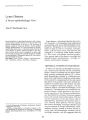
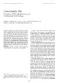
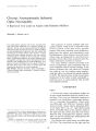
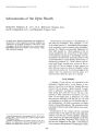
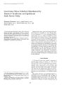
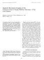
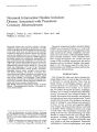
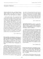
Show Journal of Clinical Neuro-ophthalmology 7(4): 219-222, 1987. Schwannoma of the Optic Sheath Richard K. Simpson, Jr., M.D., Ph.D., Richard L. Harper, M.D., Joel B. Kirkpatrick, M.D., and Benjamin Cooper, M.D. {, 1987 Raven Press, ltd., New York An optic nerve sheath Schwannoma was surgically removed from a child. Magnetic resonance imaging was valuable for the surgical localization of the tumor. This is thought to be the first reported case of such a tumor. Key Words: Optic nerve sheath-Schwannoma-Magnetic resonance imaging. From the Departments of Neurosurgery (R.K.S., R.L.H.), Pathology (J.B.K.), and Neurology (B.C.), Baylor College of Medicine, Houston, Texas. Address correspondence and reprint requests to Dr. R. K. Simpson, Jr. at Department of Neurosurgery, Baylor College of Medicine, One Baylor Plaza, Houston, TX 77030, U.S.A. 219 Schwannomas (neurinoma or neurilemoma) of the orbit are uncommon. They comprise -1-6% of all orbital tumors (1). Intraorbital schwannomas most frequently occur in patients with neurofibromatosis (1). Patients are generally diagnosed in their fifth decade (1). There appears to be no sexual predilection (2). Occurrence of this tumor in the pediatric age group is exceedingly rare (2). Intraorbital schwannomas generally present with visual impairment, ocular motility dysfunction, or disfiguring proptosis (3). These tumors can become quite large, causing bony erosion of the orbital walls (4). Neural origins of these tumors are often obscure, however, most are thought to arise from ciliary branches (3). To the knowledge of the authors, this case represents the first report of a schwannoma within the optic nerve sheath. CASE REPORT A healthy ll-year-old boy was admitted to the Methodist Hospital with a I-year history of progressive visual loss in his left eye. Initially this was treated with eye glasses. Approximately 3 weeks prior to admission, he began to complain of difficulty seeing faces with his left eye. He also complained of mild intermittent headaches and scotoma. On the night prior to admission, he could only perceive dim light with his left eye. He had no symptoms in his right eye. He denied other ocular disturbances. He had no personal or family history of neurofibromatosis. The remainder of his past medical history and family history were unremarkable. Examination of his cranial nerves revealed normal vision in his right eye but only light perception in his left eye. Direct pupillary responses were normal on the right and sluggish on the left. Consensual pupillary responses were normal on the left and sluggish on the right. A left MarcusGunn reaction was observed. Funduscopic exami- 220 R. K. SIMPSON, JR. ET AL. nation showed left optic atrophy. No papilledema was observed. Ocular motility was normal. There was no proptosis and no vertical or horizontal displacement of the globe. The remainder of his neurological examination and his general physical examination were normal. There was no evidence of neurofibromatosis. A computed tomogram (CT) of his head revealed an acutely angulated and dilated left optic nerve near the orbital apex (Fig. lA). There was also some enlargement of the optic foramen. A magnetic resonance image (MRI) of his head, using a T1 weighted pulse sequence, showed a widened left optic nerve in the distal optic canal at the apex of the orbit (Fig. 16). A discrete mass was not visualized. However, separation of the optic sheath from the optic nerve adjacent to the angulation was seen. This suggested an extra-axial mass within the sheath. A T2 weighted pulse sequence revealed an abnormal increase in signal intensity from the intraorbital portion of the left optic nerve (Fig. lC). This signal suggested an engorged optic sheath secondary to CSF blockade. A cerebral arteriogram was normal. The patient was taken to the operating room where a left frontotemporal craniotomy was performed. The orbital roof was removed overlying the optic canal using the microscope and air drill. The optic sheath appeared normal. The sheath was incised in its midline from the intracranial foramen through the annulus of Zinn and into the orbital apex (Fig. 2). A small elevation along the inferomedial border of the optic nerve was visualized through the arachnoid. This appeared to cause optic nerve angulation -3-4 mm posterior to the annulus of Zinno The optic nerve was abnormally widened, with considerable vascular engorgement evident immediately above the mass. Upon opening the arachnoid, a mobile, well-encapsulated, reddish-black mass was found. The tumor was in the optic canal extending into the apex of the orbit. The optic nerve appeared normal proximal and distal to the lesion. The tumor (-3 x 5 x 7 mm) was totally excised, leaving the optic nerve and its pia intact and free of angulation and compression. Examination of the removed tissue, by light microscopy, showed conclusively that the neoplasm FIG. 1. Computed tomographic (CT) evaluation revealed an angulated left optic nerve and slightly enlarged optiC foramen (A). T1 weighted magnetic resonance images reveal exaggerated displacement of the left optic sheath from the nerve (B). High signal intensity was recorded from the left optic nerve in T2 weighted images (C). OPTIC SHEATH TUMOR 221 FIG. 2. Intraoperative photograph (top) with drawing (bottom) demonstrates the orbital fat (a), suture through the severed annulus and levator (b), the tumor (c), the optic nerve (d), and the carotid artery (e). was a schwannoma. Fusiform nuclei forming palisades around anuclear zones of distinct fibrillarity (Verocay bodies) were seen (Fig. 3). Antoni A and B tissue patterns were observed. Examiantion of the ultrastructure revealed prominent basement lamina surrounding tumor cells. This patient's postoperative course was uneventful. On the day following surgery, visual acuity testing showed that light perception in his left eye was unchanged. However, on the day of discharge, 6 days later, he could detect forms and perceive gross movements. Three weeks postoperatively, he could count fingers placed in his inferior nasal field. DISCUSSION Only 55 cases of solitary, intraorbital schwannoma have been clearly documented (3). Such tumors can occur in 1.5-18% of patients with neurofibromatosis (1). However, intraorbital schwannomas are exceedingly rare in the absence of this disease (1). Infrequently, cases of these tumors have been described in the pediatric age group (2). The present case is the youngest patient known to have had an intraorbital schwannoma. Schwannomas generally arise from sensory nerves (3,5). They may originate from sympathetic twigs traveling with either motor nerve branches or blood vessels (2). Although intracranial schwannomas most commonly arise from the vestibulocochlear nerve, they have been found in association with cranial nerves I, III, IV, V, VII, IX, and XII (6-11). Intracerebral origins have also FIG. 3. Light microscopy revealed morphological features consistent with schwannoma. Hematoxylin and eosin. x 248. I Clill Nellro-opiltilal",o[, Vol. 7. No.4. 1987 222 R. K. SIMPSON, JR. ET AI. been described (12). Intraorbital schwannomas arising from the globe are highly uncommon (4). Infraorbital, supraorbital, and lacrimal nerve origins have been reported (3). However, ciliary branches are thought to be the most common intraorbital site of origin (13). There has been no case of such a tumor originating from within the optic sheath. Early anatomical investigations reported that schwannomas cannot occur in the optic nerve (4). Indeed, previous studies have suggested that the term schwannoma should not be applied to optic nerve pathology (4). However, sympathetic nerves, which are accompanied by Schwann cells, innervate the vasculature of the orbit (14). This also includes unmyelinated fibers innervating the central retinal artery (14). The possibility of an optic nerve schwannoma was recognized in the early part of this century (4). However, a definitive example is not available. A putative optic nerve schwannoma has been described in India, but the pathological description closely resembles a glioma (15). Clinical features of this tumor lack distinctive diagnostic characteristics (13). Radiological studies, however, are diagnostically helpful if the intraorbital lesion is relatively large. CT of the orbit has become the procedure of choice for the evaluation of intraorbital masses (3). Most schwannomas are imaged as well-defined, rounded masses that moderately enhance with intravenous contrast (13). Because this patient's tumor was small, a discrete mass was not visualized by CT. Only displacement of the optic nerve was seen. Although MRI also failed to image a well-defined mass, the exact location of the extraaxial tumor was determined by accurate resolution of the optic sheath and nerve. The microscopic appearance is the same as for schwannomas of peripheral nerve origin, including Antoni A and B patterns, Verocay bodies, and thickened hyalinized vessles (2,16). These characteristics distinguish schwannomas from other intraorbital tumors, such as optic nerve gliomas or neurofibromas (17). An optic nerve glioma generally causes diffuse enlargement, not simply displacement of the nerve. Cellular polarity, microcystic changes, and Rosenthal fibers characteristic of optic nerve glioma were not seen in this case. The tissue also lacked the diffuse, patternless proliferation of Schwann cells within a loose collagen matrix indicative of a neurofibroma. Although optic nerve meningiomas can cause nerve displacement, characteristic nests of menin- I UIIJ gothelial cells, whorls, and psammoma bodies were not evident in the specimen (17). Intraorbital schwannomas are benign solitary masses (4). The prognosis for intraorbital schwannoma is favorable. However, if visual loss has occurred, functional recovery may be limited (2). These tumors can recur after prolonged asymptomatic intervals (16). In general, visual loss is a late and often minor manifestation of an intraorbital schwannoma (3). Because of the tumor location, visual loss was the presenting symptom in the present patient. This case clearly emphasizes the need for careful evaluation of progressive visual loss in children. Vision can be preserved or visual loss can be limited by early recognition and surgical treatment of small intraorbital tumors. REFERENCES 1. Konrad EA, Thiel HJ. Schwannoma of the orbit. Ophthalmologica 1984;188:118-27. 2. Schmitt E, Spoerri O. Schwannomas of the orbit. Acta Neurochir 1980;53:79-85. 3. Cantore G, Ciappetta P, Raco A, Lunardi P. Orbital schwannomas: Report of nine cases and review of the literature. Neurosurgery 1986;19;583-8. 4. Standal B. Neurinoma of the orbit. Acta Ophthalmol 1950; 28:49-70. 5. Revilla AG. Neurinomas of the cerebellopontine recess. Bull Johns Hopkins Hosp 1947;80:254-96. 6. Fink LH, Early CB, Bryan RN. Glossopharyngeal schwannomas. Surg NeuroI1978;9:239-45. 7. Kansu T, Ozcan OE, Ozdirim E, Onol B, Gurcay O. Neunnoma of the oculomotor nerve. Case report. J Clin NeuroophthalmoI1982; 2:271-2. 8. King JS. Trochlear nerve sheath tumor. Case Report. JNeurosurg 1976;44:245-7. 9. Lilliequist B, Thulin CA, Tovi 0, Wiberg A, Ohman J. Neunnoma of the labyrinthine portion of the facial nerve. Case report. J Nenwsurg 1972;37:105-9. 10. Schisano G, Olivecrona H. Neurinomas of the gasserian ganglion and trigeminal root. J Neurosurg 1960;17:306-22. 11. Scott M, Wycis HT. Intracranial neurinoma of the hypoglossal nerve. Successful removal. Case report. J Neurosurg 1949;6:333-6. 12. Ghatak NR, Norwood CW, Davis CH. Intracerebral schwannoma. Surg NeuroI1975;3:45-7. 13. Schields JA, Kapustiak J, Arbizo V, Augsburger JJ, Schnitzer RE. Orbital neurilemmoma with extension through the superior orbital fissure. Arch Ophthalmol1986; 104:871-3. 14. Anderson DR, Hoyt WF. Ultrastructure of intraorbital porhon of human and monkey optic nerve. Arch Ophthalmol 1969;82:506-30. 15. Kulkarni RG, Satalkar VY, Bhawthankar AW. Neurilemmoma of the optic nerve. Indian JOphthalmoII980;28:95-6. 16. ChIsholm lA, Polyzoidis K. Recurrence of benign orbital neurilemmoma (schwannoma) after 22 years. Canad JOphthalnIOI1982; 17:271-3. 17. Burger, PC, Vogel FS. Surgical pathology of the nervous system and its coverings. 2nd ed. New York: Wiley and Sons. 1982;84-106,292-301,663-99. |