| OCR Text |
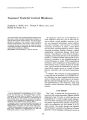
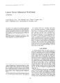
Show Journal of Clinical Neuro-ophthalmology 7(3): 125-128, 1987. Third Nerve Palsy as a Presenting Sign of Acquired Immune Deficiency Syndrome Michael V. Antworth, M.D., and Roy W. Beck, M.D. © 1987 Raven Press, Ltd., New York The case of a 29-year-old former parenteral drug abuser who presented with a 3rd nerve palsy and contralateral ataxia is reported. The patient was found to have a positive HLTV-III titer and acquired immune deficiency syndrome was diagnosed. Computed tomography demonstrated two ring-enhancing lesions in the brain which were presumed to be due to Toxoplasma gondii. Key Words: Acquired immune deficiency syndromeThird Nerve Palsy-Toxoplasmosis. From the Departments of Ophthalmology (M.V.A., RW.B.), Neurology (RW.B.), and Neurosurgery (RW.B.), University of South Florida College of Medicine, Tampa, Florida. Address correspondence and reprint requests to Dr. R W. Beck at Department of Ophthalmology, University of South Florida College of Medicine, Tampa, FL 33612, U.S.A. 125 Acquired immune deficiency syndrome (AIDS) is characterized by profound depression of the cellular immune system (1,2). Patients with AIDS usually present with lymphadenopathy, disseminated Kaposi's sarcoma, or an opportunistic infection (3-6). Ocular manifestations of AIDS include cotton wool spots, cytomegalovirus retinitis, conjunctivitis, keratitis, and conjunctival Kaposi's sarcoma (7-10). However, ocular complaints as the initial manifestation of AIDS are rare (11). Neurologic complications in AIDS are frequent and may be due to toxoplasma gondii, progressive multifocal leukoencephalopathy, lymphoma, subacute encephalitis, and peripheral neuropathy (12-14). We present a patient who developed an oculomotor palsy as the initial manifestation of AIDS. CASE REPORT A 29-year-old man was in his usual state of health when he developed diplopia after being struck on the left side of the face with a heavy object. He was dazed but did not lose consciousness. The diplopia persisted, and 1 week later he was seen in an emergency room. Limitation of up gaze in the left eye was observed and a possible blow out fracture of the orbit was diagnosed. His pupils were noted to be equal in size and reactivity. Two weeks later the patient was seen in the ophthalmology clinic because ptosis of the left upper lid had developed. A history of parenteral cocaine abuse 2 years previously was elicited. Homosexual relations were denied. On ophthalmic examination visual acuity was 20/25 and the visual field was full in both eyes. The right pupil was 4 mm and briskly reactive both directly and consensually, whereas the left pupil was 8 mm and nonreactive. There were 6 126 M. V. ANTWORTH AND R. W. BECK mm of left ptosis and an inability to adduct, depress, or elevate the left eye (Fig. 1). The right lids and eye movements were normal. Funduscopic and slit lamp examinations were unremarkable. General physical and neurologic examinations revealed a slightly lethargic thin man with a temperature of 100.3°F. He exhibited a wide-based gait with a tendency to drag his right foot. His strength was equal bilaterally in upper and lower extremities. He had positive drift of the right upper extremity and showed poor finger-nose coordination on the right side. Oral lesions suggestive of candidiasis were noted. There was no nuchal rigidity. A hematocrit was 40.2% and white blood cell count 2,800 with 66% polymorphonuclear cells, 24% lymphocytes, and 10% mononuclear cells. Computed tomography (CT) detected two ring enhancing lesions with surrounding edema: a 1.5 em area in the left thalamus extending downward into the midbrain (Fig. 2) and a 0.75 em area in the right parietal area. Subsequent laboratory studies included a nonreactive VORL and negative blood, urine, and sputum cultures. A serum toxoplasmosis IgG titer was greater than 1:400 and an HTLV-III antibody titer was positive. A lumbar puncture was not performed. A stereotactic brain biopsy of the midbrain lesion was performed but was nondiagnostic. Four days later a right third nerve palsy was noted in addition to the left third nerve palsy. A presumptive diagnosis of toxoplasmosis was made and he was treated with a 6 week course of sulfadiazine (500 mg q.i.d.), pyramethamine (25 mg q.d.), leukovorin (10 mg q.d.), and dexamethasone (14 mg q.i.d.). There was eventual resolution of the ring- FIG. 1. Clinical photograph shows left ptosis, exo1,-;:,,..,;,,,. and hypertropia There was an inability to ado •• ,-'e-'::l!'" the left eye. enhancing lesions and edema on CT. When he was transferred to an extended care facility, there had been no change in the third nerve palsies. Further follow-up is not available. DISCUSSION Much of the morbidity and mortality of AIDS may be attributed to the neurologic sequelae of the disease. Two large series of AIDS patients found that neurologic symptoms occurred in 30-39% (14,15). Approximately 10% of patients develop neurologic symptoms prior to AIDS being diagnosed (16). Autopsy studies have demonstrated central nervous system involvement in 73% of patients dying with AIDS (15). Toxoplasma gondii is one of the more prevalent opportunistic central nervous system infections in the immunocompromised patient (17). The inability to mount a cell-mediated response to this obligate intracellular protozoan may result in encephalitis, often with multiple necrotic abscesses. Histologically, these focal areas of involvement show large necrotized granulomas with thin capsules and little inflammation (18). Computed tomography may detect lesions that are ring enhancing, solid enhancing, or even nonenhancing with focal edema. Approximately 50% of these lesions are located in the basal ganglia. Other sites include the white matter- gray matter interface and the white matter itself (14,15,19,20). It is important to note that Levy et al. reported two cases in which magnetic resonance imaging detected lesions not seen on CT (15). A positive biopsy whether open or needle is certainly an aid to diagnosis of toxoplasmosis. However, there are cases reported such as ours in which biopsy was negative and toxoplasmosis was a presumptive diagnosis. Levy et al. (15) have advocated biopsy of accessible lesions whereas Pitchenik et al. (21) and Elkin and Leeds (22) have recommended empirical treatment. Significant short-term improvement has been reported using pyrimethamine and sulfadiazine. This was the case with our patient. Our patient is unique in that a focal ring-enhancing abscess situated in the region of the red nucleus was responsible for his unilateral third cranial neuropathy and contralateral ataxia (Benedickt's syndrome: ipsilateral third nerve palsy with contralateral rubral tremor and ataxia). It was the third nerve lesion that led him to seek medical attention. The pattern of eye muscle impairment THIRD NERVE PALSY AS A SIGN OF AIDS 127 FIG. 2. Computed tomography scans demonstrate a ring-enhancing lesion in the left thalamus (A) extending into the midbrain (B). indicated that the nerve fascicle rather than the nucleus was affected. It is interesting to speculate on the relationship of the blow to his head and the development of the third nerve palsy. Although it is possible that the two were a chance occurrence, third nerve palsy following relatively minor head trauma has been previously reported in association with intracranial masses (23,24). Most of these cases have involved basicranial tumors, and it has been speculated that the tumor tethers the nerve, making it susceptible to a minor blow. In our case the third nerve was presumably involved within the brainstern. It is possible that the blow to the head produced downward displacement of the brainstem due to the pressure differential created above and below the foramen magnum. With this movement the fascicle of the nerve may have been damaged because of the mass effect of the abscess on the nerve. In summary this case is presented to illustrate the possibility that AIDS may prevent in a multitude of ways. The ophthalmologist when confronted with an unusual neuro-ophthalmologic finding should consider AIDS as a possible diagnosis. REFERENCES 1. Gottlieb MS, Schroff R, Schanker H, et a!. PnelmlllclfstiS carInii pneumonia and mucosal candidiasis in pre'viously healthy homosexual men: evidence of a new severe acquired cellular immunodeficiency. N Engl J Mcd 1981; 305:1425. 2. Fauci AS, Macher AM, Longo DL, et a!. Acquired immunodeficiency syndrome: epidemiologic, clinical, immunologic and therapeutic considerations. AmI Intem Med 1984; 100:92-106. 3. Masur H, Michelis MA, Greene }B, et a!. An outbreak of community-acquired Pllel/lIlocysti, carillii pneumonia: initial manifestations of cellular immune dysfunction. N Ellgi J Med 1981;305:1431-8. 4. Siegel FP, Lopez C. Hammer GS, et a!. Severe acquired immuno-deficiency in male homosexuals, manifested by chronic perianal ulcerative herpes simplex lesions. N Engl J Mcd 1981;305:1439-8. 5. Friedman-Kien AE, Laubenstein L). Rubinstein P, et a!. Disseminated Kaposi's sarcoma in heterosexual men. Ann III tern Med 1982;96:693-703. 6. Center for Disease Control Task Force on Kaposi's Sarcoma and Opportunistic Infections. Epidemiological aspects of the current out break of Kaposi's sarcoma and opportunistic infections. N Ellgll Med 1982;306:248-52. 7. Khadem M, Kalish SB, Goldsmith). et aL Ophthalmologic findings in acquired immune deficiency syndrome (AIDS). Arch Ophthalmo/1984;102:201-6. 8. Rosenberg PR, Uliss AE, Friedland GH, et aL Acquired immunodeficiency syndrome. Ophthalmic manifestations in ambulatory patients. Ophthalmology 1983;90:874-8. 9. Holland GN, Gottlieb MS, Vee RD, et aL Ocular disorders I C1111 Nt'Ilro-0l'htilillmol, Vlli. 7. No.3, 1987 128 M. V. ANTWORTH AND R. W. BECK associated with a new severe acquired cellular immunodeficiency syndrome. Am' Ophtfullmol 1982;93:393-402. 10. Holland GN, Pepose JS, Pettit TH, et al. Acquired immune deficiency syndrome. Ocular manifestations. Ophthalmology 1983;90:859-73. 11. Weiss A, Margo CE, Ledford OK, et al. Toxoplasmic retinochoriditis as an initial manifestation of the acquired immune deficiency syndrome [Letter]. Am , Ophthalmol1986; 101:248-9. 12. Wong B, Gold IWM, Brown AE, et al. Central nervous system toxoplasmosis in homosexual men and parenteral drug abusers. Ann Intern Med 1984;100:36-42. 13. Nielson SL, Petito CK, Urmacher CD, Posner JB. Subacute encephalitis in acquired immune deficiency syndrome. A post-mortem study. Am' Gin PathoI1984;82:678-82. 14. Snider WD, Simpson OM, Nielsen SL, et al. Neurologic complications of acquired immune deficiency syndrome: analysis of 50 patients. Ann NeuroI1983;14:403-18. 15. Levy RM, Bredesen DE, Rosenblum ML. Neurological manifestations of the acquired immunodeficiency syndrome (AIDS): experience at USCF and review of the literature. , Neurosurg 1985;62:475-95. 16. Bredesen DE, Messing R. Neurological syndromes her-aiding the acquired immune deficiency syndrome [Abstract]. Ann NeuroI1983;14:141, 12983. 17. Luft BJ, Brooks RG, Conley FK, et aI. Toxoplasmic encephalitis in patients with the acquired immune deficiency syndrome. lAMA 1984;252:913-7. 18. Sher JH. Cerebral toxoplasmosis [Letter]. Lancet 1983; 1:1225. 19. Whelan MA, Kricheff II, Handler M, et aI. Acquired immunodeficiency syndrome: cerebral computed tomographic manifestations. Radiology 1983;149:477-84. 20. Kelly WM, Brant-Zawadzki M. Acquired immunodeficiency syndrome: neuroradiologic findings. Radiology 1983; 149:485-91. 21. Pitchenik AE, Fischl MA, Walls KW. Evaluation of cerebral mass lesions in acquired immunodeficiency disease [Letter]. N EnglJ Med 1983;308:1099. 22. Elkin CM, Leeds NE. The diagnosis of intracranial lesions in AIDS [Letter]. lAMA 1985;253:3398. 23. Eyster EF, Hoyt WF, Wilson CB. Oculomotor palsy from minor head trauma. lAMA 1972;220:1083-6. 24. Neetens A. Extraocular muscle palsy from minor head trauma: initial sign of intracranial tumor. Neuro-ophtfullmology 1983;3:43-8. .j' .' [VBtoxoplasmosis] |