| OCR Text |
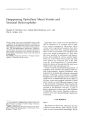
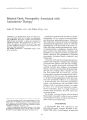
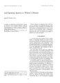
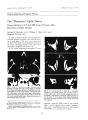
Show IOll", al of Clinical NellnH11, htha{ mo{ ogy 8111: 25- 28, 1988. Bilateral Optic Neuropathy Associated with Amiodarone Therapy Sarkis M. Nazarian, M. D., and Walter M. Jay, M. D. { 1988 Raven Press, Ltd., New York Amiodarone, an antiarrhythmic agent, has been associated with mild visual loss secondary to papillopathy and papilledema, We report a patient who developed bilateral optic neuropathy 4 weeks after initiation of amiodarone therapy, Nine months later, his vision was 20/ 50 in the right eye and 20/ 200 in the left. This report provides additional evidence that amiodarone may cause toxic optic neuropathy. Key Words: Amiodarone- Amphiphilic drugs- Toxic optic neuropathy. From the Neurology ( S. M. N.) and Ophthalmology ( W. M. j.) Services, John L. McClellan Memorial Veterans Hospital, Little Rock, Arkansas, and the Departments of eurology ( S. M. N.) and Ophthalmology ( W. M. J.), University of Arkansas for Medical Sciences, Little Rock, Arkansas, U. S. A. Address correspondence and reprint requests to Dr. Sarkis M. Nazarian at Neurology Service 127- LR, John L. McClellan Memorial Veterans Hospital, Little Rock, AR 72205, U. S. A. 25 Amiodarone hydrochloride ( Cordarone, Wyeth, Philadelphia, PA) is a recently introduced diiodinated benzofuran antiarrhythmic agent. Patients on this medication commonly develop a keratopathy characterized by pigmented, curved or whorl- like microdeposits in the lower half of the cornea ( 1). Other side effects include photosensitivity, hyperor hypothyroidism, constipation, elevation of hepatic enzymes, fine hand tremor, insomnia, and headaches. Less common reactions are pulmonary fibrosis, slate- gray skin pigmentation, peripheral neuropathy, proximal muscle weakness, and trunkal rash. Visual loss with optic disc changes has been described with amidarone. Gittinger and Asdourian ( 2) reported two patients with optic disc swelling. Their first patient developed unilateral disc swelling 8 months after initiation of amiodarone therapy, and their second patient developed bilateral optic disc swelling within 3 months of onset. The first patient had no visual loss, and the second patient experienced minimal acuity and visual field loss in one eye only. The authors chose to call their finding papillopathy rather than ischemic optic neuropathy. Fikkers et al. ( 3) described a 58- year- old man who developed pseudotumor cerebri with papilledema and irreversible visual changes while on amiodarone therapy. In a paper by Chew et al. ( 4) devoted primarily to amiodarone keratopathy, there is a single mention of ischemic optic neuropathy. This latter report has no clinical details. We describe a 56- year- old man who developed bilateral optic neuropathy within 4 weeks of onset of amiodarone therapy and had significant residual visual loss 9 months later. CASE REPORT A 56- year- old man was referred to the McClellan Memorial Veterans Hospital in November 1986 with a 2- week history of rapid and 26 5. M. NAZARIAN AND W. M. JAY painless loss of vision in his left eye. His local ophthalmologist noted that he had counting fingers vision in his left eye, a left afferent pupillary defect, and bilateral optic disc swelling. The patient had been hypertensive for 12 years. He had been on Tenormin, 25 mg daily, verapami!, 40 mg twice a day, and Minipress, 1 mg three times a day, for several years. Two months prior to admission, he developed dyspnea and atrial fibrillation. Cardiac evaluation at that time, including cardiac catheterization, was unremarkable. He was tried on various antiarrhythmics, which were discontinued because of unacceptable side effects. Six weeks prior to admission, he was started on amiodarone, 400 mg twice a day. The patient had a history of mild obstructive pulmonary disease and ulcerative colitis. He did not use tobacco or alcohol, but had been a heavy smoker until 12 years prior to admission. Pulse was 64 and regular, blood pressure 182/ 92 mm Hg. He had a grade IINI crescendo- decrescendo systolic murmur at the left lower sternal border. Carotid pulses were full. No carotid or cranial bruits were heard. Mental status and speech were normal. General neurologic exam was normal, except for mild hand tremor. Vision was 20125 in the right eye and counting fingers at 6 inches in the left. Confrontation visual field was intact in the right eye. In the left eye, a large central scotoma was present. The patient correctly identified all color plates with his right eye, and none with his left. An afferent pupillary defect was present in the left eye. Extraocular movements and intraocular pressures were normal. Slit lamp exam showed bilateral Grade I corneal epithelial deposits ( 5). Ophthalmoscopic exam revealed bilateral optic disc swelling, more pronounced on the left ( Fig. 1). The patient was admitted to the neurology service. Erythrocyte sedimentation rate was 6 mmlh. Contrast- enhanced computed tomographic scan of the head was normal. He had three lumbar punctures during his hospitalization. Opening pressures ranged from 16 cm H 2 0 to 24 cm H20. Lymphocyte counts ranged from 0 to 6. Protein ranged from 25 to 29 mgldl, and glucose values were in the normal range. Because of concern about temporal arteritis, prednisone, 80 mg daily, was begun. Amiodarone was discontinued because of possible toxic optic neuropathy. Over the next 3 days, he suffered right eye visual loss, from 20/ 20 acuity to 20/ 40. Left temporal artery biopsy performed a week 1.: .. - I,.-.,., c" j fl1ildlthern::; c! erosis. He was dis... jl\. t, 1pering predni- FIG. 1. ( Top) Right disc, ( bottom) left disc in November 1986. Bilateral disc swelling with hemorrhages is present. sone dose. His vision at time of discharge was 20/ 40 in the right eye and counting fingers at 6 ft in the left. He was seen in outpatient follow- up in March 1987. Visual acuity was 20/ 60 in the right eye and 20/ 200 in the left. Ophthalmoscopic exam now revealed bilateral optic atrophy, with severe segmental arteriolar narrowing and sheathing ( Fig. 2). He was seen again in July 1987. His visual acuity was now 20/ 50 in the right eye and 20/ 200 in the left. On Goldmann visual fields, a superotemporal island of vision remained in the left eye. In the right eye, he had a cecocentral scotoma and depression in an inferior arcuate pattern ( Fig. 3). DISCUSSION Our patient presented with bilateral optic neuropathy occurring within 4 weeks of initiation of amiodarone therapy. This temporal sequence implicates amiodarone as a possible causative agent. However, it is conceivable that the patient had bilateral non- arteritic ischemic optic neuropathy that was unrelated to amiodarone. Our patient had hypertension, which is a risk factor for ischemic optic BILATERAL OPTIC NEUROPATHY AND AMIODARONE 27 FIG. 2. ( Top) Right disc, ( bottom) left disc 4 months after start of amiodarone. Bilateral optic atrophy with arteriolar narrowing and sheathing is present. neuropathy ( 6,7). Bilateral ischemic optic neuropathy can present initially with visual loss in one eye and asymptomatic optic disc edema in the fellow eye ( 8). The fellow eye then develops visual loss days to months later. Second eye involvement in bilateral ischemic optic neuropathy develops within a month or less in 19( 7.:, ( 9) to 23% ( 10) of patients. Like other drugs that cause corneal deposits in a vortex pattern, amiodarone is a cationic amphiphilic drug ( 11). These drugs have the common property of possessing hydrophilic and hydrophobic groups in close proximity to each other. This property allows them to penetrate Iysosomes and to bind irreversibly to polar lipids. The resulting complexes accumulate within lysosomes and can be seen with electron microscopy as lamellated or crystalloid inclusion bodies. Pathologic studies have demonstrated that the peripheral neuropathy seen with amiodarone is a toxic axonal ( 12) or demyelinative ( 13) neuropathy with osmiophilic lysosomal inclusion bodies in Schwann cells and capillary endothelial cells. Perhexiline, an amphiphilic antianginal drug, has been reported to cause papilledema with visualloss ( 14) and asymptomatic disc swelling ( 15). Two other amphiphilic drugs, iodochlorhydroxyquin and diiodohydroxyquin, have been associated with subacute myelo- optic neuropathy ( 16) and with optic atrophy ( 17), respectively. Amiodarone causes the same pathologic changes in various neural tissues as that caused by other amphiphilic drugs. As some of these medications have toxic effects on the optic nerve, it is possible that amiodarone may cause optic nerve damage. To date, the evidence that this occurs remains circumstantial. Further studies are needed to establish an unequivocal relationship between amiodarone therapy and optic neuropathy. FIG. 3. Goldmann visual fields obtained 9 months after onset of visual symptoms. 28 S. M. NAZARIAN AND W. M. JAY REFERENCES 1. Harris L. McKenna WJ, Rowland E, Krikler OM. Side effects and possible contraindications of amiodarone use. Am Hearl I 1983; 106: 916- 23. 2. Gittinger jW, Asdourian GK. PapillopathY caused by amiodarone. Arch 0l'hthalll/( l/ 1987; 105: 3- 19- 51. 3 Fikkers BG, Bogousslavsky j, Regli F, Glasson S. Pseudotumor cerebri with amiodarone. I NClIrol NClIroslIrg Psych; all'll 1986;- 19: 606. - I. Ch'ew E, Ghosh M, McC'llloch C Amiodarone- induced cornea verticillata. ('/ 1/ ') l'hIII< 1II110/ 1982; 17: 96- 9. 5. Orlando RG, Dangel lVII:, Schaal SF. Clinical experience and grading of amiodarone keratopathy. 0l'hthaimoiosy 198-- 1; 91: 118- 1- 7 6. Repka MX, Savino PJ, Schatz Nj, Sergott RC Clinical profile and lung- term implications of anterior ischemic \) ptic neuropathy. Am I 0l'htll< 1/ l/ 1ol 1983; 96:-- 178- 83 7. Guyer DR, Miller NR. Auer CL, Fine SL. The risk \) f cerebrl~ vascular and cardiovascular disease in patients with anterior ischemic optic neuropathl'. Ard, 0l'hllwill/ ol 1985; 103: 1136-- 12 8. Hayreh 55. Ankrior ischemic optic neuropathl'. V. Optic disc edema an earlv sign. Arch 0l'hthalll/( l11981; 99: 1l130-- IO. 9. Ellenberger C jr, Keltner JL. Burde RM. Acute optic neuropathy in older patients. Arch NellroI1973; 28: 182- 5. 10. Hayreh 55, Podhajsky P. Visual field defects in anterior ischaemic optic neuropathy. Doc Ophthalmol Proc Series 1979; 19: 53- 71. 11. D'Amico OJ, Kenyon KR. Drug- induced lipidoses of the cornea and conjuncti\' a. Jilt OphtlwlmoI1981; 4: 67- 76. 12 Pellissier JF, Pouget J, Cros D, De Victor B, Serra trice G, Toga M. Peripheral neuropathy induced by amiodarone chlorhydrate- a clinicopathological study. / Nellrol SCI 1984; 63: 251- 66 13. jacobs JM, Costa- Jussa FR. The pathology of amiodarone neurotoxicity. II. Peripheral neuropathy in man. Brain 1985; 108: 753- 69. H. Gibson JM, Fielder AR. Garner A. Millac P. Severe ocular side effects of perhexilene maleate: Case report. Br / Ophthalmo/ 1984; 68: 553- 60. 15 Atkinson AB, McAreayey D, Trope G. Papilloedema and hepatic dysfunction apparently induced by perhexiline maleate ( Pexid) Sr Heart / 1980;- 13: 490- 1. 16. Selbv G. Subacute myelu- optic neuropathy in Australia. Lance! 1972; 1: 123- 5. 17. Behrens MM. Optic atrophy in children after diiodohYdroquin therapy. lAMA 197-- 1; 228: 693- 4. |