| OCR Text |
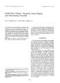
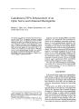
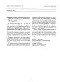
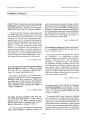
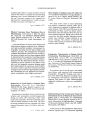
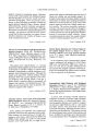
Show Tournai or Clinical Neurv-" l'htlullOl/ ology 9(~ I: 83- 97. 1% 9. Current Review Interventional Neuroradiology in Neuro- ophthalmology Eddie Kwan, M. D., Grant B. Hieshima, M. D., Randall T. Higashida, M. D., Van V. Halbach, Samuel M. Wolpert, M. D. Endo\' ascular n ur IradlOlogic pro-" Jure is the treatment ~ f hoice for patient with symptomatic direct carotid c \' E~ rnou fistulae ( CCF) and dural arteriovenous fi tulae ( DA \' F) that failed manual carotid artery/ jugular velll compre sion. Preservation of visual function and the prevention of cata trophic intracranial hem rrhage are the prime therapeutic objectives. The choice of tran arterial versus transvenous approach is dictated by the pathophysiology, pattern of venous drainage, and the risklbenefit ratio in each patient. In 180 patients with CCF, 95% have been cured with a combination of manual compression, transarterial or transvenous embolization, while 3% have significant complication. In pateints with DAVF, transarterial and transvenous embolization has resulted in clinical cure in 77 and 90%, respectively; the complication rate is between 4-- 5%. Intra- aneurysmal embolization with silicone detachable balloons in its present stage of development is an acceptable alternative for patients with intracranial aneurysms not otherwise amenable to standard neurosurgical clipping. In aneurysms with a well- defined neck, aneurysm thrombosis with preservation of flow in the parent vessel has been accomplished in 90% of the patients treated. The complication rate in patients presenting with mass effect and subarachnoid hemmorrhage is 5 and 14%, respectively. Key Words: Carotid cavernous fistula- Dural arteriovenous fistula- Embolization- Interventional neuroradiology- Intracranial aneurysm. From the Section of NeuroradioJogy, University of California at San Francisco Medical Center, San Francisco, California ( E. K., G. B. H., R. T. H., and V. V. H.), and the Section of Neuroradiology, New England Medical Center. Boston, Massachusetts ( EX and S. M. W.), U. S. A. Address correspondence and reprint requests t~ Dr. E. Kwan, Section of NeuroradioJogy, New England MedICal Center, Box 88, Boston, MA 02111, U. S. A. 83 < 91989 Raven Press, Ltd" New York M. D., and Interventional neuroradiology is an evolving subspecialty that concentrates on the endovascular therapy of CNS vascular lesions. These endovascular techniques represent a logical refinement of those developed for diagnostic neuroangiography. In selective instances, interventional neuroradiology offers an alternative therapy for patients with vascular lesions otherwise not amenable to standard surgical techniques ( 1- 4). Over the last decade, with advances in real- time digital subtraction angiography ( 5) as well as the refinement of coaxial catheter systems, new embolic agents, and balloon technologies ( 6), interventional neuroradiology has progressed rapidJy. Furthermore, the clinical experience that has accumulated over the same period has altered our understanding of the pathophysiology of these vascular lesions and, as a result, the indications for treatment. The purpose of this communication is to describe the management of three entities that are pertinent to the practice of neuro- ophthalmology and interventional neuroradiology. This is by no means a complete list of lesions amenable to endovascular intervention, but the lesions are sufficiently common to be of interest to most practitioners of ophthalmology, neurology, and neurosurgery. The three entities are direct carotid cavernous fistulae ( CCFs), dural arteriovenous fistulae involving the cavernous sinus, and intracranial aneurysms. For each entity, the pathophysiologic condition, the indication for treatment, the risks involved, and the results to be expected will be discussed. While some of the information is 84 E. KWAN ET AL. widely accepted within our discipline, other concepts represent our own bias based on cumulative observations and experience over the past 15 years. In our institutions. the majority of neurointerventional procedures are performed with the patient awake but sedated. The only exceptions are children under the age of 12, in whom the benefit of general anesthesia outweighs the advantage of the frequent neurological monitoring possible with local anesthesia. A transfemoral approach is usually employed using specialized coaxial catheter systems. Ln adults. a 7- 7.3 French catheter is inserted into the femoral artery; the distal inner coaxial catheter that is superselectively navigated into the intracranial circulation for delivery of embolic agents varies from 2 to 3 French. Catheters of intermediate caliber are also deployed coaxially to facili tate selective ca theteriza tion. In selected cases. direct puncture of the cervical carotid artery may be necessary. In cases of CCFs and dural arteriovenous fi tulae, when the arterial approach is not possible due to prior surgical ligation or as a result of trauma, transvenous catheterization is a viable option ( 7- 9). Rarely, an intraoperative approach may be necessary to gain access to vascular lesions for embolization. Because thromboembolic complications are related to catheter manipulation and procedure times, unless the patient is actively bleeding, all intracranial arterial procedures are carried out with systemic heparinization, and anticoagulation is reversed with i. v. protamine sulfate prior to catheter removal. Anticoagulation is not used for the transvenous procedures. High- quality biplane fluoroscopy and real- time digital subtraction angiography with road- mapping capabilities are indispensable for superselective catheterization of intracranial vessels and continuous monitoring during the delivery of embolic agents. Most procedures require an average of 3- 5 h. Ischemic pain may occur for 2- 4 days following embolization of the external carotid artery territory; progressive thrombosis following balloon embolization of giant aneurysms may also be associated with severe headache and surrounding edema. Such discomfort can be Significantly palliated by tapering doses of corticosteroids and narcotics over several days. DIRECT CCF A CCF r pre ents ( II") ilbnnrmal communication b, : ., -" ! ,, · \, l;, i :, rlny dl1d the cavern-ous sinus. It is most often the sequela of posttraumatic tearing of the cavernous carotid artery. An associated basilar skull fracture is common. Other causes include direct penetrating injuries, rupture of intracavemous aneurysms ( Fig. I), collagen vascular deficiency, direct surgical trauma, and fibromuscular dysplasia. Most CCFs are high- flow, high- pressure lesions with an abrupt onset of symptoms including orbital bruits, retro- orbital pain, chemosis, restricted ocular motion, proptosis, and progressive visual loss. The symptoms and prognosis of CCF are related to its size, duration, location, and, most importantly, the route and adequacy of venous drainage. In a review of 122 cases of direct CCF, Halbach et al ( 10) identified the following angiographic and clinical features that are associated with poor clinical outcome: the presence of a carotid pseudoaneurysm or a cavemous sinus giant varix associated with epistaxis and subarachnoid hemorrhage, respectively; venous drainage from the fistula through the cortical veins; rapid visual loss; rapidly progressive proptosis associated with thrombosis of the superior ophthalmic vein; and cerebral ischemia secondary to vascular steal through the fistula. We believe in urgent treatment for patients with any of these features. A common misconception is that a CCF with a loud bruit and a dilated superior ophthalmic vein requires urgent treatment to preserve visual function. A dilated superior ophthalmic vein does not produce significant visual symptoms as long as the vein is adequately decompressed by the angular vein, the frontal vein, and the inferior and superior petrosal sinuses. If the superior ophthalmic vein is not decompressed, as may occur when the venous walls thicken following long- term arterial pressure and turbulent flow, rapid visual deterioration may occur ( Fig. 2). A clinical clue is decrease in the intensity of the bruit. Furthermore, if thrombosis of the superior ophthalmic vein occurs in those patients with superimposed hypoplasia or prior thrombosis of the petrosal sinuses, the shunted arterial blood may then be decompressed only via the cortical, retinal, and ciliary veins. Most ocular symptoms and sequelae in CCF can be directly or indirectly related to venous congestion. Delayed visual impairment in CCF is usually attributable to corneal ulceration, secondary glaucoma, and retinal hypoxia ( 11). Factors contributing to retinal hypoxia in CCF include a drop in effective ophthalmic artery pressure secondary to the " steal phenomenon" across the fistula, an elevation of episcleral venous pressure secondary to venous outflow restriction, and neovaseular prolif- INTERVENTlONAL NEURORADIOLOGY 85 c D FIG. 1. From an 83- year- old woman with spontaneous left- sided bruit, right- sided chemosis, and proptosis of 4 months' duration. Left common carotid arteriogram, anteroposterior ( AP) ( A) and lateral ( 8) projections, showing ruptured left cavernous carotid aneurysm ( small arrow), early opacification of the left sphenoparietal sinus ( arrowhead), the circular sinus ( black open arrow), the right cavernous sinus ( white open arrow), the right superior ophthalmic vein ( large arrow), and the pterygoid plexus ( angled arrow). Note the venous occlusive disease in the left inferior ophthalmic vein ( curved arrow). ( e): Left common carotid arteriogram, AP view, taken following navigation of a partially inflated balloon ( small arrows) into a ruptured aneurysm. The left sphenoparietal sinus is no longer opacified, and shunting of contrast material into the right cavernous sinus ( large arrow) has decreased significantly. ( D) and ( E): These are AP ( D) and lateral ( El projections. With further inflation of the detachable balloon, the fistula ( white arrow) is completely closed. ( F): Lateral skull x- ray film following embolization; the ballon overlaps the dorsum sella. Due to incomplete mixing, metrizamide ( black arrow) occupies the dependent portion of balloon, while radiolucent 2- hydroxylethyl- methacrylate ( white arrow) occupies the nondependent portion. I CIi" NeIlTO · oplltllalmol. Vol. 9. No. 2. 1989 86 E. KWAN ET AL. c E FIG. 2. From a 43- year- old man with a traumatic left carotid cavernous fistula of 6 months' duration. An outside angiogram performed 3 months prior to admission demonstrated anterior drainage to the superior ophthalmic vein and posterior drainage to the inferior petrosal sinus. The patient had minimal ocular symptoms in spite of loud bruit. One day prior to balloon embolization, the patient awoke with severe chemosis and proptosis and a soft bruit. ( A) and ( 8): Left internal carotid arteriogram, lateral projection, early ( A) and late ( B) arterial phases. Note the shunting of contrast material into the left superior ophthalmic vein ( arrow). the inferior ophthalmic vein ( arrowhead), and multiple engorged ciliary veins ( small arrows). ( C): Anteroposterior veiw, left internal carotid arteriogram. Neither the inferior petrosal sinus nor the frontal or angular veins are opacified. Engorged ciliary veins and severe chemosis are due to thrombosis of orbital venous outflow pathway. ( 0): Postembolization left internal carotid angiogram, fistula ( arrow) closed with one silicone balloon filled with 0.15 ml of metrizamide ( E): Postembolization lateral skull x- ray film. Contrast materialfilled balloon ( arrows) is deformed by septation within the cavernous sinus. eration on the iris and the angle of the anterior chamber, resulting in aqueous outflow restriction. TREATMENT The preservation of visual function and the prevention of catastrophic intracranial hemorrhages are the prime therapeutic objectives in the treatment ( If r · · ~ tient with CCF. While elevated intra- ' 0 rnpl) rarily corrected with medical therapy and lateral canthotomy, definitive treatment must be aimed at closing the fistula and maintaining an adequate ocular arteriovenous pressure gradient. Different forms of treatment are possible. Any procedures that lower arterial pressure without a concomitant reduction in venous or intraocular pressure will further compromise the ocular perfusion. This explains the dismal results in patients treated with carotid ligation and/ or trapping procedures. INTERVENTIONAL NEURORADIOLOGY 87 Manual Compression Manual compression of the cervical carotid artery and jugular vein is indicated in patients with slow- flow CCF and without significant vi ual impairment. It is contraindicated in patients with angiographic evidence of cortical venous drainage from the fistulae, a hypersensitive carotid sinus, and atherosclerotic stenosis of the carotid bifurcation. Patients are instructed to locate the pul e of the cervical carotid artery with the hand contralateral to the site of the CCF; by applying gradually increasing pressure, the carotid artery and jugular vein are compressed until the palpable pulsation or the audible retro- orbital bruit is obliterated. Compression should be maintained for 10- 15 sand repeated three to four times per hour. The contralateral hand is used for compression to guard against transient cerebral ischemia during therapy; if weakness develops unknown to the patient, the compressing hand will fall, and treatment will automatically terminate. In our ~ xperience, the time required for complete closure of CCF with compression therapy varies from 15 min to 3 months. A CCF closure with compressive therapy can be explained on the basis of simultaneous arterial hypotension and venous hypertension; this results in a transient decrease of pressure gradient across the fistula, thereby promoting cavernous sinus thrombosis. Our experience over the past 14 years showed that 3% of CCFs closed spontaneously ( 12), while an additional 17% closed with intermittent external compression of the carotid artery and jugular vein ( 13). Transarterial Embolization In patients with rapidly progressive visual loss, cortical venous hypertension, pseudoaneurysms, or enlarged varices, intra- arterial embolization with detachable balloons and preservation of flow in the parent carotid artery is currently the treatment of choice. Our standard detachable balloon measures 1.5 by 4 mm uninflated and can be maximally inflated to 0.5 cc. The balloon is attached to the tip of a 2/ 4/ 7.3 French coaxial catheter system. For complex vascular anatomy, the 2 Fre~~ h polyethylene catheter can be configured to faClhtate entrance into the fistula. Small CCFs can often be closed with one detachable balloon; multiple balloons are necessary to pack dilated cavernous sinus varices associated with chronic high- flow CCFs. Closure of chronic high- flow CCFs is often performed in stages to prevent complications secondary to normal perfusion pressure breakthrough in the brain ( 14). After correct positioning of the balloon is confirmed fluoroscopically, to prevent future balloon deflation, metrizamide within the balloon is partially exchanged for HEMA ( 2- hydroxylethylmethacrylate), a hydrophilic polymer that solidifies over 45 min at body temperature. Real- time digital subtraction angiography is performed during test occlusion of the fistula to assess patency of the parent internal carotid artery. Repositioning of the balloon may be necessary before detachment if the balloon partially herniates into the parent vessel or if it blocks the venous outflow without totally occluding the fistula. Detachment is usually accomplished by traction alone. In selected cases, a second nondetachable balloon is inflated transiently within the cavernous internal carotid artery to stabilize the first balloon during detachment. Following fistula closure, CCFs can reopen by slight shifting of the balloon axes. A large recurrent CCF may require additional balloon embolization, while small residual CCFs can be treated by intermittent carotid artery and jugular vein compression or embolization via the transvenous approach ( Fig. 3). The formation of a pseudoaneurysm at the site of an embolized CCF can be secondary to shifting or deflation of the balloon ( if it is filled with contrast material alone). Transvenous Embolization While the vast majority of CCFs can be closed by transarterial embolization, this approach is not possible in a patient with an occluded carotid artery secondary to trauma or prior ligation, vessel transsection, severe stenosis or redundancy of the internal carotid artery, or a fistula whose orifice is smaller than the deflated balloon. Transvenous embolization is an alternative for such circumstances. The most accessible route is via the inferior petrosal sinus following a transfemoral approach. Both the ipsilateral and the contralateral cavernous sinuses via the circular sinus can be reached by this technique with a 2.7 French Tracker microcatheter and steerable guide wire. Superior ophthalmic vein catheterization is utilized less commonly because it requires surgical exposure; severe kinking of this vessel at the superior orbital fissure subjects the patient to risks of vessel injury and retro- orbital hemorrhage in acute CCFs when the wall of the superior ophthalmic vein has not hypertrophied. Embolic agents for J Gin Neuro- ophtlullmol. Vol. 9. No. 2. 1989 ss . " WA ET AL. o G INTERVENTIONAL NEURORADlOLOGY 89 transvenous embolization include platinum wire/ coils, stainless steel minicoils, isobutyl- 2- cyanoacrylate or N- butylcyanoacrylate ( both liquid adhesives), silk suture, and a detachable silicone balloon. The latter is used sparingly because navigation of the silicone balloon via th transvenous approach is difficult due to multiple partitions within the cavernous sinus. Liquid adhesive is injected only after the fistula flow is first slowed sufficiently with coils or balloons and usually with precautions to avoid reflux into the carotid artery. Reflux of the embolic agent from the cavernous sinus into the carotid artery during embolization is a real risk when the cavernous sinus has undergone subtotal thrombosis, thereby reducing the pressure gradient across the fistula. If the major venous drainage is occluded prior to complete closure of the fistula, aggravation of the symptoms can occur. Another potential complication of this approach is diversion of arterialized venous blood to cortical vein when the other venous drainage pathways are occluded due to occasional difficulty in the precise placement of embolic agents at the fistula orifice. Our success rate in closing CCFs via the transvenous approach is greater than 809c ( 8). Surgery Surgical exposure to the cavernous sinus followed by direct packing of the sinus with thrombogenic material ( 15,16) or direct injection of an embolic agent remains an alternative when standard endovascular techniques fail. Results Over the past 14 years, using a combination of cervical carotid artery/ jugular vein compression and transarterial and transvenous embolization, we have succeeded in closing more than 95% of the 180 CCFs we have treated. The most common .. morbidity related to treatment is transient cranial nerve paralysis ( 12%). The rate of the occurrence of more severe complications, including stroke, intracranial hemorrhage, and subarachnoid hemorrhage, is 3%. CAVERNOUS SINUS DURAL ARTERIOVENOUS FISTULA ( DAVF) A DAVF represents an abnormal communication between the dural branches of the internal and external carotid arteries and the cavernous sinus. Bilateral arterial and venous involvement are common. A DAVF often occurs spontaneously in postmenopausal women, but it can be associated with sinusitis, cavernous sinus thrombosis ( 17), trauma, and pregnancy. In general, the initial symptoms are similar to those of the CCFs but are often insidious and less severe. Closure can occur spontaneously or following diagnostic angiography ( 18,19). Nevertheless, a DAVF is not a benign entity, as it can result in severe visual loss if treated inappropriately. The arteriovenous shunt in a DAVF is characterized by low flow, in contradistinction to the high- flow and high- pressure shunt encountered in CCF. Similar to CCF, the severity of ocular symptoms correlates with an inadequacy of venous decompression from the cavernous sinus. Ocular symptoms increase when there is posterior venous thrombosis or distal superior ophthalmic vein obstruction, thereby shifting " arterialized" blood to the small veins of the orbit, which cannot accommodate the flow. Therefore, the most severe symptoms often occur in the very- slow- flow shunts, which caUSe marked venous hypertension in the small orbital veins ( Fig. 4). TREATMENT Therapeutic options in patients with DAVF include carotid artery/ jugular vein compression, FIG. 3. From a 28- year- old man with posttraumatic right- sided carotid cavernous fistula ( CCF). ( A) and ( 8): Right vertebral arteriogram with simultaneous right common ca. rotid compression, ~ arly ( A) a~ d midarterial ( B) pha. ses. Note the shunting of contrast material from the posteno~ ca~ ernous carotid arte~ Into the cavernou. s SI~ US ( arrowhead) and the superior ophthalmic vein ( arrow). ~ C): Right Internal carotid arten~ gram. anteroposten. or VI~ W demonstrates early opacification of both cavernous sinuses ( arrows), the. angular vein ( ope~ ar~ ow),. the . lnfenor petrosal sinus ( arrowhead). and the facial vein. ( curved arrow) on t~ e left sl~ e. (~): Postembolizatlon fight Internal carotid angiogram. lateral view. Note the minimal encroachment Into the Junction of. the preca~ er~ ous an~ cavernous carotid arteries by a detachable ballon ( small arrows). ( E): Two days follOWing embolization, brUits recurred. Right internal carotid arteriogram, lateral vj~ w. Opacificat! on of the r. ight inferior petr~ sal sinus ( open arrows) is due to a slight shifting of the b~ lIoon ~ XIS an. d reope~ lng of the f1stul~. ( F): Followln~ transvenous embolization of the right inferior petrosal SinUS With platrnum COIls ( arrows). artenovenous shuntrng decreased significantly. ( G): The right CCF closed completely after 7 days of carotid artery/ jugular vein compression. I Clin Neuro" Ophthalmol. Vol. 9. No. 2, 1989 ..~ o E " J FIG. 4. From a 33- year- olo woman with rne mSlo'IOUS Ofl:: it:( ur let( penor" brtdl swelltflg, laken following .:: l motor vehlcre accid& 1l! .' 3 }' ears ago. ( A} and ( 8"= U>. ft inlern!'! l carotid arteriogram. lateral ( A) and anteroposterior ( AP) ( B) vIews showing rapid shunting of contrast material into the left superior ophthalmic vein ( large arrow) via small dural feeders ( small arrow) from the mferolateral trunk of the left cavernous internal carotid artery. Decompression of the superior ophthalmic vein by angular and facial veins ( open arrows) accounted for minimal ocular symptoms. ( C) and ( D): Left external carotid arteriogram. lateral ( e) and AP ( D) views demonstrate shunting of contrast material into the superior ophthalmic vein ( large arrow) via multiple dural arterial feeders ( small arrows), from the distal internal maxillary artery. ( E): This superselective angiogram of the left middle menmgeal artery with a microcatheter ( arrOWhead), lateral projection. delineates one component of the dural arteriovenous ( AV) shunt ( open arrow). ( F) and ( G): Postembollzatlon left external carotid ( F) and teft internal carotid ( G) arteriograms, lateral view. after 0.2 ml of an N- butyl cyanoacrylate and Pantopaque mixture was mjected into the petrosal branch of the left middle menmgeal artery. A cast of liquid adhesive ( small arrows) is located within the subtotally thrombosed superior ophthalmic vein. Severe chemosis and a progressive decline In visual acuity occurred within hours following embolization. ( H): A repeat left external carotid arteriogram, lateral projection, performed 4 h after embolization shows a persistent dural shunt ( arrow). a subtotally thrombosed superior ophthalmic vein ( open arrow), and engorged ciliary vems ( small arrows) The combination of a patent AV shunt and venous outflow restriction was responsible for the rapid decline of visual acuity. ( I) and ( J): Taken following a repeat embolization of the accessory meningeal artery with a liqUid adhesive. these arteriograms. lateral projections of the left external artery carotid ( I). and the lefllnternal carotid artery ( J), demonstrate total obliteration of the dural AV shunt. INTERVENTIONAL NEURORADIOLOGY 91 transarterial embolization, transvenous embolization, and combined embolization/ surgical resection. The choice of any technique has to be weighed against the risklbenefit ratio in each individual patient. Manual Compression In patients who are highly motivated and without cortical venous drainage or a recent decline in visual acuity, intermittent carotid artery/ jugular vein compression is the first treatment modality. This maneuver alone has resulted in complete cure in 30% of our patients ( 20). Contraindications and methodology are similar to those outlined in the treatment of CCF. Patients with cortical venous drainage are excluded from compression therapy based on the theoretical risk of shunting of blood from the cavernous sinus with a further elevation of cerebral venous pressure resulting in a venous infarct or parenchymal hemorrhage. Transarterial Embolization In symptomatic patients who have failed carotid artery/ jugular vein compression, embolization is the next logical step. Selective and superselective catheterization of bilateral internal and external carotid branches are mandatory to document pedides feeding the fistula, to delineate hazardous extracranial to intracranial collaterals, and to evaluate the venous drainage pathway. The choice of transarterial versus transvenous embolization depends on a full appreciation of the anatomical and hemodynamic information derived from diagnostic angiography. Transarterial embolization is an established treatment modality. With the advent of microcatheters, steerable microguide wires, and real- time digital subtraction angiography with roadmapping capability, superselective catheterization of small dural arterial feeders from the external carotid artery can be routinely accomplished ( Fig. 4). We have also managed to superselectively catheterize small dural branches originating from the internal carotid artery. Depending on anatomical and hemodynamic constraints, embolic materials include polyvinyl alcohol particles ( 200- 300 / Lm) or liquid adhesives such as isobutyl- 2- cyanoacrylate or N- butyl- cyanoacrylate, hypertonic glucose, and collagen. Detachable balloons are not appropriate due to the small caliber of the arteriovenous communication in DAVF. The major limitation of transarterial embolization is the inability to consistently embolize arterial feeders originating from the internal carotid artery without simultaneously subjecting patients to the risk of embolic reflux into intracranial circulation. In addition, transarterial embolization can result in the proximal occlusion of arterial feeders without thrombosis of the shunt, thereby producing a transient " angiographic cure" but also stimulating recruitment of other vascular pedicles that are often more difficult or hazardous to embolize. While liquid adhesive is potentially curative due to its ability to permeate the nidus of the DAVF, a drawback to its use is that it can result in occlusion of venous outflow pathways prior to total obliteration of the shunt, thereby further aggravating ocular symptoms. The embolic agent can also result in infarction of normal tissue or in permanent cranial nerve palsies. Following liquid adhesive embolization, patients often experience local pain, nausea, vomiting, and low- grade fever. Polyvinyl alcohol particulate embolization, although not as permanent as liquid adhesive, can often result in a clinical cure. Transvenous Embolization Historically, transvenous embolization of DAVF was initially attempted in patients who failed transarterial embolization or in whom there was no readily accessible arterial route follOWing prior embolization and/ or surgical ligation. Our recent success with this approach indicates it may be the technique of choice in the treatment of the cavernous sinus DAVF ( 9). The major advantage of transvenous embolization is that it replaces the often tedious superselective embolization of multiple arterial feeders by direct placement of thrombogenic agents within the cavernous sinus, thereby promoting sequential thrombosis of the fistula and the venous outflow pathway. This procedure is performed via a percutaneous femoral vein approach; navigation through the inferior petrosal sinus is facilitated by a coaxial microcatheter and steerable guide wires. Both the ipsilateral and contralateral cavernous sinuses can be superselectively catheterized by this technique ( Fig. 5). Navigation through the inferior petrosal sinus and the basilar venous plexus is facilitated by the roadmapping capability of real- time digital subtraction angiography follOWing arterial contrast injection. In several instances, we managed to traverse the inferior petrosal sinus that was not angiographically visible ( and presumably thrombosed) with microcatheters and steerable guide wires. The potential dangers of transvenous embolization are perforation of the sinus and reflux of the embolic agent into the internal carotid artery I Gill Nfllro- ophlhalmol. Vol. 9. No. 2. 1989 \.. ~ B " ' 0' -.-. c . i: 1 I) FIG. 5. From an 82- year- old woman with a 4month histo ry of left retro- o rbital headache. glaucoma. and bilateral chemosis, ( A) A right common carotid arteriogram. lateral view, demonstrates dural shunts supplied by the meningohypophyseal and inferolateral trunks of the right internal carotid artery opacifying the right cavernous sinus ( arrows) and the right inferior petrosal sinus ( curved arrows), ( B): Left com- _ ~, mon carotid arteriogram. anteroposterior ( AP) view. Note the rapid arteriovenous shunting from the dural branches of the left cavernous internal carotid artery into both superior ophthalmic veins ( small arrows), the right cavernous si nus ( large arrow), the circular sin us ( open arrow), and the right inferior petrosal sinus ( curved arrows). ( C): A left cavernous sinus venog ram. AP projection, via superselective catheterization of the right inferior petrosal sin~ and the circular sinus ( small arrows), The right ( large arrow) and left ( open arrow) superior ophthalmic veins are opacified. ( D): Left common carotid arteriogram, AP vi w. Left- sided dural shunts are obliterated following injection of 0.2 ml of an N- butyl- cyanoacrylate and Pantopaque mixture into the posterior left cavernous si us. ( E): Right common carotid arteriogram, AP view, obtained immediately after ( 0). Note the persistent dural shunts into the right inferior petrosal sinus ( arrows). ( F) a d ( G): Following placement of three ptatinum coils ( arrows) into the posterior right cavernous sinus and the inferior petrosal sinus via a microcatheter, these right c mmon carotid arteriograms, AP ( F) and lateral ( G) projections. show complete thrombosis of the dural shunts within minutes after embolization. INTERVENTIONAL NEURORADIOLOGY 93 via the dural branches. Commonly utilized embolic agents via the transvenous approach include platinum wires/ minicoils, silk sutures, and liquid adhesives; the latter are used when the former fail to close the fistula completely. RESULTS In a series of 22 patients who underwent transarterial embolization in our institutions, complete cure occurred in 17, while improvement occurred in four; there were three transient deficits and one permanent stroke related to therapy ( 20). With improved technology and experience, the complication rate should decrease in the future. The cure rate following the transvenous approach is close to 90%, with a complication rate of 5%. When the DAVF is subtotally occluded by embolization, further thrombosis can be induced by carotid artery/ jugular vein compression. ANEURYSM The current treatment of choice for most ruptured or symptomatic intracranial aneurysms is surgical clipping. Aneurysms with neuro- ophthalmological implication include those located at the cavernous carotid artery, the carotid ophthalmic junction ( Fig. 6), the anterior communicating artery, the posterior communicating artery, the supraclinoid carotid artery, and the tip of the basilar artery ( Fig. 7). Most giant aneurysms involving the cavernous internal carotid artery, the carotid ophthalmic junction, and the midbasilar artery are not accessible to surgical clipping. The absence of a definable neck and the presence of calcified mural thrombi within a giant aneurysm further increase the technical difficulty of neurosurgical dipping. Currently, our selection criteria for intravascular balloon embolization of aneurysms include patients who failed surgical dipping ( Fig. 8), patients with aneurysms that are surgically inaccessible, and patients with preexisting medical conditions that make them poor surgical risks ( 2). If an aneurysm has a definable neck, then intra- aneurysmal balloon placement with preservation of flow in the parent vessel represents an ideal end result. A contraindication for balloon embolization is in an aneurysm with a fresh mural thrombus, due to the inherent risks of distal embolization. Severe headache and sometimes cranial nerve palsies often closely correlate with acute intra- aneurysmal thrombosis; in such situations, balloon embolization may be postponed for several weeks to allow clot lysis or organization. We have also observed extensive vasogenic edema surrounding giant aneurysms that are undergoing acute thrombosis on computed tomography and magnetic resonance imaging in the brain. If the aneurysm has no neck, the preservation of flow in the parent vessel is impossible. Proximal occlusion of the parent artery or trapping of the aneurysm are the only available options ( 1,21). With proximal occlusion, at least two balloons are detached, the distal one being as close to the aneurysm as technically possible to prevent filling of the aneurysm through collateral channels. The proximal balloon prevents cephalad migration of the distal balloon, which may occur with constant arterial pulsation. With the trapping procedure, the location of balloons is dictated by the necessity of maintaining the patency of vital branches originating from the parent vessel. In patients in whom proximal occlusion is a possible therapeutic option, to test the collateral reserve of the circle of Willis, a test occlusion of the parent vessel under systemic anticoagulation is performed prior to balloon embolization. A double- lumen, nondetachable balloon catheter is positioned in the parent artery and inflated to produce occlusion. Continuous neurological and hemodynamic monitoring is carried out, and the occlusion is maintained for 30 min or until a neurological deficit develops. In patients who do not tolerate test occlusion, an extracranial- to- intracranial bypass procedure is performed to augment flow into the ischemic territory; this will then be followed by another test occlusion. The balloon used for intravascular embolization is constructed of silicone and has an internal selfsealing miter valve within the neck to prevent deflation after detachment. For proximal occlusion or the trapping procedure, it is sufficient to fill the silicone balloon with nonionic contrast material such as metrizamide ( 200 mg iodine/ ml). When intra- aneurysmal balloon placement with preservation of parent arterial flow is the therapeutic end point, metrizamide within the silicone balloon has to be partially exchanged for HEMA ( 22) prior to detachment. The HEMA, a hydrophilic liquid, when cross- linked and activated will polymerize into a solid within 40 min at body temperature. The volume of the balloon will thus be maintained indefinitely, thereby preventing recanalization of the aneurysm when the balloon shell deteriorates. The silicone shell of the balloon is semipermeable; therefore, it is crucial not to substitute other nonionic contrast material for metrizamide because such agents have a much higher osmolality when compared with metrizamide of an identical J Clin Neuro- ophthalmol. Vol. 9. No. 2. 1989 94 E. KWAN ET AL. II iodine concentration. Filling the balloon with a hyperosmolar contrast material will result in volume expansion over time. The advent of real- time digital subtraction angiography with roadmapping capability is one of the most important technological breakthroughs to pr<"' jd,, , ! T'-" :"." , f ..! · ;" ihi:-. high- risk proce- FIG. 6. From a 27- year- old woman with two previous unsuccessful surgical clippings of a left carotid ophthalmic junction aneurysm. ( A) and ( B): Left internal carotid arteriograms. AP ( A) and lateral ( 8) projections. show an ovoid carotid ophthalmic junction aneurysm. Note the close proximity of the aneurysm neck to the origin of the ophthalmic artery ( arrow). ( C): Left common carotid arteriogram. lateral view, taken following the detachment of one balloon within the carotid ophthalmic junction aneurysm. Contrast stasis is noted along the periphery of the aneurysm ( small arrows). ( 0): Left internal carotid arteriogram. lateral view. taken 2 days following balloon embolization. Note the complete thrombosis of the left carotid ophthalmic junction aneurysm and preservation of the flow in the ophthalmic artery ( arrow). ( E): Postembolization lateral skull x- ray film, Metrizamide/ 2- hy' droxylethyl- methacrylate- filled balloon ( arrows) overlaps the anterior clinoid process. dure. Roadmapping allows the image of the parent vessel and the aneurysm to be fixed on the TV monitor while the balloon is navigated into the aneurysm under fluoroscopic control. With this technique, the balloon can be inflated to exactly match the dimensions of the aneurysm. The potential complications of this procedure are premature de- INTERVENTIONAL NEURORADIOLOGY Il 95 FIG. 7. From a 58- year- old man. taken 2 weeks following a subarachnoid hemorrhage. ( A) and ( B): Right vertebral arteriograms. anteroposterior ( AP) ( A) and lateral ( B) projections. demonstrate a basilar tip aneurysm. ( C) and ( 0): Right vertebral arteriograms. AP ( e) and lateral ( D) projections. taken 5 months following detachment of three balloons within a basilar tip aneurysm. The aneurysm is totally thrombosed while the flow is preserved in both proximal posterior cerebral arteries ( arrows). ( E): Plain skull x- ray film. Towne projection, shows three metrizamide/ 2- hydroxylethylmethacrylate- filled balloons ( arrows) within a thrombosed aneurysm. J Gill Neuro · oplJlhlllmoi, Vol. 9. No. 2. 1989 96 E. KWAN ET AL. c tachment of the balloon; underinflation, which results in an incomplete thrombosis of the aneurysm; and overinflation, which may result in aneurysm rupture or balloon rupture during polymerization of HEMA. The latter can result in a massive stroke. This last complication can be prevented by careful observation of the balloon contilL" "-"':" 1" hip"'~" ( 1;,. ' r",;'- e'py teo detect abnormal FIG. 8. From a 56- year- old woman, taken 1 week following a subarachnoid hemorrhage. ( A) and ( 8): Right internal carotid arteriograms, lateral ( A) and anteroposterior ( AP) ( B) projections, show a giant distal internal carotid aneurysm ( open arrows) and a smaller right middle cerebral bifurcation aneurysm ( arrows), Both aneurysms were explored. The middle cerebral aneurysm was clipped successfully; the neck of the distal carotid aneurysm extended into the cavernous carotid artery and therefore was not surgically clippable. ( C) and ( D): Right internal carotid arteriograms. lateral ( C) and AP ( 0) projections, taken following clipping of a middle cerebral artery aneurysm and balloon embolization of a distal internal carotid aneurysm. E: Postembolization plain skull x- ray film. AP projection, shows two aneurysm clips ( white open arrow) and two metrizamide/ 2- hydroxylethyl- methacrylate- filled balloons ( arrows). stress on the balloon shell. The maximum inflation volume of our balloon is 0.5 cc. For a giant aneurysm, multiple balloons are required to fill the lumen, Depending on the dimension of the aneurysm as well as the geometry of the neck and of the parent vessel, detachment can be accomplished either by gentle traction or with the aid of a second balloon ( detachable or nondetachable) to prevent INTERVENTIONAL NEURORADIOLOGY 97 shifting during detachment. The nondetachable balloon can also be used for repositioning the primary balloon after detachment. When an aneurysm has a well- defined neck, intra- aneurysmal placement of balloon( s) with preservation of flow in the parent vessel is successful in 90% of our cases. The neck of the balloon must not project into the parent vessel, as this can become a potential nidus of thrombus formation and result in distal embolization. From animal data, we have extrapolated 6 months to be the minimal period following embolization before a dot becomes fibrosed within the aneurysm. Currently, balloon embolization of aneurysms is limited by a mismatch between the variable configurations of the aneurysms and the available balloons. However, if the balloon( s) occupies greater than 85% of the aneurysm volume, then complete thrombosis is likely ( Fig. 6C and D). With proximal occlusion, aneurysm thrombosis has been attained in more than 90% of the cases we have performed. Following embolization, patients are instructed to avoid all antiplatelet medication for at least 6 months. In cases with proximal occlusion, blood pressure should be closely monitored to prevent hypotension and cerebral ischemia. RESULTS The mortality and morbidity of intra- aneurysmal balloon embolization in patients with mass effect are 5%; in patients who have subarachnoid hemorrhage, the mortality and morbidity are 14%. One potential side effect of proximal artery occlusion in the treatment of an unclippable aneurysm is the development of an aneurysm elsewhere in the circle of Willis secondary to altered intracranial hemodynamics. CONCLUSION Our results indicate interventional neuroradiologic procedures are the treatment of choice for patients with CCFs and DAVFs in the cavernous sinus region. Balloon embolization in its present stage of development offers an acceptable alternative for patients with intracranial aneurysms not otherwise amenable to standard surgical clipping. It is crucial for the referring physicians and the patients to appreciate that all interventional neuroradiology techniques are experimental, altho~ gh the success rate is very acceptable. In an evolvmg specialty, innovative techniques will inevitably be adopted as new information and technology be-corne available. Continued advances in balloon and coil design together with an accumulation of clinical experience will undoubtedly improve the safety of embolization procedures in the future. REFERENCES L Fox AJ, Vinuela F, Pelz DM, et al. Use of detachable baJloons for proximal artery occlusion in the treatment of undippable cerebral aneurysm. J Neurosurg 1987; 66: 40- 6. 2. Halbach W, Hieshima GB, Higashida RT. Treatment of intracranial aneurysms by balloon embolization. Sem Interventional Radiol 1987; 4: 261- 8. 3. Serbinenko FA. Balloon catheterization and occlusion of major cerebral vessels. J Neurosurg 1974; 41: 125-- 45. 4. Debrun GM, Lacour P, Vinuela F, et a!. Treatment of 54 traumatic carotid cavernous fistulas. J Neurosurg 1981; 55: 678- 92. 5. Foley KT, Cahan LD, Hieshima GB. Intraoperative angiography using a portable digital subtraction unit. J Neurosurg 1986; 64: 816- 8. 6. Hieshima GB, Grinnell VS, Mehringer CM. A detachable balloon for therapeutic transcatheter occlusions. Radiology 1981; 138: 227- 8. 7. Manelfe Co Berenstein A. Treatment of carotid cavernous fistulas by venous approach: report of one case. J NeuroradioI1980; 7: 1>- 9. 8. Halbach VV, Higashida RT, Hieshima GB, et al. Transvenous embolization of direct carotid cavernous fistulas. AJNR 1988; 9: 741- 7. 9. Halbach W, Higashida RT, Hieshima GB, et aJ. Transvenous embolization of dural fistulas involving the cavernous sinus. AJNR ( in press). 10. Halbach W, Hieshima GB, Higashida RT, et al. Carotid cavernous fistulae: indications for urgent treatment. AJNR 1987; 8: 627- 33. 11. Sanders MD, Hoyt WF. Hypoxic ocular sequelae of carotid cavernous fistulae. Br J Ophthalmol 1969; 53: 82- 97. 12. Goto K, Hieshima GB, Higashida RT, et al. Treatment of direct carotid cavernous fistulae. Acta Radiol ( Suppl) Stockh ( 1986); 369: 576- 9. 13. Higashida RT, Hieshima GB, HaJbach W. et aJ. Closure of carotid cavernous sinus fistulae by external compression of carotid artery and jugular vein. Acta Radiol ( Suppll Stockh ( 1986); 369: 580- 3. 14. Halbach VV, Higashida RT, Hieshima GB, et al. Normal perfusion pressure breakthrough occurring during treatment of carotid and vertebral fistulas. AJNR 1987; 8: 751- 6_ 15. Hosobuchi Y. Electrothrombosis of carotid cavernous fistula. J Neurosurg 1975; 42: 76- 85. 16. Mullan S_ Treatment of carotid- cavernous fistulas by cavernous sinus occlusion. I Nellrosurg 1979; 50: 131- 44. 17. Seeger JF, Gabrielsen TO, Giannotta SL, et al. Carotid cavernous sinus fistulae and venous thrombosis. AJNR 1980; 1: 141- 8. 18. Grove AS. The dural shunt syndrome. Ophthalmology 1984; 91: 31- 43. 19. Vinuela F, Fox AI. Debrun GM, et al. Spontaneous carotidcavernous fistulas: clinicaL radiological and therapeutic considerations_ J Nellrosllrg 1984; 60: 976- 84. 20. Halbach W, Higashida RT, Hieshima GB, et al. Dural fistulas involVing the cavernous sinus: results of treatment in 30 patients. Radiology 1987; 163: 437- 42. 21. Berenstein A, Ransohoff J, Kupersmith M, et a1. Transvascular treatment of giant aneurysms of the cavernous carotid and vertebral arteries. SlIrg NeuroI1984; 21:>- 12. 22. Goto K, Halbach W, Hardin CW, Higashida RT, Hieshima GB. Permanent inflation of detachable balloons with a low Viscosity hydrophilic polymerizing system. Radiology 1988; 169: 787- 90. I Clin Nellro- ophthalmol, Vol. 9, No. 2, 1989 |