| OCR Text |
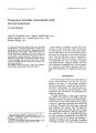
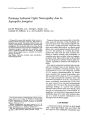
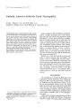
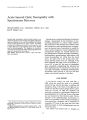
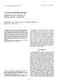
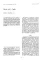
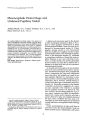
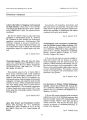
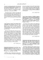
Jou17Ul1 of Clinical Neuro- ophthalmology 9( 1): 55- 59, 1989. Clinicopathological Correlation Metastatic Malignant Meningioma Michael L. Slavin, M. D. © 1989 Raven Press, Ltd., New York The onset of a rapidly progressive abducens and trigeminal neuropathy, third- order neuron Horner's syndrome, and decreased lacrimation clinically suggest a malignant lesion at the base of the middle cranial fossa, commonly a metastatic process, A case is reported in which computed tomography and magnetic resonance imaging failed to image the lesion but a bone scan clearly depicted the abnormal area, A malignant meningioma ( en plaque) was evident on biopsy, and pulmonary metastases later ensued. Common histological patterns of meningioma ( often thought of as a benign tumor) include meningothelial, fibrous, and transitional types. The association of cellular atypia, nuclear pleomorphism, marked mitoses, and brain invasion warrants the designation of malignant meningioma. The incidence of malignancy in meningioma ranges from 2 to 10% with reported metastases occurring in 0.1% Key Words: Meningioma- Malignant meningiomaSixth nerve palsy- Trigeminal neuropathy- Homer's syndrome, From the Division of Neuro- ophthalmology, Department of Ophthalmology, Long Island Jewish Medical Center, New Hyde Park, and School of Medicine, Health Sciences Center, State University of New York at Stony Brook, Stony Brook, New York, U. S. A. Address correspondence and reprint requests to Dr. M. Slavin at Department of Ophthalmology, Long Island Jewish Medical Center, New Hyde Park, NY 11042, U. S. A. 55 Meningioma, often considered a benign tumor, may typically result in chronic progressive visual loss or diplopia when it compresses anterior visual pathways, or ocular motor nerves III, IV, or VI in the superior orbital fissure, cavernous sinus, or base of the middle cranial fossa. Aggressive clinical behavior of meningioma in a minority of cases may be correlated with the finding of " atypical" or malignant features on histopathology. I report a patient in whom there was rapid progression of trigeminal and abducens cranial neuropathy, dry eye, and oculosympathetic palsy, due to a malignant meningioma at the base of the middle cranial fossa. Pulmonary metastases, a rare occurrence ( 1), rapidly ensued. CASE REPORT A 71- year- old healthy woman developed numbness in the right lip and cheek that progressed to the right mandible and forehead in December 1984. Skull x- ray films with tomography were stated to be normal. Six months later, she developed dryness of the right nostril, and noted a 12- lb weight loss. Two months after this, horizontal diplopia ensued. On neuro- ophthalmic examination, visual acuity and visual fields were normal. Motility was normal except for limited abduction of the right eye ( 20% of normal). In addition there was moderate right superficial punctate keratopathy, diminished tear production ( Schirmer's test), and an absent corneal reflex, Funduscopic examination was unremarkable. Neurologic examination revealed a dense sensory disturbance in the VI, V2, and V3 branches of the trigeminal nerve. The motor division of V3 ( muscles of mastication) was normal. Results of computed tomography ( CT) of the brain and nasopharynx were unremarkable except for enlargement of the right foramen ovale ( Fig. 1) 56 M. L. SLAVIN FIG. 1. Computed axial tomography through the base of the middle cranial fossa. Note the normal oval shape of the left foramen ovale ( arrow), which transmits the mandibular division of the trigeminal nerve, but the enlarged right foramen ( arrowhead), and questionable hyperostosis of the bones of the right middle cranial fossa. Magnetic resonance ( MR) imaging, workup for occult malignancy, multiple lumbar punctures, nasopharyngoscopy, and biopsies of the nasopharynx were unrevealing. During the next month, a partial right third- order neuron oculosympathoparesis ( Horner's syndrome) developed and the abduction defect was total. A repeat CT scan after injection of intrathecal metrizamide was unremarkable. Bone scan, however [ single photon emission computed tomography ( SPECT)] revealed increased uptake in the bones at the base of the right middle cranial fossa, especially in the squamous portion of the temporal bone ( Fig. 2). A right middle cranial fossa craniotomy was performed on February 17, 1986 and showed an infiltrating dural mass, which was incompletely resected. Some portions of the biopsy were consistent with meningioma due to a typical syncytial pattern of tumor cells ( vide infra) and the finding of psammoma bodies, whereas other areas showed 11l.:. rkcd nuclt~ M pleomorphism and multi- " ," ,', ~ ;',' c' 1, 1989 pIe mitotic figures ( Fig. 3) compatible with atypical or malignant meningioma. Despite the institution of radiotherapy, the patient developed a total right third- nerve palsy. A follow- up chest x- ray film 2 months later showed multiple nodular densities, compatible with metastatic disease ( Fig. 4). Bronchoscopy revealed tissue identical histopathologically to the central nervous system tumor. The patient died in December 1986 and no postmortem was performed. COMMENT The clinical features in this case were consistent with an extradural lesion at the base of the middle cranial fossa, involving cranial nerve V ( VI- V3) or the gasserian ganglion resulting in sensory disturbance of the face and absent corneal reflex; the greater superficial petrosal nerve causing decreased lacrimation; the pericarotid sympathetic plexus resulting in a third- order neuron Homer's syndrome; and cranial nerve VI. Although neither CT nor MR imaging demonstrated a definitive soft- METASTATIC MALIGNANT MENINGIOMA 57 LP 1s 11.' UY~ r_ ~ Il"" Illl ,)'! 1. A FIG. 2. After intravenous injection of 20 MCi of technetium- 99m, a single photon emission computed tomography study with cross- sectional views of the skull base [ axial ( left), coronal ( center), right parasagittal ( right)] revealed discrete foci of increased tracer activity in the squamous portion of the temporal bone ( arrows). tissue or bony abnormality ( MR imaging does not image bone), bone scan ( SPECT) with the capability to depict axial, coronal, and sagittal sections was invaluable in identifying the lesion. The tentative diagnosis was osteoblastic metastases, although other bony abnormalities such as fibrous dysplasia, and Paget's disease were considered. The diagnosis of " benign" ( garden variety) meningioma en plaque was withheld due to the rapidly progressive clinical course. Meningioma, derived from meningothelial arachnoid cells, is a tumor that is attached to dura mater and usually compresses adjacent brain rather than invading it. Hyperostosis of neighboring bone often seen on radiologic studies may indicate either actual tumoral invasion of bone or may represent a nontumoral osteoblastic reaction. Meningioma may show well- circumscribed borders and appear as a sessile or pedunculated mass. An en plaque meningioma refers to a tumor that grows flat along the dural surface, with a softtissue component that may be difficult to identify radiologically ( as in our case). Three histologic patterns of meningioma predominate: meningothelial or syncytial, fibrous, and transitional ( mixed) ( 2- 4). The meningothelial type consists of large epithelial- like cells with indistinct borders, which form a uniformly lobulated pattern. A whorl pattern of the cells with centrally located areas of calcification known as psammoma bodies is virtually pathognomonic of meningioma. The fibrous variety, on the other hand, shows spindle cells in a streaming pattern that are typically separated by a network of collagen and reticulin fibers. The transitional type shares features with both of the above entities. It should be noted that any of the above pattern when associated with cellular atypia, nuclear pleomorphism, a marked number of mitotic figures, focal necrosis, or invasion of border tissues may be designated as a malignant meningioma, and may behave in an aggressive clinical fashion, including metastasis. There are two additional patterns, papillary ( 5) and angioblastic ( 5) meningioma, which typically behave as malignant lesions and are accordingly associated with marked cellular atypia histopathologically. Other primary dural tumors such as meningosarcoma and hemangiopericytoma should not be confused with malignant meningioma. Hemangiopericytoma is a highly cellular tumor with spindle- shaped cells and an abundance of capillaries. In my patient, the dearth of capillaries was inconsistent with a diagnosis of angioblastic meningioma or hemangiopericytoma. Of course, a metastatic lesion to the meninges of the base of the skull should always be considered. The reported incidence of malignancy in meningioma ranges from 2 to 10% ( 6,7). The occurrence of distant metastases from meningioma, which is usually to the lungs ( 1), liver, and cerebrospinal pathways is reported in - 0.1% of cases ( 8). Jaaskelainen and associates ( 9) in 1986 studied 936 primary intracranial meningiomas and their recurrences. Ninety- four percent were histologically benign whereas 4.7% were deemed atypical and 1.0% were anaplastic. The benign tumors ( after an apparent complete gross removal of tumor neurosurgically) showed a 5- year recurrence rate of 3% but was found to be 21% at 25- year follow- up. A 38 and 78% 5- year recurrence rate, however, was seen for the atypical and anaplastic varieties, respectively. In another study by Jaaskelainen and I Cli" Neuro- ophthalmoi, Vol. 9, No. 1, 1989 58 M. L. SLAVIN u.;: S. HIstopathology of malignant meningioma. Hematoxylin and eosin. x 16 ( top), x32 ( bottom). Note the typical psammoma body ( arrow) amid pleomorphic tumor cells with marked nuclear atypia. A mitotic figure ( arrowhead) is seen. associates ( 10), average tumor doubling time was found to be 415 days ( range 138- 1,035 days) for benign meningioma versus 178 days ( range 34-- 551 days) in the atypical variety and 205 days ( range 3(}... 472 days) in anaplastic types. A highly maIig- ; .';", ., 1,. '. ~ j., ,.,..' I\}. l. 1. 1989 nant tumor with a doubling time of - 1 month, for example, would grow to 16 times its original size in just a 4- month period of observation. Jaaskelainen also found similar recurrence rates in anaplastic varieties despite postoperative radiotherapy. The METASTATIC MALIGNANT MENINGIOMA 59 FIG. 4. Chest x- ray film shows multiple, noncalcified pulmonary nodules compatible with metastatic disease. stated preoperative clues to malignancy on CT in their study included inhomogeneity of enhancement of the tumoral mass after intravenous contrast injection ( perhaps indicating focal necrosis), " mushrooming" or nodular growth of the lesion ( probably indicative of invasion of brain tissue), and a consistent lack of associated calcification. Benign lesions, on the other hand, harbored calcified areas in 28% of cases. REFERENCES 1. Kepes JJ, MacGee EE, Vergara G, SiJ R. Malignant meningioma with extreme pulmonary metastases. Kansas Med Soc J1971; 72: 312- 6. 2. Kepes JJ. Meningiomas: Biology, Pathology and Differential Diagnosis. New York: Masson Publishing USA, 1982. 3. Boldrey E. The meningiomas. In: Minckler J, ed. Pathology of the Nervous System Vol. 2. New York: McGraw- HilL 1971: 2125- 44. 4. Earle KM, Richany SF. Meningiomas--- a study of the histology, incidence, and biologic behavior of 243 cases from the Frazier- Grant collection of brain tumors. Med Ann DC 1969; 38: 353-- 8. 5. Ludwin SK, Rubinstein LJ, Russell OS. Papillary meningioma: a malignant variant of meningioma. Cancer 1975; 36: 1363- 73. 6. Turner GA, Craig W McK, Kernohan JW. Malignant meningiomas- a clinical and pathologic study. Surgery 1942; 11: 81- 100. 7. Tytus JS, Lasersohn JT, Reifel E: The problem of malignancy in meningiomas. J Neurasurg 1967; 27: 551- 7. 8. Strong RR, Tobi 0, Nordenstam H. Meningioma with intracraniaL cerebellar and visceral metastases. J Neurosurg 1964; 21: 109~ 1102. 9. Jaaskelainen J, Haltia M, Servo A. Atypical and anaplastic meningiomas: radiology, surgery, radiotherapy, and outcome. Surg Neural 1986; 25: 233- 42. 10. Jaaskelainen J, Haltia M, Laasonen E, Wahlstrom T, Valtonen S. The growth rate of intracranial meningiomas and its relation to histology. An analysis of 43 patients. Surg NeuroI1985; 24: 165- 72. JClin Neuro- ophthalmol. Vol. 9. No. 1, 1989 |