| OCR Text |
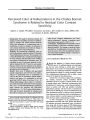
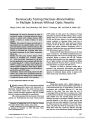
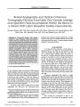
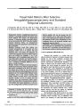
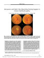
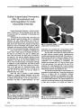
Show PHOTO ESSAY Amaurosis and Optic Disc Blanching During Upgaze in Graves Ophthalmopathy Loren S. Seery, MD, Renzo A. Zaldı ´ var, MD, and James A. Garrity, MD Abstract: A 60- year- old man with worsening Graves ophthalmopathy complained of complete loss of vision in the left eye on upgaze (‘‘ gaze- evoked amaurosis'' [ GEA]). Examination disclosed conges-tive orbital signs bilaterally. Upon upgaze, intraocular rose from 18 to 35 mm Hg in the left eye and visual acuity fell in that eye from 20/ 25 to no light perception. Evidence of optic nerve dysfunction and a swollen optic disc were present in the affected eye, and CT showed enlarged extraocular muscles with apical orbital compression of the optic nerve. Fundus photography performed during GEA disclosed blanching of the optic disc surface vessels in the affected eye. Surgical orbital decompression allevi-ated the congestive features and optic neuropathy and eliminated the GEA. This is the second case of GEA to be reported in Graves ophthalmopathy and the first to document blanching of optic disc vessels. The presumed mechanism of GEA in this patient is optic nerve ischemia, perhaps provoked by an elevation in intraocular pressure in a congested optic disc. ( J Neuro- Ophthalmol 2009; 29: 219- 222) FIG. 1. Minimally swollen right optic disc ( A) and markedly swollen left optic disc ( B) in a patient with Graves ophthalmopathy. During upgaze ( C), visual acuity in the left eye falls from 20/ 25 to no light perception (‘‘ gaze- evoked amaurosis''), and there is complete blanching of optic disc vessels of the left eye. Two minutes after the eyes have returned to primary gaze ( D), visual acuity returns to 20/ 25 in the left eye and normal vascularity is evident on the optic disc. Department of Ophthalmology, Mayo Clinic, Rochester, Minnesota. Address correspondence to James A. Garrity, MD, Mayo Clinic, 200 First Street SW, Rochester, MN 55905; E- mail: garrity. james@ mayo. edu J Neuro- Ophthalmol, Vol. 29, No. 3, 2009 219 A 60- year- old man noted simultaneous onset of hyper-thyroidism and orbital swelling. He was treated with radioactive iodine 1 month later, followed by oral replace-ment thyroid hormone. The orbital swelling was pro-gressive such that 60 mg/ day prednisone was started and tapered over 1 month. The orbital manifestations improved, only to worsen when the prednisone was tapered. Having been a heavy cigarette smoker, he had stopped completely 3 months later. Our initial examination, 8 months after discontinu-ation of prednisone, disclosed a best- corrected visual acuity of 20/ 25 in each eye. The pupils were of normal size and reactivity and color vision ( by Ishihara plates) was normal. The eyelids were mildly edematous and erythematous, and the conjunctiva and episclera were injected over the rectus muscle insertions. Each caruncle was mildly chemotic, as was the lateral conjunctiva bilaterally. Exophthalmometry was 25 mm bilaterally with moderate nontender resistance to retropulsion. Diplopia was present at the extremes of gaze. Ocular ductions were normal in adduction, mildly reduced in abduction, and severely reduced in supraduction bilaterally. There was some discomfort with eye movement. Each optic disc appeared normal with a small cup. There were no choroidal folds. There was no evidence of pretibial myxedema or thyroid acropachy. No therapy was given. When we reexamined him 2 months later, he reported more painful eye movements and complete loss of vision in the left eye when he looked upward. The visual loss began with peripheral constriction and progressed to no light perception in the left eye. With return to primary gaze, the visual recovery began from the center and would be complete within 30 seconds. Best- corrected visual acuity was 20/ 25 in both eyes but there was a trace left afferent pupillary defect while the eyes were in primary position. He could identify all Ishihara color plates with either eye but was more hesitant when viewing with the left eye. There was now more evidence of congestion and injection of the conjunctiva in both eyes. Exophthalmometry had increased to 27 mm bilaterally. Ophthalmoscopy disclosed choroidal folds extending across each fovea. The right optic disc was mildly swollen ( Fig. 1A); the left optic disc was massively swollen ( Fig. 1B). In primary gaze position, the intraocular pressure was 18 mmHg bilaterally. In upgaze and during the visual loss, it was 33 mmHg in the right eye and 35 mmHg in the left eye as measured by a Perkins tonometer. The left optic disc blanched markedly during the visual loss, a feature confirmed with fundus photography ( Fig. 1C). It resumed its baseline swollen appearance 2 minutes after the patient's eyes had returned to primary position ( Fig. 1D). Automated visual fields showed nerve fiber bundle defects that were more pronounced in the inferior visual field in both eyes and in the left than the right eye ( Fig. 2). Orbital CT showed symmetric enlargement of all the rectus muscles with apical crowding bilaterally ( Fig. 3). The patient underwent bilateral transantral orbital decompression. The gaze- evoked amaurosis ( GEA) ceased almost immediately. When he was reexamined 6 weeks later, visual acuity was 20/ 20 in each eye. There was a 5- mm reduction in proptosis. Bilateral medial rectus recession led to resolution of the diplopia. Follow- up visual field examination showed marked improvement in both eyes ( FIG. 4). Our patient manifested GEA, an uncommon symptom in which transient visual loss occurs when the affected eye is voluntarily moved into eccentric gaze. The GEA in the left eye consisted of a reduction of vision to no light perception and occurred in upgaze, when the intraocular pressure rose from 18 to 35 mmHg. During the GEA, the optic disc surface vessels, but not the retinal arteries, blanched, indicating a diminution in blood flow to the optic nerve. Our patient manifested massive optic disc edema and optic nerve dysfunction in the affected eye ( afferent pupil defect and visual field loss) attributed to apical orbital compression from enlarged extraocular muscles. After surgical orbital decompression, all manifestations, in-cluding the GEA, resolved. In most reported cases, GEA has occurred in associa-tion with an intraorbital process ( 1), but it has been reported in Graves ophthalmopathy only once ( 2). The other reported case was a letter to the journal editor regarding a 62- year- old smoker with thyroid eye disease and bilateral GEA occurring on upgaze. This patient did not have evidence of optic neuropathy and was treated with corticosteroids, which resulted in resolution of the amaurosis. FIG. 2. Visual fields performed at diagnosis show nerve fiber bundle defects. 220 q 2009 Lippincott Williams & Wilkins J Neuro- Ophthalmol, Vol. 29, No. 3, 2009 Seery et al First described 30 years ago ( 3), GEA usually results from intraconal orbital masses such as optic nerve sheath meningiomas ( 3- 8) and cavernous hemangiomas ( 9- 11) and less commonly from retained metallic foreign body ( 12), orbital metastasis from renal cell carcinoma ( 13), traumatic hemorrhagic cyst ( 3), optic nerve glioma ( 6,7), granular cell myoblastoma ( 7), and neurofibromatosis type II ( 14). There are a few reported cases of GEA due to pseudotumor cerebri ( 15,16) and with extraconal processes such as a juvenile nasopharyngeal angiofibroma ( 17), medial wall fracture ( 17), and osteoma ( 18). The mechanism of GEA is thought to be ischemic ( 8). Wright et al ( 3) reported absence of retinal arterial blood flow on fluorescein angiography in a single patient with GEA from an optic nerve meningioma. In a case of GEA from a varix with hemorrhage, Knapp et al ( 4) demonstrated a complete absence of the arterial waveform in the central retinal artery during eccentric gaze that caused a decrease in vision to no light perception. FIG. 4. Visual fields performed 3 months after surgical orbital decompression show marked improvement. The gaze- evoked amaurosis had also disappeared. FIG. 3. Precontrast coronal ( top) and axial ( bottom) CT performed at diagnosis shows bilateral enlargement of all the extraocular muscles with optic nerve compression at the orbital apex. 221 Gaze- Evoked Amaurosis J Neuro- Ophthalmol, Vol. 29, No. 3, 2009 Our patient is unique in that we were able to demonstrate photographically a blanching of the optic nerve surface vessels but not the retinal vessels during GEA. Presumably the increased intraocular pressure associated with upgaze exceeded the intraocular perfusion pressure, leading to reversible optic nerve ischemia. Our patient with GEA in Graves ophthalmopathy had evidence of optic nerve dysfunction. In the majority of the reported patients with GEA, baseline optic nerve dysfunction is present. In many of these patients, an afferent pupillary defect became more prominent in the eccentric gaze position that resulted in amaurosis. GEA has rarely been reported in patients with normal optic nerve function ( 1,2). REFERENCES 1. Orcutt JC, Tucker WM, Mills RP, et al. Gaze- evoked amaurosis. Ophthalmology 1987; 94: 213- 18. 2. Bremner FD, Sanders MD, Stanford MR. Gaze evoked amaurosis in dysthyroid orbitopathy. Br J Ophthalmol 1999; 83: 501. 3. Wright JE. Primary optic nerve meningiomas: clinical presentation and management. Trans Sect Ophthalmol Am Acad Ophthalmol Otolaryngol 1977; 83: 617- 25. 4. Knapp MEP, Flaharty PM, Sergott RC, et al. Gaze- induced amaurosis fromcentral retinal artery compression. Ophthalmology 1992; 99: 238- 240. 5. Hampton GR, Krohel GB. Gaze- evoked blindness Ann Ophthalmol. 1983; 15: 73- 6. 6. Jakobiec FA, Depot MJ, Kennerdell JS, et al. Combined clinical and computed tomographic diagnosis of orbital glioma and meningioma. Ophthalmology 1984; 91: 137- 55. 7. Bradbury PG, Levy IS, McDonald WI. Transient uniocular visual loss on deviation of the eye in association with intraorbital tumours. J Neurol Neurosurg Psychiatry 1987; 50: 615- 19. 8. Oohira A, Kubo R. Ocular blood flow defect in gaze- evoked amaurosis. Nippon Ganka Gakkai Zasshi 1999; 103: 56- 60. 9. Manor RS, Ben Sira I, Odel JG, et al. Amaurosis fugax at downward gaze. Surv Ophthalmol 1987; 31: 411- 16. 10. Brown GC, Shields JA. Amaurosis fugax secondary to presumed cavernous hemangioma of the orbit. Ann Ophthalmol 1981; 13: 1205- 09. 11. Tsai RK, Chen JY, Wang HZ. Gaze- evoked amaurosis caused by intraconal cavernous hemangioma: a case report. Kaohsiung J Med Sci 1997; 13: 324- 27. 12. Danesh- Meyer HV, Savino PJ, Bilyk JR, et al. Gaze- evoked amaurosis produced by intraorbital buckshot pellet. Ophthalmology 2001; 108: 201- 06. 13. Mezer E, Gdal- On M, Miller B. Orbital metastasis of renal cell carcinoma masquerading as amaurosis fugax. Eur J Ophthalmol 1997; 7: 301- 04. 14. Smith L, Kriss A, Gregson R, et al. Gaze evoked amaurosis in neurofibromatosis type II. Br J Ophthalmol 1998; 82: 584- 85. 15. O'Duffy D, James B, Elston J. Idiopathic intracranial hypertension presenting with gaze- evoked amaurosis. Acta Ophthalmol Scand 1998; 76: 119- 20. 16. Pascual J, Combarros O, Berciano J. Gaze- evoked amaurosis in pseudotumor cerebri. Neurology 1988; 38: 1654- 55. 17. Otto CS, Coppit GL, Mazzoli RA. Gaze- evoked amaurosis: a report of five cases. Ophthalmology 2003; 110: 322- 26. 18. Wilkes SR, Trautmann JC, DeSanto LW, et al. Osteoma: an unusual cause of amaurosis fugax. Mayo Clin Proc 1979; 54: 258- 60. 222 q 2009 Lippincott Williams & Wilkins J Neuro- Ophthalmol, Vol. 29, No. 3, 2009 Seery et al |