| OCR Text |
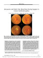
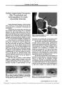
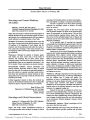
Show ORIGINAL CONTRIBUTION Retinal Angiography and Optical Coherence Tomography Disclose Focal Optic Disc Vascular Leakage and Lipid- Rich Fluid Accumulation Within the Retina in a Patient With Leber Idiopathic Stellate Neuroretinitis Hirokuni Kitamei, MD, PhD, Yasuo Suzuki, MD, PhD, Mitsuo Takahashi, MD, Satoshi Katsuta, MD, Hideo Kato, MD, Masahiko Yokoi, MD, PhD, and Manabu Kase, MD, PhD Abstract: A 52- year- old woman with clinical features of Leber idiopathic stellate neuroretinitis ( LISN) underwent retinal fluorescein and indocya-nine green angiography that revealed lipid- containing fluid leakage from a single arteriole in the superficial nerve fiber layer of the optic disc. The fluid expanded gradually into the upper half of the optic disc and the adjacent peripapillary retina. Optical coherence tomography ( OCT) demonstrated fluid accumulation in two separate subretinal spaces and in the outer nuclear- plexiform layer, which extended from the optic disc margin to the fovea. These angiographic and OCT findings support the hypothesis that LISN develops from focally increased permeability of an optic disc surface arteriole from which lipid- rich fluid flows through the outer nuclear- plexiform layer space to pool in these retinal areas. ( J Neuro- Ophthalmol 2009; 29: 203- 207) L eber idiopathic stellate neuroretinitis ( LISN) presents with unilateral visual loss, macular star, and optic disc edema. It is believed to result from increased permeability of vessels on the optic disc, followed by the appearance of a macular star after resolution of optic disc edema ( 1). Recent studies using optical coherence tomography ( OCT) have revealed an accumulation of fluid in the subretinal space and in the outer plexiform layer ( 2,3). The path of this fluid to those loci remains unknown. We report a case of LISN in which OCT and retinal angiography with fluorescein and indocyanine green indicated that a lipid- rich exudate escaped from a single arteriole on the optic disc and flowed directly into the outer nuclear- plexiform layer space. A serous component seeped beneath the neurosensory retina. CASE REPORT A 52- year- old woman reported a 1- week history of decreased vision in the right eye. Best- corrected visual acuity was 20/ 300 in the right eye and 20/ 15 in the left eye. A relative afferent pupillary defect was present in the right eye. Goldmann perimetry detected a cecocentral scotoma in the right eye, and no visual field defect in the left eye. Slit lamp examination showed no abnor-malities. Ophthalmoscopic examination showed that the optic disc in the right eye was edematous and hyperemic in its upper half, which had a cloudy surface ( Fig. 1A). Serous retinal detachment was observed in the upper papillomacular region adjacent to the optic disc, where small scattered yellow exudates and linear retinal hemor-rhages were seen. There were faint opacities with vitreous cells in the posterior vitreous above the lower margin of the optic disc. Fluorescein angiography ( FA) detected a single leaking arteriole on the optic disc ( Fig. 2A), from which hyperfluorescent fluid expanded slowly over the upper half of the optic disc and within the peripapillary retina over a 9- minute course after dye injection. There appeared to be no leakage of dye from the other retinal vessels or the retinal pigment epithelium. Indocyanine green angiography ( IA) also revealed clear leakage from the same arteriole observed on FA, and the dye spread over the optic disc over a 10- minute course after injection ( Fig. 2B). The choroidal circulation showed no abnormalities. Neither FA nor IA showed leakage from capillaries on the optic disc surface. OCT revealed an accumulation of fluid in the subretinal space at the foveal and peripapillary regions Department of Ophthalmology ( HK, YS, MT, SK, MY, MK), Teine Keijinkai Hospital, Sapporo, Hokkaido, Japan; and Megumino Eye Clinic ( HK), Eniwa, Hokkaido, Japan. Address correspondence to Hirokuni Kitamei, MD, PhD, Department of Ophthalmology, Teine Keijinkai Hospital, Maeda 1- 12, Teike- ku, Sapporo, Hokkaido 006- 0811, Japan; E- mail: hiromekita@ yahoo. co. jp J Neuro- Ophthalmol, Vol. 29, No. 3, 2009 203 and in the outer nuclear- plexiform layer space in the papillomacular region ( Fig. 3A), similar to a pattern reported previously ( 3). To evaluate the relationship between the subretinal space and fluid space in the outer nuclear- plexiform layer and the margin of the optic disc, OCT was performed around the optic disc ( Fig. 4). The optic nerve fibers were remarkably swollen at the upper portion of the optic disc ( Fig. 4A). Subretinal detachment was present temporal to the intermediary tissue of Kuhnt of the optic disc and was particularly extensive at the superotemporal peripapillary region, whereas the neuro-sensory retina nasal to the disc was not detached. A fluid space was also observed between the outer plexiform layer and the outer nuclear layer just at the margin of the optic disc ( arrow in Fig. 4BC). There were no abnormalities in the left eye. FIG. 1. A. Fundus photography of the right eye during our first examination shows optic disc edema and serous retinal detachment with linear retinal hemorrhages and yellowish exudates at the upper papillomacular region. B. One week later, the optic disc edema has lessened and radiating lipid- rich exudates have appeared. C. One month later, the optic disc edema and serous retinal detachment have resolved, and hard exudates have formed a partial macular star. FIG. 2. A. Retinal fluorescein angiography of the right eye performed at our first examination shows leakage from a single arteriole in the superficial layer of the optic disc during the arterial stage and slow diffusion of fluid over the optic disc and within the peripapillary retina. B. Retinal indocyanine green angiography performed at our first examination shows leakage from the same arteriole and slow expansion of the dye ( Numbers indicate time, in minutes and seconds, after injection of dye.) 204 q 2009 Lippincott Williams & Wilkins J Neuro- Ophthalmol, Vol. 29, No. 3, 2009 Kitamei et al Results of brain MRI were negative. Serologic tests detected no abnormalities suggestive of viral infection or autoimmune disease. The patient was treated with 250 mg/ day methyl-prednisolone intravenously for 3 days, followed by 60 mg/ day oral prednisolone, which was eventually tapered. Seven days after treatment was begun, best- corrected visual acuity in the right eye had improved to 20/ 40. Optic disc edema in that eye had lessened and posterior vitreous opacities had disappeared. Yellow exudates with a radiating pattern had appeared nasal to the fovea ( Fig. 1B). OCT at that time showed that fluid in the outer nuclear- plexiform layer space had largely been absorbed, whereas subretinal detachment was clearly observed ( Fig. 4B). Yellow exudates appeared, caused by a high OCT reflex signal indicated by a red color ( Fig. 4B). One month later, best- corrected visual acuity was 20/ 20 in the right eye, coincident with disappearance of the subfoveal detachment ( Fig. 4C). Optic disc edema had also completely resolved at this time. The yellow hard exudates were still present, showing a " macular star figure" nasal to the fovea ( Fig. 1C). DISCUSSION Ophthalmoscopic investigations in the present patient showed unilateral visual loss with optic disc edema and serous retinal detachment, followed by a macular star figure, features typical of LISN, an inflammatory optic neuropathy that is believed to develop from increased permeability of vessels on the optic disc ( 3- 5). In our patient, FA revealed massive leakage of dye from a single FIG. 3. A. Optical coherence tomography ( OCT) performed at our initial examination shows the anatomic transition of the subretinal and outer nuclear- plexiform spaces. A subretinal space ( large arrow) has appeared at the foveal and peripapillary regions. An outer nuclear- plexiform layer space ( small arrow) is located from the margin of the optic disc to the fovea. B. One week later, OCT shows that the outer nuclear- plexiform space has disappeared, but the subretinal space ( large arrow) is still present. The macular star figure is indicated by the red color signal ( arrowheads). C. One month later, the subretinal space has completely disappeared. ( The right column cursor indicates where the retina was scanned.) 205 Leber Idiopathic Stellate Neuroretinitis J Neuro- Ophthalmol, Vol. 29, No. 3, 2009 arteriole that was located superficial to the prelaminar region of the optic disc. Capillaries surrounding the arteriole showed no leakage. Another prominent feature was the length of time it took for the fluid to spread over the upper half of the optic disc and then within the peripapillary retina. This slow diffusion of fluorescein dye implied that the fluid escaping from the arteriole was highly viscous. IA showed the same leakage pattern. Because indocyanine green binds com-pletely to both low and high lipoproteins in the blood, whereas about 80% of fluorescein is bound to albumin with the rest remaining unbound ( 6,7), the leaking fluid must have contained lipid- rich components that are larger than water or albumin molecules. Optic disc edema with slow leakage of indocyanine green dye from a single arteriole, but not from capillaries, is clearly a different process from that seen in an acute attack of typical optic neuritis. These OCT findings provide important evidence that fluid accumulates in the subretinal space and in the outer nuclear- plexiform layer space near the intermediary tissue of Kuhnt. They suggest that the lipid- rich fluid leaking from a single arteriole, which in our patient was located in the superficial optic nerve fiber layer in the optic disc, had flowed directly into the outer nuclear- plexiform space near the optic disc margin and then seeped into the subretinal space. These two destinations may have developed because of the selective property of the external limiting membrane, which allows water but not lipids and proteins to pass through it ( 7). The serous component of the fluid had seeped through the external limiting membrane from the outer nuclear- plexiform layer and had accumulated beneath the neurosensory retina, whereas lipid- rich and protein- rich exudates had remained in the outer nuclear- plexiform space. In fact, follow- up investigations with OCT showed FIG. 4. Optical coherence tomography ( OCT) of the retinal peripapillary region performed at our initial examination shows swollen fibers in the upper optic disc ( A), and the appearance of an outer nuclear- plexiform space ( arrow) at the optic disc margin in the middle ( B) and lower regions ( C). A subretinal space is evident near the disc in each column. 206 q 2009 Lippincott Williams & Wilkins J Neuro- Ophthalmol, Vol. 29, No. 3, 2009 Kitamei et al that fluid accumulation in the outer nuclear- plexiform space had stopped significantly sooner than that in the subretinal space. The findings in our patient are in good agreement with the speculations of Gass ( 8). REFERENCES 1. Casson RJ, O'Day J, Crompton JL. Leber's idiopathic stellate neuroretinitis: differential diagnosis and approach to management. Aust NZ J Ophthalmol 1999; 27: 65- 9. 2. Ando R, Shinmei Y, Nitta T, et al. Central serous retinal detachment detected by optical coherence tomography in Leber's idiopathic stellate neuroretinitis. Jpn J Ophthalmol 2005; 49: 547- 58. 3. Stewart MW, Brazis PW, Barrett KM, et al. Optical coherence tomography in a case of bilateral neuroretinitis. J Neuroophthalmol 2005; 25: 131- 3. 4. Wade NK, Levi L, Jones MR, et al. Optic disk edema associated with peripapillary serous retinal detachment: an early sign of systemic Bartonella henselae infection. Am J Ophthalmol 2000; 130: 327- 34. 5. Matsuda A, Chin S, Ohashi T. A case of neuroretinitis associated with long- standing polyarteritis nodosa. Ophthalmologica 1994; 208: 168- 71. 6. Yoneya S, Saito T, Komatsu Y, et al. Binding properties of indocyanine green in human blood. Invest Ophthalmol Vis Sci 1998; 39: 1286- 90. 7. Marmor MF. Mechanisms of fluid accumulation in retinal edema. Doc Ophthalmol 1999; 97: 239- 49. 8. Gass JD. Diseases of the optic nerve that may simulate macular disease. Trans Sect Ophthalmol Am Acad Ophthalmol Otolaryngol 1977; 83: 763- 70. 207 Leber Idiopathic Stellate Neuroretinitis J Neuro- Ophthalmol, Vol. 29, No. 3, 2009 |