| OCR Text |
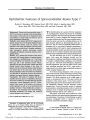
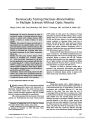
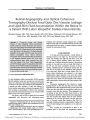
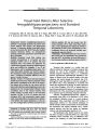
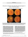
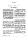
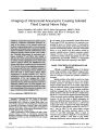
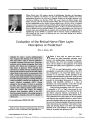
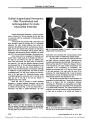
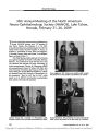
Show ORIGINAL CONTRIBUTION Two Patients With Spinocerebellar Ataxia Type 7 Presenting With Profound Binocular Visual Loss Yet Minimal Ophthalmoscopic Findings Matthew J. Thurtell, MBBS, FRACP, J. Alexander Fraser, MD, Elisa Bala, MD, Robert L. Tomsak, MD, PhD, Vale ´ rie Biousse, MD, R. John Leigh, MD, and Nancy J. Newman, MD Abstract: Two patients with genetically confirmed spinocerebellar ataxia type 7 ( SCA7) presented with progressive visual loss. Examination disclosed sub-stantial visual acuity loss, central scotomas, and marked dyschromatopsia. Ophthalmoscopic abnor-malities were subtle, with only mild retinal artery attenuation and minimal foveal region pigmentary abnormalities. Both patients had slow saccades and partially limited ductions, although neither reported diplopia. One patient had obvious extremity and gait ataxia, but the other had only an unsteady tandem gait. Results of electroretinography ( ERG) were abnormal in both patients. These cases illustrate that SCA7 may present with profound visual loss yet minimal ophthalmoscopic findings and sometimes minimal ataxia. The clues to diagnosis are the abnor-mal color vision, retinal artery attenuation, abnormal eye movements, and a family history of similar manifestations, which may have gone undiagnosed. Full- field or multifocal ERG will always disclose photoreceptor dysfunction. Genetic testing is now available to confirm the diagnosis. ( J Neuro- Ophthalmol 2009; 29: 187- 191) S pinocerebellar ataxia type 7 ( SCA7) is a rare neuro-degenerative disease caused by a CAG triplet repeat expansion in the SCA7 gene on chromosome 3 ( 1- 3), encoding for a protein called ataxin 7 ( 4). It is inherited in an autosomal dominant fashion, and sporadic cases are reported rarely ( 5). SCA7 characteristically produces pro-gressive ataxia. Its common ophthalmologic manifestations include visual loss from photoreceptor degeneration and ophthalmoplegia ( 6,7). We describe two unrelated patients with SCA7 who had visual loss with only subtle ophthalmoscopic signs. Electroretinography ( ERG) disclosed photoreceptor dys-function. Both patients had slow saccades and partial limita-tion of ocular ductions. We emphasize the importance of assessing eye movements and obtaining ERG for timely diagnosis of this disorder. CASE REPORTS Case 1 A 12- year- old African- American boy presented with progressive painless bilateral visual loss over several years. His family had also noticed that his eyes became ‘‘ crossed'' when he looked laterally, but he denied diplopia or any other neurologic symptoms. His mother and twin sister had similar manifestations. Best- corrected visual acuity was 20/ 200 in the right eye and 20/ 300 in the left eye. He could identify only the control Ishihara plate bilaterally. Confrontation visual fields revealed bilateral central scotomas. Results of external and anterior segment examinations were normal. Ophthalmos-copy revealed equivocally pale discs and attenuated retinal arteries. The foveal light reflexes were absent, and there was subtle pigmentary mottling in the foveal region ( Fig. 1). Departments of Neurology ( MJT, RJL) and Ophthalmology ( EB, RLT), University Hospitals Case Medical Center, Cleveland, Ohio; Daroff- Dell'Osso Ocular Motility Laboratory ( MJT, RLT, RJL), Louis Stokes Department of Veterans Affairs Medical Center, Cleveland Ohio; and Departments of Ophthalmology ( JAF, VB, NJN), Neurology ( VB, NJN), and Neurological Surgery ( NJN), Emory University School of Medicine, Atlanta, Georgia. This study was supported in part by a departmental grant ( Department of Ophthalmology) from Research to Prevent Blindness, Inc., New York, and by core grants ( P30- EY06360, Department of Ophthalmology) from the National Institutes of Health ( NIH), Bethesda, MD. Dr. Newman is a recipient of a Research to Prevent Blindness Lew R. Wasserman Merit Award. Drs. Leigh and Thurtell are supported by NIH Grant EY06717, by the Department of Veterans Affairs, and by the Evenor Armington Fund. Supplemental digital content is available for this article. Direct URL citations appear in the printed text and are provided in the HTML and PDF versions of this article on the journal's Web site ( www. jneuro- ophthalmology. com). Address correspondence to Nancy J. Newman, MD, Neuro- ophthal-mology Unit, Emory Eye Center, 1365- B Clifton Road NE, Atlanta, GA 30322; E- mail: ophtnjn@ emory. edu J Neuro- Ophthalmol, Vol. 29, No. 3, 2009 187 FIG. 1. Case 1. Retinal artery attenuation, subtle pigmentary mottling in the foveal region, and equivocal temporal optic disc pallor are evident. FIG. 2. Case 1. Precontrast T1 axial ( A), sagittal ( B), and coronal ( C) MRI shows cerebellar atrophy, most apparent in the vermis, and minimal brainstem atrophy. FIG. 3. Case 1. Multifocal electroretinogram from the right eye ( left panel) and left eye ( right panel). Trace arrays ( top) and field plots ( bottom) demonstrate severely attenuated central responses with an absent foveal peak. 188 q 2009 Lippincott Williams & Wilkins J Neuro- Ophthalmol, Vol. 29, No. 3, 2009 Thurtell et al Pupil examination was normal. Ocular motor examination revealed normal alignment in primary gaze position, slow horizontal saccades, and limited abduction of each eye that could be partially overcome with the doll's head maneuver. ( Video, Supplemental Digital Content 1, http:// links. lww. com/ WNO/ A3). Tandem gait was unsteady, and results of the remainder of the neurologic examination were unremarkable. Humphrey visual fields confirmed bilateral central scotomas. Brain MRI revealed cerebellar atrophy ( Fig. 2). Multifocal ERG showed severely attenuated foveal re-sponses ( Fig. 3). Genetic testing revealed an increased CAG repeat number of 65 ( normal < 18, borderline 19- 36) in one SCA7 allele, confirming the diagnosis of SCA7. Case 2 A 21- year- old Caucasian woman presented with bilateral visual loss and gait ataxia, both progressive since age 14 years. By age 18 years, she had developed a wide-based gait and, over the following 3 years, increasing clumsiness of her upper limbs. She took no regular medica-tions and denied alcohol consumption. Similar manifes-tations were present in her mother, in whom multiple sclerosis had been diagnosed, and in her mother's male cousin. Best- corrected visual acuity was 20/ 100 in both eyes. She could identify only the control Ishihara plate with each eye. Confrontation visual fields revealed bilateral central scotomas. Results of external and anterior segment exam-inations were unremarkable. Ophthalmoscopic examination revealed normal optic discs and slightly attenuated retinal arteries. The foveal light reflexes were absent, but there were no pigmentary changes ( Fig. 4). Pupillary examination was normal. Ocular alignment was normal in primary gaze position. Horizontal and vertical saccades were slow, and there was reduced abduction and supra-duction that could be partially overcome with the doll's eye maneuver ( Video, Supplemental Digital Content 2, http:// links. lww. com/ WNO/ A4). She had mild bilateral ptosis, facial weakness, head titubation, and appendicular, truncal, and gait ataxia. Deep tendon reflexes were symmetrically brisk, but plantar responses were flexor. Fundus autofluorescence was normal ( Fig. 5). Humphrey and Goldmann visual fields showed bilateral central scotomas. Brain MRI showed brainstem and cerebellar atrophy ( Fig. 6). Full- field ERG showed abnor-malities consistent with severe cone dysfunction and moderate rod dysfunction. Genetic testing revealed an increased CAG repeat number of 56 in one SCA7 allele, confirming the diagnosis of SCA7. DISCUSSION In contrast with the other autosomal dominant spinocerebellar ataxias, SCA7 is characterized by pro-gressive irreversible bilateral visual loss due to a degen-erative retinopathy that initially affects cone photoreceptors but progresses toward a cone- rod dystrophy phenotype ( 8,9). Early in the disease, visual acuity is decreased and there are central scotomas. Peripheral vision and night FIG. 4. Case 2. Subtle retinal artery attenuation is the only evident abnormality. FIG. 5. Case 2. Ocular fundus autofluorescence is normal. 189 Visual Loss in SCA7 J Neuro- Ophthalmol, Vol. 29, No. 3, 2009 vision are relatively preserved ( 6,10). Dyschromatopsia is usually severe and may be detected years before symptom onset in affected individuals ( 6). The retinopathy classically results in granular pigmentary changes in the foveal region ( 6,10), occasionally producing a ‘‘ bull's eye'' appearance ( 11), often with associated secondary optic disc pallor ( 6,10). However, these ophthalmoscopic abnormalities are not usually present until late in the course of the disease. Indeed, it has been noted that the fundus may initially appear normal, even after substantial visual acuity loss has occurred ( 6), potentially causing a diagnostic delay. Despite noting visual loss for several years, our 2 patients had only subtle retinal artery attenuation and absent foveal light reflexes ( Fig. 1 and 4). In our Case 1, who had more severe visual loss, there was very subtle pigmentary mottling in the foveal regions and equivocal temporal optic disc pallor. Our reviewof fundus photographs from previously reported patients with SCA7, in whom the ocular fundi were con-sidered normal, suggests that subtle abnormalities, such as retinal arterial attenuation, were actually present ( 12). A prospective study of individuals with the SCA7 mutation is required to further delineate the sequence of ophthalmo-scopic changes in relation to symptom onset and visual function. Pigmentary changes in the foveal region may be more evident on fluorescein angiography ( 11). Retinal thinning that is more extensive than suggested by ophthalmoscopy can be detected with optical coherence tomography ( OCT) ( 11). The role of fundus autofluorescence photography ( 13), a noninvasive imaging technique for topographical mapping of lipofuscin in the retinal pigment epithelium, has not been evaluated in SCA7. Lipofuscin is a fluores-cent pigment that accumulates in the retinal pigment epithelium cells as a consequence of photoreceptor degradation ( 13). Although lipofuscin accumulation can be seen with a variety of inherited and acquired retinal diseases, fundus autofluorescence was normal in our Case 2, suggesting that it might not be a useful tool for detecting retinal degeneration in the early stages of SCA7. The presence of photoreceptor disease can be con-firmed with ERG. Although full- field ERG is widely available and is abnormal once ophthalmoscopic changes have become apparent ( 10), multifocal ERG may be more sensitive for detecting foveal dysfunction in the early stages of the disease ( 11,12). Indeed, the multifocal ERG can be grossly abnormal even when ophthalmoscopic changes are absent or subtle ( 12), as in our Case 1. Ocular motor abnormalities are common in SCA7. Slowing of saccades is an early sign, suggesting involve-ment of the brainstem reticular formation ( 10,14), and is later followed by progressive ophthalmoplegia. The ophthalmoplegia is often partially overcome with vestibular stimulation in the earlier stages of the disease, but later becomes consistent with an external ophthalmoplegia ( 10). In both of our patients, there were no symptoms, such as diplopia, to suggest ocular motor dysfunction. Ocular motor abnormalities that are commonly seen in association with spinocerebellar degeneration, such as downbeat, gaze-evoked, and rebound nystagmus, dysmetric saccades, and impaired smooth pursuit ( 15), were not present in either of our patients, consistent with previous reports ( 6,7,10). Unfortunately, there are few reports of quantitative eye movement recordings in SCA7 ( 10,14). Signs of cerebellar dysfunction can be found in almost all patients with SCA7 as the disease progresses ( 7), but may be subtle or absent in the early stages, as in our Case 1. Cerebellar and brainstem atrophy is often seen on MRI ( 10,14) but can also be subtle and easily overlooked. REFERENCES 1. Gouw LG, Kaplan CD, Haines JH, et al. Retinal degeneration characterizes a spinocerebellar ataxia mapping to chromosome 3p. Nat Genet 1995; 10: 89- 93. 2. Benomar A, Krols L, Stevanin G, et al. The gene for autosomal dominant cerebellar ataxia with pigmentary macular dystrophy maps to chromosome 3p12- p21.1. Nat Genet 1995; 10: 84- 8. FIG. 6. Precontrast T1 axial ( A) and sagittal ( B) and coronal FLAIR ( C) MRI show marked cerebellar and brainstem atrophy. 190 q 2009 Lippincott Williams & Wilkins J Neuro- Ophthalmol, Vol. 29, No. 3, 2009 Thurtell et al 3. Holmberg M, Johansson J, Forsgren L, et al. Localization of auto-somal dominant cerebellar ataxia associated with retinal degeneration and anticipation to chromosome 3p12- p21.1. Hum Mol Genet 1995; 4: 1441- 5. 4. Kaytor MD, Duvick LA, Skinner PJ, et al. Nuclear localization of the spinocerebellar ataxia type 7 protein, ataxin- 7. Hum Mol Genet 1999; 8: 1657- 64. 5. Stevanin G, Giunti P, Belal GD, et al. De novo expansion of inter-mediate alleles in spinocerebellar ataxia 7. Hum Mol Genet 1998; 7: 1809- 13. 6. Gouw LG, Digre KB, Harris CP, et al. Autosomal dominant cerebellar ataxia with retinal degeneration: clinical, neuropathol-ogic, and genetic analysis of a large kindred. Neurology 1994; 44: 1441- 47. 7. David G, Durr A, Stevanin G, et al. Molecular and clinical corre-lations in autosomal dominant cerebellar ataxia with progressive macular dystrophy ( SCA7). Hum Mol Genet 1998; 7: 165- 70. 8. Aleman TS, Cideciyan AV, Volpe NJ, et al. Spinocerebellar ataxia type 7 ( SCA7) shows a cone- rod dystrophy phenotype. Exp Eye Res 2002; 74: 737- 45. 9. Michalik A, Martin JJ, Van Broeckhoven C. Spinocerebellar ataxia type 7 associated with pigmentary retinal dystrophy. Eur J Hum Genet 2004; 12: 2- 15. 10. Enevoldson TP, Sanders MD, Harding AE. Autosomal domi-nant cerebellar ataxia with pigmentary macular dystrophy: a clinical and genetic study of eight families. Brain 1994; 117: 445- 60. 11. Ahn JK, Seo JM, Chung H, et al. Anatomical and functional char-acteristics in atrophic maculopathy associated with spinocerebellar ataxia type 7. Am J Ophthalmol 2005; 139: 923- 25. 12. Hugosson T, Granse L, Ponjavic V, et al. Macular dysfunction and morphology in spinocerebellar ataxia type 7 ( SCA 7). Ophthalmic Genet 2009; 30: 1- 6. 13. Schmitz- Valckenberg S, Holz FG, Bird AC, et al. Fundus autofluo-rescence imaging: review and perspectives. Retina 2008; 28: 385- 409. 14. Oh AK, Jacobson KM, Jen JC, et al. Slowing of voluntary and involuntary saccades: an early sign in spinocerebellar ataxia type 7. Ann Neurol 2001; 49: 801- 4. 15. Leigh RJ, Zee DS. The Neurology of Eye Movement. 4th ed. New York: Oxford University Press; 2006. 191 Visual Loss in SCA7 J Neuro- Ophthalmol, Vol. 29, No. 3, 2009 |