| OCR Text |
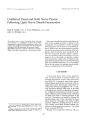
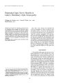
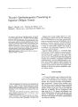
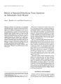
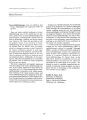
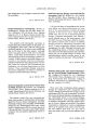
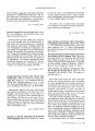
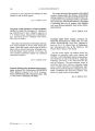
Show jOllrnal 0/ Clime" l Nellro- 0l'htlwllll" loSY 12( 2): 116-- 12[ 1, 1992. Bilateral Internuclear Ophthalmoplegia in a Patient with Wernicke's Encephalopathy Monica A. De La Paz, M. D., Sophia M. Chung, M. D., and John A. McCrary III, M. D. 1;; 1992 Raven Press, Ltd., New York The most common cause of bilateral internuclear ophthalmoplegia is multiple sclerosis, Wernicke's encephalopathy has been reported as a cause of unilateral internuclear ophthalmoplegia but not of bilateral internuclear ophthalmoplegia, In this report, we present the case of a patient with a history of alcohol abuse and acute onset of bilateral internuclear ophthalmoplegia whose clinical course and diagnostic studies are most consistent with a diagnosis of Wernicke's encephalopathy, Key Words: Internuclear ophthalmoplegia- Wernicke's encephalopathy- Alcoholism From the Department of Ophthalmology ( M. A. D.), Massachusetts Eye and Ear Infirmary. Department of Ophthalmology, Boston. Massachusetts; Bethesda Eye Institute ( S. M. C.), Saint Louis University Medical Center, Saint Louis. Missouri; and Cullen Eye Institute a. A. M.), Baylor College of Medicine, Houston. Texas. U. s. A. Address correspondence and reprint requests to Dr. Sophia M. Chung. Bethesda Eye Institute. Saint Louis University Medic~ 1 C.' nlt'f, 3ASS Vi<; la A\' enut', Saint LouIs. 11.1063110. U. S. A. Internuclear ophthalmoplegia is caused by a lesion of the medial longitudinal fasciculus and manifests as impaired adduction of the eye ipsilateral to the side of the lesion, with jerk nystagmus of the abducting eye. Vertical nystagmus usually evoked by upgaze is a frequent finding. The most common cause of bilateral internuclear ophthalmoplegia is multiple sclerosis ( 1,2). Other causes include ischemia from basilar arterial disease, vasculitides, brainstem malignancies, syphilis, Arnold- Chiari malformation, trauma, and overdose of exogenous agents, such as narcotics ( 3- 8). An ocular motor pattern resembling internuclear ophthalmoplegia has been described in myasthenia gravis ( 9). Unilateral internuclear ophthalmoplegia has been well described in association with Wernicke's encephalopathy ( 1, 10). The association of Wernicke's encephalopathy with bilateral internuclear ophthalmoplegia has, to the best of our knowledge, not been described. In this report, we describe the case of a patient with bilateral internuclear ophthalmoplegia whose clinical presentation and diagnostic studies are most consistent with a diagnosis of Wernicke's encephalopathy. CASE REPORT A 29- year- old white man with a long history of alcohol abuse was in his usual state of health until 2 weeks before presentation, when he noted the onset of horizontal diplopia. The episode occurred after a drinking binge, at which time the patient lost consciousness and awoke with horizontal diplopia and divergent gaze. Prior ocular history was unremarkable, and there were no complaints related to other neurological findings. The past medical history was remarkable for gastritis. There was a history of intravenous cocaine abuse. The last use of cocaine was several days before the onset of diplopia. 116 DIVERGENT GAZE IN WERNICKE'S ENCEPHALOGRAPHY 117 On examination, the patient was a slightly drowsy, thin white man. Visual acuity of both eyes was 20/ 20. There was mild ptosis of both eyes, with normal function of both levator palpebrae. The pupils were sluggishly reactive to light. There was no light- near dissociation or afferent pupillary defect. A variable angle alternating exotropia from 30 to 90 prism diopters ( PO) was present. Downbeat nystagmus was present in primary gaze. There was evidence of a bilateral internuclear ophthalmoplegia ( Fig. 1), with horizontal nystagmus in abduction and marked impairment of adduction of both eyes. Adduction was improved with convergence. Lateral rectus function was full in both eyes. Upbeat nystagmus occurred with elevation and downbeat nystagmus with downgaze. Confrontation visual fields were normal. Funduscopic examination was remarkable for bilateral juxtapapillary myelinated nerve fibers. The remainder of the ocular examination was normal. The patient was not oriented to place, person, or time. There was impairment of short- term memory, and he confabulated. The other cranial nerves were normal. The motor and sensory examination and deep tendon reflexes were normal. No Babinski sign was present. Cerebellar examination revealed decreased rapid alternating movements and a wide- based ataxic gait. The remainder of the physical examination was within normal limits. Hematological studies revealed a macrocytosis without anemia. The erythrocyte sedimentation rate was 15 mm/ h. Serum chemistries showed evidence of abnormal liver function consistent with the chronic abuse of alcohol. The ANA was negative. The rheumatoid factor was 28 IU/ mI. and the RPR was nonreactive. The patient was admitted for further evaluation. Intravenous thiamine followed by intramuscular supplementation was administered, in addition to supplemental folate and overall nutritional support. A lumbar puncture was performed. The opening pressure was normal, and the cerebrospinal fluid was clear and colorless, with 2 WBClmm3 . The glucose level was 67 mgldt and the protein was 61 mgldl. The spinal fluid electrophoretic study was normal. Specifically, no oligoclonal bands were detected. Special stains revealed no organisms, and cultures were sterile. Cerebrospinal fluid cytology showed no evidence of malignant cells. An HIV antibody test was negative. A Tensilon test was performed with no improvement in the ocular motility. A computed tomography ( CT) scan of the brain with contrast showed no lesions. Subsequently, magnetic resonance imaging ( MRI) of the brain was performed. Highintensity signals on both spin density and T2weighted scans were present in the mesencephalon anterior to the aqueduct extending along both medial longitudinal fasciculi ( Figs. 2 and 3). There was no mass effect, and no other lesions were noted. Thiamine supplementation was begun at the time of admission. Orientation and drowsiness improved significantly within the first 24 hours; the ataxia improved within the second 24 hours of the hospitalization. Symptoms of alcohol withdrawal were treated with oral chlordiazepoxide. Over the next 2 weeks, the ocular motility examination of the patient improved. The amplitude and frequency of the vertical nystagmus diminished. There was improvement in the intermittent exotropia, which ranged from 10 to 60 PD. The adduction weakness was unchanged. The patient was discharged from the hospital after 2 weeks; he FIG. 1. Motility series of patient demonstrating bilateral internuclear ophthalmoplegia. There is exotropia in all positions of gaze and marked adduction deficit of both eyes. I Clin Neuro- ophthalmol. Vol. 12. No. 2. 1992 118 M. A. DE LA PAZ ET AL. FIG. 2. This T2- weighted MR scan taken at the level of the mesencephalon demonstrates the periaqueductal gray matter lesion ( arrow). failed to return for follow- up, although an emergency room physician noted divergent gaze 3 months after hospitalization. DISCUSSION We present a patient with a history of alcoholism who developed acute onset of diplopia secondary to bilateral internuclear ophthalmoplegia, with additional features of downbeat nystagmus in primary gaze, vertical nystagmus, and cerebellar dysfunction. The MRI findings correlate the clinical features with bilateral lesions of the medial longitudinal fasciculi. Bilateral internuclear ophthalmoplegia is the most common ocular motor manifestation of multiple sclerosis ( 1). In the patient under discussion, numerous features of the case make the diagnosis of multiple sclerosis less likely. By history, there are no prior episodes of neurological symptoms, although it is possible that this may be a primary event. The cerebrospinal fluid has been shown to be abnormal in 90% of clinically definite cases of multiple sclerosis ( 11, 12). In the case under discussion, no oligoclonal bands were present. Furthermore, downbeat nystagmus in the primary position is only rarely associated with multiple sclerosis ( 13,14). FIG. 3. This spin- density MR scan demonstrates extension of the disease process into the medial longitudinal fasciculi ( arrow indicates right fasciculUS). MRI has been shown to be superior to CT in the diagnosis of multiple sclerosis ( 15,16). White matter lesions adjacent to the lateral ventricles are the most common finding on MRI in patients with multiple sclerosis ( 17,18). The plaques are best seen on the T2- weighted scans ( 16,19). Our patient did not reveal periventricular changes on MRI. Despite clinical evidence of brainstem and cerebellar pathology, lesions in these areas are not often demonstrated with MRI ( 17). One study, however, found that three patients with ocular motor disturbances secondary to multiple sclerosis had MRI studies demonstrating lesions of the brainstem with areas of prolonged T1 and/ or T2 ( 20). These patients had at least one additional periventricular white matter lesion. The history and clinical evaluation reasonably excluded other etiologies of bilateral internuclear ophthalmoplegia, including Arnold- Chiari malformation, basilar arterial disease, meningitis, vasculitides, brainstem malignancies, syphilis, and narcotic overdose. Wernicke's encephalopathy is a metabolic disease of the central nervous system caused by a nutritional deficiency of thiamine, which usually occurs in malnourished alcoholic patients ( 21). The most common neurological features associated with Wernicke's encephalopathy are disturbance of mentation and consciousness, paralysis of eye DIVERGENT GAZE IN WERNICKE'S ENCEPHALOGRAPHY 119 movements, and ataxic gait as demonstrated by our patient. Eye movement disorders include weakness of abduction, gaze- evoked nystagmus, horizontal and vertical gaze palsies, primary position upbeat and downbeat nystagmus, and internuclear ophthalmoplegia ( 10,22,23). Bilateral internuclear ophthalmoplegia has not been described in association with Wernicke's encephalopathy. Four patients reported by Schiffter ( 24) to have bilateral internuclear ophthalmoplegia caused by Wernicke's encephalopathy are not compatible with our patient as they had bilateral failure of abduction of the eyes, not adduction failure. Thiamine deficiency in Leigh's disease, a necrotizing hemorrhagic encephalomyelopathy in patients without a history of alcoholism or malnutrition, has been shown to cause bilateral internuclear ophthalmoplegia ( 25). The presence of the classic triad of impaired ocular motility, ataxia, and disturbance of mentation followed by prompt improvement in the clinical findings after thiamine supplementation is diagnostic of Wernicke's encephalopathy. It is well documented that improvement in abducens paresis may occur within hours after administration of thiamine and is usually complete within days ( 6,23), whereas nystagmus may persist for years despite adequate thiamine replacement ( 10). What remains unclear is the rate of response of internuclear ophthalmoplegia to thiamine. Postmortem studies describe destructive changes adjacent to the third and fourth ventricles and the aqueduct of Sylvius ( 26,27). Histopathological features of lesions in Wernicke's encephalopathy include varying degrees of parenchymal necrosis with neuron loss and astrocytic proliferation and, in some cases petechial hemorrhages. These pathological findings, however, do not explain the rapid reversal of ocular palsies in response to thiamine administration; instead, a biochemical deficit is implied, rather than a structural lesion. Pathological studies support this interpretation by the lack of significant damage of the ocular motor nuclei seen at autopsy in patients with Wernicke's disease ( 27). The destructive changes seen may also explain the slow and incomplete recovery of the nystagmus, ataxia, and memory. In the patient we have presented, the primary position downbeat nystagmus, mild confusional state, and ataxic gait followed by the improvement in the clinical findings after thiamine supplementation are highly suggestive of a diagnosis of Wernicke's encephalopathy. The incomplete resolution of the bilateral internuclear ophthalmoplegia upon thiamine supplementation, although not typical of Wernicke's encephalopathy, may be explained on the basis of destructive lesions involving both medial longitudinal fasciculi, as suggested by the MRI findings. There are only a few reports in the literature that describe the neuroimaging findings of patients with Wernicke's encephalopathy. CT scanning has revealed bilateral low- density thalamic lesions ( 28); nonenhancing hypodense areas around the cerebral aqueduct were noted in one patient who presented in a stuporous state with absent oculocephalic responses, with minimal improvement after thiamine and nutritional supplementation ( 29). MRI findings include increased signal in the dorsal medial nuclei of the thalami on T2- weighted images. Another study found that MRI scans on three of five patients demonstrated signal hyperintensities surrounding the third ventricle and aqueduct on T2- weighted images ( 30). These hyperintensities showed a " double wing" configuration that resembled that of the patient under discussion. In addition, the intensity of these regions was noted to decrease on follow- up studies, with consequent third ventricular and aqueductal dilation. However, exact correlation between the type of ocular motor deficit and MRl abnormality was not indicated in the article. MRl is considered to be superior to CT scanning in Wernicke's encephalopathy because of improved clarity of lesions ( 31,32). In summary, we believe that the clinical course of this patient with bilateral internuclear ophthalmoplegia and a history of alcoholism is most consistent with a diagnosis of Wernicke's encephalopathy. The case is of interest because we have not found previous reports of bilateral internuclear ophthalmoplegia in association with Wernicke's encephalopathy. In addition, the acute MRl findings in a patient with Wernicke's encephalopathy are readily demonstrated, indicating that MRI scanning can play a valuable role in establishing the diagnosis of this potentially fatal disease. The possibility that alcohol abuse resulting in nutritional compromise and Wernicke's encephalopathy may be the cause of bilateral internuclear ophthalmoplegia must be recognized. Prompt, appropriate replacement of depleted thiamine by parenteral administration is essential for the best neurologic outcome as Wernicke's encephalopathy represents a neurologic emergency. REFERENCES 1. Smith JL, Cogan DC. Internuclear ophthalmoplegia. Are · view of 58 cases. Arch OphthalmoI1959; 61: 687. JClin Neuro- ophthalmol. Vol. 12, No. 2. 1992 120 M. A. DE LA PAZ ET AL. 2. Cogan DG. Internuclear ophthalmoplegia, typical and atypical cases. Arcll Ophthalmol 1970; 84: 583. 3. Minor RH, Kearns TP, Millikan CH, Siekert RG, Sayre GP. Ocular manifestations of occlusive disease of the vertebralbasilar arterial system. Arch Ophthalmol 1959; 62: 112. 4. Cogen MS, Kline LB, Duvall ER. Bilateral internuclear ophthalmoplegia in systemic lupus erythematosus. J Clill Neuro-[ 1phtlltllmol 1987; 7: 69. 5. Cogan DC, Wray SH. Internuclear ophthalmoplegia as an early sign of brainstem tumors. Neurology 1970; 20: 629. 6. Woody RC, Reynolds JD. Association of bilateral internuclear ophthalmoplegia and myelomeningocele with Arnold- Chiari malformation, type ii. J Clin Neuro- ophthalmol 1985; 5: 124. 7. Keane JR. Traumatic internuclear ophthalmoplegia. J Clin Nt'uro- ophtlltllmol 1987; 7: 65. 8. Barret LG, Vincent FM, Arsac PL, Debru JE, Faure JR. internuclear ophthalmoplegia in patients with toxic coma. Frequency, prognostic value, diagnostic significance. J Toxicol- Clin TO. 1: icoI1983; 20: 373. 9. Jay WM, Nazarian SM, Underwood DW. Pseudo- internuclear ophthalmoplegia with downshoot in myasthenia gravis. J Clin Neuro- ophthalmol 1987; 7: 74. 10. Cogan DG, Victor M. Ocular signs of Wernicke's disease. Arch OplJthalmol 1954; 51: 204. 11. Johnson KP, Nelson BJ. Multiple sclerosis: diagnostic usefulness of cerebrospinal fluid. Am! Nezlro/ 1977; 2: 425. 12. Thompson EJ. Multiple sclerosis, immunological and biochemical diagnosis. Br Med Bull 1977; 33: 28. 13. Masucci EF, Kurtzke JF. Downbeat nystagmus secondary to multiple sclerosis. Ann Ophthalmo/ 1988; 20: 347. 14. Halmagyi GM, Rudge P, Gresty MA, Sanders MD. Downbeating nystagmus. A review of 62 cases. Arch Neuro/ 1983; 40: 777. 15. Sheldon JL Siddharthan R, Tobias L Sheremata WA, Soila K. Viamonte M. MR imaging of multiple sclerosis: comparison with clinical and CT examinations in 74 patients. Am / NeuroradioI1985; 6: 683. 16. Gebarski 55, Gabrielsen TO, Gilman 5, Knake JE, Latack n, Aisen AM. The initial diagnosis of multiple sclerosis: clinical impact of magnetic resonance imaging. Ann Neurol 1985; 17: 469. 17. Grenman R, Aantaa E, Katevuo K, Kormano M, Panelius M. Otoneurological and ultra low field MRl findings in multiple sclerosis patients. Acta Otolaryllgol 1988; 449: 77. 18. Jacobs L. Kinkel WR, Polachini I. Kinkel RP. Correlations of nuclear magnetic resonance imaging, computerized tomography, and clinical profiles in multiple scleroSIS. Neurology 1986; 36: 27. 19. Slamovits TL. Gardner TA. Neuroimaging in neuro- ophthalmology. Ophthalmology 1989; 96: 555. 20. Bogousslavsky L Fox AJ, Carey LS, Vinitski 5, Bass B, Noseworthy JH, Ebers GC, Barnett HJM. Correlates of brainstem ocular motor disorders in multiple sclerosis. Arch Neurol 1986; 43: 460. 21. Blass jP, Gibson GE. Abnormality of a thiamine- requiring enzyme in patients with Wernicke- Korsakoff syndrome. N Engl J Med 1977; 297: 1367. 22. Victor M, Adams RD, Collins GH. The Wernicke- Korsakoff syndrome: a clinical and pathological study of 245 cases, 82 with postmortem examinations. Philadelphia: Davis Co, 1971: 15- 58 23. Rush JA. Ophthalmoplegia and Wernicke's encephalopathy. Ann OphthalmoI1980; 12: 783. 24. Schiffter R. Die internuklearen ophthalmoplegien. Klinische analyse von 25 krankheitsfallen. Nervenarzt 1975; 46: 116. 25. Delgado G, Gallego J, Tunon T, Zarranz jJ, Villanueva JA. Necrotising haemorrhagic encephalopathy in an adult. ? Leigh's disease. / Neural Neurosurg Psychiatr 1987; 50: 224. 26. Phillips GB, Victor M, Adams RD, Davidson CS. A study of the nutritional defect in Wernicke's syndrome: the effect of a purified diet. thiamine, and other vitamins on the clinical manifestations. / Clin Inllest 1952; 31: 859. 27. Riggs HE. Boles RS. Wernicke's disease: a clinical and pathological study of 42 cases. Quart J Stud Alcohol 1944; 5: 361. 28. McDowell JR, LeBlanc HJ. Computed tomographic findings in Wernicke- Korsakoff syndrome. Arch Neural 1984; 41: 453. 29. Yokote K. Miyagi K, Kuzuhara 5, Yamanouchi H, Yamada H. Wernicke encephalopathy: follow- up study by CT and MR. J Comput Assist Tomogr 1991; 15: 835. 30. Donnal JF, Heinz ER, Burger Pc. MR of reversible thalamic lesions in Wernicke syndrome. AJNR 1990; 11: 893. 31. Gallucci M, Bozzao A, Splendiani A. Masciocchi C, Passariello R. Wernicke encephalopathy: MR findings in five pa-tients. A/ NR 1990; 11: 887. . 32. Victor M. MR in the diagnosis of Wernicke- Korsakoff syndrome. A/ NR 1990; 11: 895. |