| OCR Text |
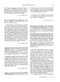
Show © 1992 Raven Press, Ltd., New York Neuro- ophthalmic Findings in Progressive Supranuclear Palsy Deborah I. Friedman, M. D., Joseph Jankovic, M. D., and John A. McCrary III, M. D. We studied 104 patients with progressive supranuclear palsy ( PSP), 38 of whom were examined by both a neurologist and a neuro- ophthalmologist. Neuroophthalmic findings that may help differentiate PSP from Parkinson's disease include vertical supranuclear ophthalmoparesis and fixation instability. Eyelid abnormalities, particularly lid retraction, blepharospasm, and " apraxia" of eyelid opening and closure, were important distinguishing signs. Although downgaze palsy is felt to be the clinical hallmark of PSP, upgaze and downgaze were equally affected at the time of diagnosis in our patients. Key Words: Progressive supranuclear palsy- Ocular motor- Blepharospasm- Eye movements- Eyelid abnormalities. From the Departments of Neurology and Ophthalmology, SUNY Health Science Center, ( D. l. F.), Syracuse, New York, and the Departments of Neurology ( J. j.) and Ophthalmology ( J. A. M.), Baylor College of Medicine, Houston, Texas, U. S. A. This work was presented in part at the American Academy of Neurology, Chicago, April B .... 19, 1989. Address correspondence and reprint requests to Dr. Deborah l. Friedman, Department of Neurology, SUNY Health Science Center, 750 East Adams Street, Syracuse, NY 13201, U. S. A. Progressive supranuclear palsy ( PSP) was described 25 years ago by Steele, Richardson, and Olszewski ( 1,2) as a distinct clinicopathological entity. The characteristic features include ( a) age at onset after 55 years, usually in the seventh decade; ( b) parkinsonian signs, including bradykinesia, postural instability, and axial rigidity; ( c) pseudobulbar signs, such as dysarthria, dysphagia, and pseudobulbar affect, and ( d) extraocular movement abnormalities, particularly vertical gaze paresis ( 3,4). Since the symptoms of PSP overlap with those of Parkinson's disease ( PO), PSP patients are often misdiagnosed as having PO, particularly in the early stages. The distinction between the two entities is important, as PSP patients show minimal response to pharmacologic therapy and tend to deteriorate more rapidly than PO patients. The diagnosis of PSP is based on observation of clinical symptoms and signs and can only be confirmed at autopsy. We evaluated 104 patients with PSP to further characterize the spectrum of neuroophthalmic findings in this disorder ( 4,5). METHODS AND PATIENTS Medical records of 104 patients from the Baylor College of Medicine Parkinson's Disease Center and Movement Disorders Clinic, Department of Ophthalmology, and the Veterans Administration Medical Center Neurology Service were reviewed. All patients satisfied the clinical criteria for PSP as defined above. The same neurologist OJ) examined all patients; 38 were also examined by a neuroophthalmologist OM, OF). Ninety patients had computed tomography ( CT) or magnetic resonance ( MR) scans to exclude other neurologic disorders. The motor and functional disability was rated on the modified Unified Parkinsonism Rating Scale ( 6). Overall disability was based on the Hoehn and Yahr 0 to 5 scale. 104 PROGRESSIVE SUPRANUCLEAR PALSY 105 RESULTS TABLE 1. Progressive supranuclear palsy: neuro- ophthalmic features in 38 patients Comprehensive neuro- ophthalmic evaluation was performed on 38 patients, and the results are reported here. There were 20 men and 18 women. The average age at onset of symptoms was 67 years. The average duration of symptoms prior to diagnosis was 4.4 years. The average Hoehn and Yahr disability score at diagnosis was 3.9, indicating bilateral disease with severe disability and the capacity to walk or stand unassisted. Twenty patients were seen only once. The other 18 patients were followed for an average duration of 30 months. Parkinson's disease was the initial diagnosis in 16 patients. The ocular findings are summarized in Table 1. Subjective complaints included blurred vision, diplopia, eye irritation or discomfort, and photophobia. Limitation of extraocular motility was present in all patients. Vertical gaze was much more impaired than horizontal gaze during all stages of the disease, with upgaze and downgaze affected equally ( Table 2). Abnormal (" cogwheel") smooth pursuit, hypometric saccades, and slow saccades were common findings. Fixation instability was observed, including square- wave jerks, nystagmus, and flutter- like oscillations. Internuclear ophthalmoplegia was also present. Numerous abnormalities of eyelid function were 12 5 11 19 15 11 3 6 1 2 1o 8 13 16 Number of patients ( follow- up) 2 2 7 11 16 2o 10 13 13 14 13 5 31o Number of patients ( at diagnosis) Degree of limitation Downgaze palsy None Mild Moderate Severe Complete Upgaze palsy None Mild Moderate Severe Complete Lateral gaze palsy None Mild Moderate Severe Complete Unknown Subjective Visual Complaints Our patients had various subjective visual complaints, but rarely complained specifically of their motility problem. Eye pain or irritation was experienced by 12 patients, presumably on the basis of blepharitis and dry eyes, which are consistent features of PSP. Dry eyes and blepharitis likely result from inadequate tear film produced by the decreased blink rate. Photophobia, seen in four patients, is another common manifestation of dry eyes. Blurred vision ( 17 patients) was the most common complaint and can occur with a variety of abnormalities seen in PSP. Dry eyes, motility abnormalities leading to " incomplete" diplopia ( disparate images not totally separated), fixation instability, and impaired convergence are some of the features of PSP that may contribute to blurred vision. Diplopia was experienced by 13 patients at some time, but an obvious motility problem was not always demonstrable at the time of examination. Unilateral or bilateral monocular diplopia due to dry eyes was a common cause of diplopia. Other findings that contributed to diplopia include internuclear ophthalmoplegia and convergence insuffi-found, particularly " apraxia" of eyelid opening and closure, and blepharospasm. Eyelid retraction, contributing to the characteristic stare of PSP, was present in all patients. Ptosis was not observed. TABLE 2. Progressive supranuclear palsy: Motility impairment in 38 patients DISCUSSION 17 13 12 4 38 37 37 28 23 19 14 11 5 4 2 4 11 8 6542 Number of Findings patients Symptoms Blurred vision Diplopia Eye pai n or irritation Photophobia Signs Lid retraction Downgaze palsy Upgaze palsy Abnormal smooth pursuit Lateral gaze palsy Hypometric saccades " Apraxia" of lid opening Blepharospasm Reflex Spontaneous Both Unknown Internuclear ophthalmoplegia Square- wave jerks Slow saccades Nystagmus " Apraxia" of lid closure Ocular flutter I Clin Neuro- ophthalmol, Vol. 12. No. 2, 1992 106 D. I. FRIEDMAN ET AL. ciency. In a series of 44 patients reported by Pfaffenbach et al. ( 7), 11 complained of diplopia, 21 had blurred vision, 17 had eye irritation, and 6 had light sensitivity. One of these patients had a history of divergence insufficiency with intermittent diplopia 5 years before any other neurologic problems began. A trial of prism spectacles was not helpful in these patients. Ocular Motility Abnormal ocular motility remains one of the diagnostic hallmarks in PSP. Although cases of pathologically confirmed PSP with normal eye movements exist ( 8,9), virtually all PSP patients have some degree of ocular motor impairment. Limitation of vertical gaze usually occurs before horizontal gaze is affected. Patients rarely notice a vertical gaze palsy per se, but their caretakers will complain of messy eating habits and tripping over furniture. Reading also becomes difficult, partially due to the ophthalmoparesis, as well as convergence insufficiency. The problem may be further exacerbated by bifocal spectacles, and patients usually benefit from single- vision reading and distance glasses. Base down prisms may sometimes be helpful to effectively raise the environment into the patient's visual field. In our series, 2 patients had full downward gaze, 2 had mild impairment, 7 had moderate limitation, 11 had less than 10 degrees of movement, and 16 had complete voluntary downgaze paresis; all patients had abnormal vertical motility at the time of diagnosis. Downgaze and upgaze were affected equally. Lateral gaze was less impaired; 14 patients had full horizontal movements, 13 had mild limitation, S had moderate impairment, 3 had less than 10 degrees of movement, and 1 had complete lateral gaze palsy ( Table 2). Classically, the vertical palsy can be at least partially improved by oculocephalic maneuvers. As the condition worsens, these maneuvers are no longer effective, and complete external ophthalmoplegia is not uncommon in the final stages. Autopsy studies of PSP brains indicate involvement of several areas of the brainstem that are important for ocular motility ( 1,10- 12). The superior colliculus shows demyelination, neuronal loss, and gliosis, especially in the deep layers. In humans, the superior colliculus likely plays a role in the initiation and control of saccadic eye movements, visual orientation tracking, and binocular vision ( 13). Loss of cholinergic neurons has been demonstrated in the midbrain nuclei that mediate vertical gaze, including the rostral insterstitial nu-r ( lin Neuro- ophthalmol, Vol. 12, NO. 2, 1992 cleus of the medial longitudinal fasciculus, the interstitial nucleus of CajaL and the superior colliculus ( 14). Demyelination has been described in the medial longitudinal fasciculus ( MLF). Neuronal loss and neurofibrillary tangles in the oculomotor, trochlear, and abducens nuclei have been reported, and demyelination of the ocular motor nerves as they exit the neuraxis has been described ( 12). Limited extraocular movements, fixation instability, lid retraction, abnormal saccades, and other clinical features similar to PSP have been reported in a patient with bilateral putamenal hemorrhages ( 15). Thus, evidence indicates that the limitation of motility is nuclear, fascicular, and supranuclear. Saccadic and Pursuit Eye Movements Smooth pursuit was impaired in 28 patients and was usually replaced by " cogwheel" pursuit movements ( 16). Hypometric saccades and slow saccades were common. Square- Wave Jerks Square- wave jerks ( SWJ) are small amplitude ( 0.5- 5 degrees) refixational eye movements. Careful observation is needed for detection of SWJ by the naked eye. Due to the retrospective nature of our study, the number of patients with SWJ ( 8) may be artificially low. Troost and Daroff ( 17) found SWJ in all eight patients analyzed with infrared reflection technique. Breuer et al. ( 18) observed SWJ in three patients using electrooculography. SWJ are associated with cerebellar dysfunction, Huntington's disease, Friedreich's ataxia, and dementia; they may be caused by disruption of areas that influence saccadic control ( i. e., cortex, cerebellum, superior colliculus, basal ganglia), or by direct injury to omnipause neurons in the brainstem gaze centers. SWJ may be partly due to demyelination and an unusual form of " grumose" degeneration that is observed in the dentate nucleus in PSP ( 19,20). Square- wave jerks, hypometric saccades, and slow saccades may be largely responsible for complaints by PSP patients of blurred vision and inability to read. Disorders of gaze- stabilizing mechanisms ( SWJ, nystagmus, flutter- like oscillations) were present in 15 patients. Internuclear Ophthalmoplegia Eleven patients had unilateral internuclear ophthalmoplegia ( INa), a finding that has been em- PROGRESSIVE SUPRANUCLEAR PALSY 107 phasized by others. Mastaglia and Grainger ( 21) found INa in 4 of 13 cases of PSP, 3 of which were bilateral. Maher and Lees ( 22) reported INa in 3 of 52 cases. We previously described INa in 3 of 16 cases ( 23). Since convergence is characteristically impaired in PSP, the designation of Cogan's " anterior" or " posterior" type INa is of little localizing value. Correlative neuropathologic studies are not available for patients displaying an INa during life, but Blumenthal and Miller ( 12) described demyelination in the MLF. Steele ( 13) also observed demyelination in this tract; however, demyelination can be secondary to involvement of the ocular motor nuclei. Given the extensive upper brainstem and tegmentum pathology in PSP, the MLF is probably affected by the underlying neurodegenerative process. Infarction of the MLF is a common cause of INa in elderly patients. Of the 11 patients with INa, one had evidence of a multi- infarct state on MR scan, and another had a lucency in the rostral midbrain that may have accounted for this finding. Blepharospasm and Eyelid Abnormalities Decreased blink rate is common in PSP, giving rise to the typical staring facial expression. Golbe and co- workers ( 24) observed a blink rate of 3 per minute in patients with PSP, which is markedly slower than the average blink rate seen in Parkinson's disease ( 12.5 per minute) and normal controls ( 15.7 per minute). These authors found an increase in blink rate with a visual task. Significant blepharospasm occurred in 11 of our patients, either spontaneous ( 4), reflex ( 5), or both ( 2). Pfaffenbach ( 7) observed blepharospasm in 8 of 44 patients. David et al. ( 25) described two cases with reflex blepharospasm, and Maher and Lees ( 22) noted blepharospasm in 4 of 52 patients. Golbe et al. ( 24) observed visually disabling blepharospasm in 6 of 38 patients. Lepore and Duvoisin ( 26) reported blepharospasm and " apraxia" of eyelid opening in a patient with " atypical Parkinson's disease" whose clinical presentation seemed consistent with PSP. The frequent occurrence of blepharospasm in PSP is consistent with the increased association of other focal dystonias in this disorder, including torticollis, facial dystonia, and limb dystonia ( 4). Bilateral lid closure can be produced experimentally in humans by stimulation of the cerebellum and midbrain ( 27,28), and blepharospasm has been reported with basal ganglia and rostral brainstem lesions ( 29- 31). Although the blepharospasm in PSP is likely due to upper brainstem pathology, no clinicopathological correlation has been described ( 32). Like essential blepharospasm and idiopathic cranial dystonia ( Meige's syndrome), PSPassociated blepharospasm may improve with botulinum toxin injections ( 33). Apraxia of eyelid opening would not be expected to improve with botulinum therapy. Newman et al. ( 34) described two patients with PSP and apraxia of eyelid opening who failed to improve after botulinum injections. However, Katz and Rosenberg ( 35) successfully treated a patient with apraxia of eyelid opening with supraorbital frontalis injections. We have treated a 63- year- old woman with advanced PSP whose most disabling feature was blindness due to a combination of blepharospasm and apraxia of eyelid opening. Both conditions were dramatically improved following botulinum injections, probably because the involuntary orbicularis oculi contractions were reduced, eliminating the trigger mechanism for apraxia of eyelid opening or lid freezing ( Fig. 1). Apraxia of eyelid opening was defined by Goldstein and Cogan ( 36) as a " nonparalytic motor abnormality characterized by the patient's difficulty in initiating the act of lid elevation." It is important to distinguish this entity from blepharospasm, which may be coexistent ( 37). In apraxia of lid opening, there is ( a) transitory inability to initiate lid opening; ( b) no evidence of ongoing orbicularis oculi contraction, such as lowering of the brows beneath the superior orbital margins ( Charcot's eyebrow sign of blepharospasm); ( c) vigorous frontalis contraction during periods of inability to raise the lids; and ( d) no oculomotor or ocular sympathetic nerve dysfunction and no ocular myopathy ( 26). Apraxia of eyelid opening was seen in 7 of 38 patients examined by Golbe et al. ( 24); three of these patients also had blepharospasm. Three patients also had apraxia of eyelid closure, and one had blepharospasm with apraxia of both eyelid opening and closure. Maher and Lees ( 22) observed 2 patients with apraxia of eyelid closure and 1 with apraxia of eyelid opening in their series of 52 patients. In a series of eight patients with PSP, Dehaene ( 38) observed apraxia of eyelid opening in three. Lepore and Duvoisin ( 26) summarized six cases of apraxia of lid opening and parkinsonism, PSP ( 2 patients), and Shy- Drager syndrome. Although the term is commonly used, eyelid " apraxia" is a misnomer. With true apraxia, there is an inability to perform a voluntary movement despite the preservation of normal power, sensation, and coordination. Since all of their patients had extrapyramidal involvement, Lepore and Du- J Clin Neuro- ophthalmol. Vol. 12, No. 2, 1992 108 D. I. FRIEDMAN ET AL. 1A- C FIG. 1. A patient with PSP and " apraxia" of eyelid opening triggered by spontaneous blepharospasm ( A); patient has to use her fingers to open her eyes ( B). Within 2 days of botulinum toxin injection into the orbicularis oculi and the corrugator muscles, she was able to keep her eyes open and open and close them voluntarily ( C). VOISin ( 26) argue against the term " apraxia" and suggest that the lid abnormality is due to " involuntary levator palpebrae inhibition of supranuclear origin." The transient inability to open or close the eyes is similar to the sudden transient freezing during attempted voluntary motion seen in a variety of parkinsonian disorders. Eleven of our patients had blepharospasm, 14 had apraxia of eyelid opening, and 10 had both. Apraxia of lid closure was present in four patients. Three patients had apraxia of both lid opening and closure. Lid retraction is seen in most patients with PSP, contributing to the astonished or worried facies characteristic of the disorder ( 3). A white band of sclera interposed between the lid margin and the upper corneal limbus when the eyes are directed straight ahead indicates the presence of lid retraction ( 39). Lid retraction is presumably due to involvement of the levator complex, located medially in the dorsal midbrain at the level of the third nerve nuclei. When the normal levator- superior rectus synkinesis is disrupted due to impaired upgaze, disproportionate lid elevation can result ( 40). Lid retraction and impaired upward gaze are also seen in patients with the pretectal syndrome, where the amount of lid retraction parallels the degree of upgaze limitation ( 41). Ptosis is uncommon in PSP and was not present in this group of patients. In our original group of 104 patients, ptosis was present in 4 ( 4,5). Dix et al. ( 42) reported ptosis in one of nine patients. Mendell and colleagues ( 43) observed bilateral ptosis in one case. Nystagmus Nystagmus is relatively uncommon in PSP; it was present in five of our patients. In their series of nine patients, Dix et al. ( 42) described asymmetric horizontal end- gaze nystagmus in one patient and nystagmoid jerks in another. COMMENT It is often difficult to distinguish PSP from PO, particularly in the early stages. The presence or evolution of nemo- ophthalmic signs may be helpful in confirming the diagnosis of PSP. Upgaze is often affected in PO, whereas both upgaze and downgaze are involved in PSP. In PSP, lid retraction and frontalis contraction give rise to an astonished or worried expression, in contrast to the hypomimia of PD. Focal dystonias, unrelated to medication, are more common in PSP than PD. Blepharospasm, a type of focal dystonia, was seen in almost one- third of our patients and was often associated with apraxia of eyelid opening. Gaze instability, although not specific to PSP, is often present and may help support the diagnosis. REFERENCES I. Steele Jc. Richardson Jc. Olszewski J. ProgreSSive supranuclear palsy: a heterogeneous degeneration involving the brain stem. basal ganglia and cerebellum with vertical gaze and pseudobulbar palsy. nuchal dystonia and dementia. Arch NellroI1963; 2: 47>- 86. 2. Richardson Jc. Steele J. Olszewski J. Supranuclear ophthalmoplegia. pseudobulbar palsy, nuchal dystonia and dementia. T" HZS Am : Vellrol Assoc 1963; 88: 25-- 7. 3. Jankovic J. Progressive supranuclear palsy: clinical and pharmacological update. Neural Clin 1984; 2: 47>- 86. 4. Friedman 01, Jankovic J. ProgreSSive supranuclear palsy: a quarter century ot progress. In: Appel SH. ed. Current neurology. Chicago: Year Book Medical Publishers, 1989; 9: 191218. 5. Jankovic j. Friedman 01, Pirozzo] o Fj. McCrary JA. ProgressIve supranuclear palsy: motor, neurobehavioral. and neuro- ophthalmic findings. In: Streifler M, ed. Ninth International Symposium on Parkinson's Disease. New York, Raven Press, 1990; 59: 293- 304. 6. Fahn S. Elton RL. Members of the UPDRS Development Committee. Unified Parkinson's Disease Rating Scale. In: Fahn S. Marsden CD. Caine 0, Glodstein M. eds. Recent de1' elopmCllt in Parkinson's disease. Vol. 2. New Jersey: McMIllan Healthcare Information, 1987: 153- 63. 7. Pfaffenbach DO, Layton DO, Kearns TP. Ocular manifestations in progressive supranuclear palsy. Am J Ophthalmol 1972; 74: 1179- 84. 8. Nuwer MR. Progressive supranuclear palsy despite nonnal eye movements. Arch Neural 1981; 38: 784. 9. Kleinschmidt- Demasters BK. Early progressive supranuclear palsy: pathology and clinical presentation. Gin Neuropathol 1989; 8: 79- 84. 10. Ishino H. Higashi H, Kuroda S. et al. Motor nuclear involvement in progressive supranuclear palsy. J Neural Sci 1973; 22: 235-- 44. 11. Blumenthal H, Miller C. Motor nuclear involvement in progressive supranuclear palsy. Arch Neural 1969; 20: 362- 7. 12. Steele Jc. ProgreSSive supranUclear palsy. Brain 1972' 95: 693- 704. ' PROGRESSIVE SUPRANUCLEAR PALSY 109 13. Sadun AA, Johnson BM, Smith LE. Neuroanatomy of the human visual system: Park II. Retinal projections to the superior colliculus and pulvinar. Nellro- oplltlwlmologl/ 1986; 6: 363- 70. 14. Juncos JL, Hirsch EC, Malessa S, et al. Mesencephalic cholinergic nuclei in progressive supranUclear palsy. Nellrology 1991; 41: 25- 30. 15. Hankey GJ, Stewart- Wvnne EG. Bilateral intracerebral haemorrhage presenting with supranuclear ophthalmoplegia, bradykinesia and rigidity. elill Exp Nellrol 1987; 23: 195- 9. 16. Chu FC, Reingold DB, Cogan DG, et al. The eye movement disorders of progressive supranuclear palsy. Ophthalmology 1979; 86:- t22- 8 17. Troost BT, Daroff RB. The ocular motor defects in progressive supranuclear palsy. AIIII Nellrol 1977; 2: 397- 403. 18. Breuer TJM, Wuisman PGWM, Korten Jj. Electrooculographic findings in progressive supranuclear palsy. Clill Nellrol Nellrosllrg 1987; 89: 87- 95. 19. Kida M, Koo H, Grossniklaus HE, Tomsak RL. Neuropathologic findings in progressive supranuclear palsy. A brief review with two additional case reports. JClill NellroophtJwlmol 1988; 8: 161- 70. 20. Arai N, Amano N, Iwabuchi K, et al. Three categories of the degenerative appearance of the human cerebellar dentate nucleus. A morphometric and morphological study. J Nellrol Sci 1988; 83: 129- 43. 21. Mastaglia Flo Grainger MR. Internuclear ophthalmoplegia in progressive supranuclear palsy. J Nellrol Sci 1975; 25: 303- 8. 22. Maher ER, Lees AJ. The clinical features and natural history of the Steele- Richardson- Olszewski syndrome ( progressive supranuclear palsy). Neurology 1986; 356: 1005- 8. 23. Jackson JA, Jankovic Jj, Ford J. Progressive supranuclear palsy: clinical features and response to treatment in 16 patients. AIIII Neural 1983; 13: 273- 8. 24. Golbe Ll, Davis PA, Lepore FE. Eye movement abnormalities in progressive supranuclear palsy. MOl' Disord 1989; 4: 297- 302 25. David NJ, Mackey EA, Smith JL. Further observations in progressive supranuclear palsy. Neurology 1968; 18: 349- 56. 26. Lepore FE, Duvoisin RC. " Apraxia" of eyelid opening: an involuntary levator inhibition. Neurology 1985; 35: 423- 7. 27. Ron S, Robinson DA. Eye movements evoked by cerebellar stimulation in the alert monkey. / Neural Neurosllrg Psychiatry 1973; 36: 1004- 9. 28. Nashold BS, Wilson WP, Boone E. Depth recordings and stimulation of the human brain: A twenty- year experience. In: Rasmussen 1, Marino R, eds. FllllctlOllal lieu rosll rgery. New York, Raven Press, 1979: 181- 95. 29. Keane JE, Young JA. Blepharospasm with bilateral basal ganglia infarction. Arch NellroI1985; 42: 1206- S. 30. Jankovic J, Patel Sc. Blepharospasm associated with brainstem lesions. Nellrology 1983; 33: 1237- 40. 31. Jankovic J. Blepharospasm with basal ganglia lesions. Arch Nellrol 1986; 43: 866- 8. 32. Rivest J, Quinn N, Marsden CD. Dystonia in Parkinson's disease, multiple system atrophy, and progressive supranuclear palsy. Neurology 1991; 40: 1571- 8. 33. Jankovic J, Brin MR. Therapeutic uses of botulinum toxin. N Ellgi/ Med 1991; 324: 1186- 94. 34. Newman SA, Slavin M, Phillips L. Apraxia of eyelid opening: A cause of botulinum failure. Presented at North American Neuro- Ophthalmology Society, Crested Butte, Colorado, 1988. 35. Katz B, Rosenberg JH. Botulinum therapy for apraxia of eyelid opening. Am J OpilthalmoI1987; 103: 718-- 9. 36. Goldstein JE, Cogen DG. Apraxia of lid opening. Arch Ophthalmol 1965; 73: 155- 9. 37. Jordan DR, Anderson RL, Digre KB. Apraxia of lid opening in blepharospasm. Ophthalmic Surg 1990; 21: 331- 4. 38. Dehaene I. Apraxia of eyelid opening in progressive supranuclear palsy. All/ I Neurol 1984; 15: 115- 6. 39. Miller NR. Anatomy and physiology of normal and abnormal eyelid position and movement. In: Miller MR, I'd. Walsh alld Hoyt's cilllicailleuro- ophtlwimology. 2nd I'd. Vol. 2. Baltimore: Williams & Wilkins, 1985: 945. 40. Cogan DG. Neurology or the ocular muscles, 2nd I'd. Springfield, IL: Charles C. Thomas, 1956: 143. 41. Keane JR. The pretectal syndrome: 206 patients. Neurology 1990; 40: 684- 90. 42. Dix MR, Harrison MJG, Lewis PD. ProgreSSive supranuclear palsy ( the Steele- Richardson- Olszewski syndrome): A report of 9 cases with particular reference to the mechanism of the oculomotor disorder. / Neurol Sci 1971; 13: 237- 56. 43. Mendell JR, Chase TN, Engel WK. Modification by L- dopa of progressive supranuclear palsy. Lallcet 1970; 1: 593- 4. I (/ m Nellro- ophtha/ mol, Vol. 12, No. 2, 1992 |