| OCR Text |
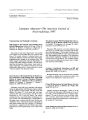
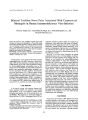
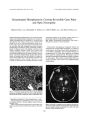
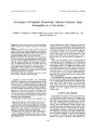
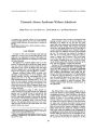
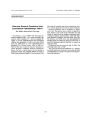
Show Journal of Neuro- Ophthalmology 19( 2): 125- 127, 1999. © 1999 Lippincott Williams & Wilkins, Inc., Philadelphia A Case of Ocular Neuromyotonia With Tonic Pupil Namir Abdulla, F. R. C. S., and Peter Eustace, F. R. C. S., F. R. c. Ophth A 48- year old woman with hypertension experienced painful oculomotor nerve palsy. After surgery for a giant aneurysm of the internal carotid artery in the cavernous sinus, phasic constrictions of the pupil developed. Two years later, this phenomenon disappeared and was replaced by intermittent involuntary cyclic spasms elevating the ptosed lid. These cyclic lid movements were not elicited with any eye movement or by increased accommodation. The pupil now manifested the pharmacologic features of a tonic pupil. The explanation for this unique case of ocular neuromyotonia is based on a misdirection phenomenon, possibly caused by ephaptic transmission. Key Words: Ephaptic transmission- Internal carotid artery aneurysm- Ocular neuromyotonia- Oculomotor nerve palsy- Tonic pupil. Ocular neuromyotonia is a rare disturbance of ocular motility, characterized by paroxysmal monocular involuntary contractions of one or more extraocular muscles supplied by the third, fourth, and sixth cranial nerves. Approximately 21 cases of ocular neuromyotonia are described in the literature. The first cases were reported by Ricker and Mertens ( 1) in 1970 and Papst ( 2) in 1972. We report a unique case of ocular neuromyotonia and tonic pupil after an episode of acute painful oculomotor nerve palsy caused by internal carotid artery aneurysm. CASE REPORT A 48- year- old woman with hypertension was first seen in March 1994 with diplopia, headache, and right ptosis. Neuro- ophthalmologic examination revealed external ophthalmoplegia with preservation of abduction and in-tortion. The pupil was dilated and unresponsive to light, with a complete ptosis of the right upper lid. Findings in examination of the remaining cranial nerves were normal. No general neurologic deficits were elicited. A computed tomographic ( CT) scan showed a large uniformly enhancing mass lesion consistent with a giant aneurysm in the right parasellar area extending superiorly. Bilateral carotid and vertebral angiograms revealed Manuscript received February 27,1998; accepted December 2,1998. From the Institute of Ophthalmology, Mater Misercordiae Hospital, Dublin, Ireland. Address correspondence and reprint request to Professor Peter Eustace, Professor of Ophthalmology, Institute of Ophthalmology, Mater Misercordiae Hospital, 60 Eccles Street, Dublin 7, Ireland. a giant aneurysm on the right side. The aneurysm appeared to arise from the internal carotid artery siphon and displaced the proximal stem of the right middle cerebral artery superiorly. No abnormality was noted on the right anterior cerebral artery or on the left carotid or vertebral basilar artery. These investigations confirmed the presence of the right internal carotid artery aneurysm. Clipping of the aneurysm was performed. In the early postoperative period, the patient experienced left- side weakness, pyrexia, and seizures. She recovered, but residual right oculomotor nerve palsy remained. Two months after surgery, at a routine outpatient follow- up, the patient was asymptomatic except for her eyes. Visual acuity was 6/ 9 in both eyes. Neuro- ophthalmologic examination showed residual complete right oculomotor nerve palsy. The pupil showed intermittent constriction against a background of a fixed, dilated pupil, unresponsive to light. This pupillary constriction was segmental and spontaneous. It was not associated with eye movement or increased accommodation. Eighteen months after surgery, this phenomenon disappeared. However, the right ptosed upper lid showed periods of intermittent elevation ( Fig. 1). The flaccid periods were longer than the tonic phases. The phasic spasms were sometimes triggered by rubbing the right lower lid. There was no constant interval between the two phases. The lid movements could not be elicited with globe movement or increased accommodation. The pupil now showed a slow reaction to light, tonic near reflex, and positive denervation hypersensitivity to 0.125% pilocarpine ( Fig. 2). This phenomenon was not associated with eye movement. Dynamic refraction showed no spasm of accommodation. DISCUSSION Ocular neuromyotonia is a rare disturbance of ocular motility. It is characterized by involuntary spontaneous contractions of extraocular muscles supplied by the third, fourth, and sixth cranial nerves. Ocular electromyograms recorded from the affected muscles show a series of high- frequency, high- voltage discharges superimposed on the muscles' normal discharge pattern, including times when the muscle would normally be inhibited. The tracings showed no myopathic characteristics ( 1,2). 125 126 N. ABDULLA AND P. EUSTACE FIG. 1. Clinical photograph showing the right ptosed upper lid during phasic spasms. The spasms can be triggered by rubbing the right lower lid. In a review of the literature, we found reports of 21 cases of ocular neuromyotonia ( 1- 11). Thirteen patients had intracranial tumors, treated with surgery and/ or radiotherapy ( 3- 5,7,8,10,11). Only one patient had a history of oculomotor nerve compression due to intracranial artery aneurysm ( 10). Fifteen patients had involvement of the oculomotor nerve ( 1- 11). Three cases with pupillary involvement were reported ( 3,10). This unique case of ocular neuromyotonia secondary to internal carotid artery aneurysm manifested as paroxysmal involuntary retraction of the upper lid which was occasionally triggered by rubbing of the lower lid. These lid movements were not associated with any eye movement. Initially, the pupil was dilated and unresponsive to light, consistent with complete oculomotor nerve palsy. Two months after ligation of the internal carotid artery aneurysm, the pupil showed intermittent spontaneous episodes of constriction. Eighteen months after surgery, the patient exhibited a tonic pupil with slow reaction to light, tonic near reflex, and positive denervation hypersensitivity to 0.125% pilocarpine. FIG. 2. Clinical photograph showing the right pupil with positive denervation hypersensitivity reaction to 0.125% pilocarpine. Ocular neuromyotonia, with its spontaneous movements, should be differentiated from cyclic oculomotor palsy, which is characterized by oculomotor paresis with regular phasic cyclic spasms. Cyclic oculomotor palsy is a rare syndrome, usually noted at birth or developing during the first year of life. Typically, it follows congenital oculomotor palsy, and the spastic phases occur at regular intervals of between 1 and 3 minutes. In the spastic phase the lid is raised, the globe moves toward the midline, the pupil constricts, and the accommodation increases. In the paretic phase, the third nerve palsy returns ( 12). The current patient does not fit this description. The oculomotor disease began much later in life. She had relatively infrequent and irregular involuntary spastic episodes limited initially to the pupil and later replaced by retraction of the ptosed lid and a tonic pupil. Oculomotor synkinesis due to aberrant regeneration has been associated with ocular neuromyotonia ( 3,10). It is clear that the cyclic movements in this patient were caused by alternate spasms and paresis of muscles innervated by oculomotor nerve. These cyclic lid movements could not be elicited with any eye movement or by increased accommodation. Thus, there was no clinical evidence of aberrant reinnervation from misdirected oculomotor nerve fibers. It proposed that ephaptic transmission accounts for the insidious transition from paresis to synkinesis that occurs in some patients by creation of an ephapse where adjacent nerve fibers were injured. Ephaptic transmission is an electrotonic, as opposed to a chemical mode of propagation between cells of fibers. It is an activation through an artificial synapse between axons with partially injured fiber tracts ( 13,14). Carbamazepine's effectiveness in some patients with ocular neuromyotonia may be because it reduces spontaneous discharges in hyperexcit-able cranial nerve fibers ( 1- 3,6- 10). It is probable that the involuntary cyclic spasms arise from the oculomotor neurones that have been damaged and then recover. The pupil may be involved because of the anatomic disposition of the third cranial nerve nucleus. Possibly through ephaptic transmission between regenerating neurones, impulses transmit through the regenerating nerve fibers that sprout across the scar to different components of the oculomotor nerve. In our patient, spasms could be sometimes triggered by rubbing the lower lid, possibly through excitation of the infraorbital nerve of the trigeminal nerve and consequently through the regenerating oculomotor nerve. The development of a tonic pupil suggests the possibility of tran-sneuronal degeneration. REFERENCES 1. Ricker VK, Mertens HG. Okulare Neuromyotonie. Klin Monatsbl Augenheilkd 1970; 156: 837^ 2. 2. Papst W. Zur differentialdiagnose der okularen neuromyotonie. Ophthalmologica 1972; 164: 252- 63. 3. Shults WT, Hoyt WF, Behrens M, Maclean J, Saul RF, Corbett JJ. Ocular neuromyotonia. A clinical description of six cases. Arch Ophthalmol 1986; 104: 1028- 34. J Neuro- Ophthalmol, Vol. 19, No. 2, 1999 OCULAR NEUROMYOTONIA WITH TONIC PUPIL 127 4. Lessell S, Lessell IM, Rizzo JF. Ocular neuromyotonia after radiation therapy. Am J Ophthalmol 1986; 102: 766- 70. 5. Salmon JF, Steven P, Abrahamson MJ. Ocular neuromyotonia. Neuro- ophthalmology 1988; 4: 181- 5. 6. Helmchen C, Dieterich M, Straube A, Buttner U. Abducens neuromyotonia with partial oculomotor paralysis. Nervenarzt 1992; 63: 625- 9. 7. Barroso L, Hoyt WF. Episodic exotropia from lateral rectus neuromyotonia: Appearance and remission after radiation therapy for a thalamic glioma. J Pediatr Ophthalmol Strabismus 1993; 30: 56- 7. 8. Newman SA. Gaze- induced strabismus. Surv Ophthalmol 1993; 38: 303- 9. 9. Frohman EM, Zee DS. Ocular neuromyotonia: Clinical features, physiological mechanisms, and response to therapy. Ann Neurol 1995; 37: 620- 6. 10. Ezra E, Spalton D, Sanders MD, Graham EM, Plant GT. Ocular neuromyotonia. Br J Ophthalmol 1996; 80: 350- 5. 11. Morrow MJ, Kao GW, Arnold AC. Bilateral ocular neuromyotonia: Oculographic correlations. Neurology 1996; 46: 264- 6. 12. Miller NR. Topical diagnosis of neuropathic ocular motility disorders. In: Walsh and Hoyt's Clinical Neuro- ophthalmology. Baltimore: William & Wilkins, 1985; 655- 7. 13. Lepore FE, Glaser JS. Misdirection revisited: A critical appraisal of acquired oculomotor nerve synkinesis. Arch Ophthalmol 1980; 98: 2206- 9. 14. Elston JS. Ocular neuromyotonia. Arch Ophthalmol 1987; 105: 24. J Neuro- Ophthalmol, Vol. 19, No. 2, 1999 |