| OCR Text |
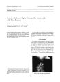
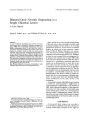
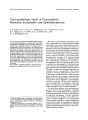
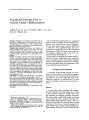
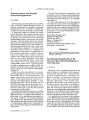
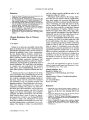
Show journal of Neuro- Ophthalmology 16( 1): 49- 54, 1996. © 1996 Lippincott- Raven Publishers, Philadelphia Acquired Esotropia Due to Arnold- Chiari I Malformation Angela R. Lewis, M. D., Lanning B. Kline, M. D., and James A. Sharpe, M. D. Objective: Diplopia is not frequently associated with Arnold- Chiari I malformation. We reviewed our cases of Arnold- Chiari I malformation in which acquired esotropia with diplopia was the main neuro- ophthalmologic finding early in the clinical course. Materials and Methods: Five patients were studied, all female, ranging in age from 17 to 36 years, who were treated by the neuro- ophthalmology service of urban teaching hospitals. Eye movement recordings using magnetic search coil technique were performed in two patients. Results: All patients reported onset of horizontal diplopia due to acquired esotropia as an initial manifestation of the Arnold- Chiari I malformation. All had full abduction of each eye. In addition, all five had gaze- evoked nystagmus, two skew deviations, and one bilateral in-ternuclear ophthalmoparesis. Oculography in two patients showed normal abducting saccadic peak velocities. This supports divergence palsy as a mechanism of acquired esotropia and provides evidence against subtle sixth nerve palsy in these patients. Four patients underwent neurosurgical decompression of their Chiari malformations, and neuro- ophthalmologic signs and symptoms improved in all. Conclusions: Acquired esotropia, often in association with other eye movement abnormalities, may be an early sign of Arnold- Chiari I malformation. This quantitative study indicates that divergence palsy is the cause of esotropia in some patients. Neurosurgical suboccipital and upper cervical decompression may lead to improvement or resolution of diplopia. Key Words: Acquired esotropia- Arnold- Chiari I malformation. Manuscript received May 5, 1995. From the Combined Program in Ophthalmology ( A. R. L., L. B. K.), Eye Foundation Hospital, University of Alabama at Birmingham, Birmingham, Alabama, U. S. A.; Division of Neurology ( J. A. S.), The Toronto Hospital, University of Toronto, Toronto, Ontario, Canada. Address correspondence and reprint requests to Dr. Lanning B. Kline, Alabama Ophthalmology Associates, 1000 South 19th Street, Birmingham, AL 35205, U. S. A. The Arnold- Chiari malformation is a congenital anomaly of the cerebellum and lower brain stem. Patients with Arnold- Chiari type I malformation are often asymptomatic until late childhood or adult life. Neuro- ophthalmologic signs and symptoms are frequently part of the presenting clinical picture ( Table 1). Indeed, after tendon reflex changes, nystagmus is the second most common neurologic abnormality ( 1). In this report we describe five patients with Arnold- Chiari type I malformation who presented with an acquired esotropia. Quantitative magnetic search coil oculography performed in two patients provides information about the mechanism of this neuro- ophthalmologic abnormality. PATIENTS AND METHODS A summary of the clinical findings in our patients is found in Table 2. All underwent complete neurologic and neuro- ophthalmologic testing. Cranial magnetic resonance ( MR) scanning established the diagnosis of Arnold- Chiari I in each case. Two illustrative case reports are presented in detail. Patient 4 A 36- year- old woman presented with progressive gait instability since early adolescence. At age 16 she developed horizontal double vision. The gait instability and diplopia gradually increased. Her visual acuity, visual fields, pupils, and fundi were normal. She had a comitant esotropia of 20 prism diopters during distance fixation, and a comitant left hypertropia of 6 prism diopters. During near fixation, there was a comitant esotropia of 4 prism diopters and a comitant left hypertropia of 6 prism diopters. Ductions in each eye were full. She 49 50 A. R. LEWIS ET AL. TABLE 1. Ocular motor abnormalities in Arnold- Chiari I malformation 1. 2. 3. 4. 5. Nystagmus downbeat sidebeat periodic alternating divergent gaze- evoked rebound positional see- saw torsional Divergence palsy Convergence spasm Internuclear ophthalmoplegia Skew deviation had gaze- evoked nystagmus on rightward, leftward, upward, and downward gaze. While fixation in primary position was stable, refixation from eccentric lateral gaze to the midposition provoked rebound nystagmus. Refixation from upgaze to midposition evoked overshoot dysmetria of sac-cades. Smooth pursuit was saccadic in all directions, and cancellation of the vestibulo- ocular reflex was impaired. Neurologic examination showed normal function of the other cranial nerves. Muscle strength and tone were normal. The patient exhibited past pointing with both arms and mild impairment of rapid alternating movements. There was moderate heel- to- shin ataxia. Tendon reflexes were normal, and both plantar responses were flexor. Sensations were intact. Her gait was broad- based and ataxic. CT showed atrophy of cerebellum, particularly the superior vermis. Sporadic cerebellar degeneration was diagnosed. Follow- up examination 18 months later showed no change in esotropia, skew deviation, or nystagmus. Her gait ataxia had worsened. Two years later, MR demonstrated a Chiari type I malformation with ectopia of the inferior cerebellum into the upper cervical cord ( Fig. 1). A posterior fossa decompression and upper cervical laminectomy were performed. The dura was tight and the foramen magnum was full, with the cerebellar tonsils displaced down to C2. An expansive duraplasty was performed. Examination seven months after surgery demonstrated comitant left hypertropia of 4 prism diopters and an exotropia of 4 prism diopters. Gaze- evoked and rebound nystagmus and gait and limb ataxia had improved. Patient 5 A 19- year- old woman complained of horizontal double vision, when viewing distant objects, for five years. She had bitemporal, episodic, pulsating headaches, consistent with migraine. Examination revealed normal visual acuity, fundi, and fields. She had a comitant esotropia of 8 prism diopters during forward, leftward, and rightward distance fixation. On near fixation there was a 4 prism diopter exophoria. Refixation saccades showed overshoot dysmetria. Smooth pursuit was saccadic. Function of the other cranial nerves was normal. TABLE 2. Summary of clinical data in patients with Arnold- Chiari I malformation Patient ( age, sex) 1 ( 17, female) 2 ( 23, female) 3 ( 24, female) 4 ( 36, female) 5 ( 19, female) Esotropia ( maximum) ET = 14 EOT = 8 comitant Rt. gaze Pri. gaze ET = 12 E = 4 ET' = 6 E' = 2ET' noncomitant ET = 4 E' = 4 comitant ET = 20 LHT = 6 E( T)' = 4 LH( T)' = 6 comitant ET = 7 RHT = 6 E' = 2 RH' = 2 comitant Lt. gaze ET = 12 ET' = 6 + rebound Nystagmus < r- <- - t 0 0 0 0 0 0 0 0 t <- 0 I nystagm I t 0 1 - - -> us I Treatment posterior fossa decompression posterior fossa decompression posterior fossa decompression posterior fossa decompression none Follow- up since surgery ( months) 52 64 80 72 Rt. ET gaze = 10 Outcome ortho 0 0 0 - ^ 0 Pri. gaze E = 2 0 <- 0 -> 0 ortho 0 « - 0 - » 0 LHT = 4 t <- 0 - » 1 Lt. gaze ET = 8 ET = esotropia; E = esophoria; HT = hypertropia; Rt. = right; Pri. = primary; Lt. = left; no nystagmus; - » = direction of nystagmus. near deviation; () = intermittent deviation; 0 / Neuro- Ophthalmol, Vol. 16, No. 1, 1996 ACQUIRED ESOTROPIA 51 / I 1 Jt3¥ r I JSJB^ L. ' w lm l > jS3sy' 1 » 5r \ 9 ItH M 7 ^ ^ -^ <^ • Mblj. ' " ^ ^ l i l- (• W*^~^ t, " M ' ( ^ ** \ £:£ * H ^ A W V, < 1 Mg'l 1 * w FIG. 1. Case 4. T1- weighted sagittal MR scan demonstrates Arnold- Chiari type I malformation with downward displacement of the cerebellar tonsils ( arrow). Motor, reflex, and sensory examinations were normal. A cranial CT scan with axial cuts through the craniocervical junction was normal. Serial follow- up examinations showed mild, progressive deterioration. At age 20 she showed mild unsteadiness during tandem walking. At age 23 she exhibited gaze- evoked nystagmus on upward, downward, rightward, and leftward fixation. At age 24 she showed gaze- evoked nystagmus, with a downbeating component on lateral gaze to right and left. By age 26 she had overshoot ocular dysmetria, saccadic smooth pursuit in all directions, and impaired cancellation of the vesti-bulo- ocular reflex. A comitant esotropia measured 7 prism diopters in forward and lateral distant fixation. There was also a right hypertropia of 6 prism diopters on rightward gaze that did not change on upward and downward fixation. At near she had a small esophoria and minimal right hyperphoria. Examination of other cranial nerves and testing of coordination and deep tendon reflexes were all normal. The patient was unable to perform tandem walking, and her gait demonstrated mild instability during rapid turns. MR demonstrated an Arnold- Chiari type I malformation with displacement of the cerebellar tonsils down to the C3 level. Because of progressive gait ataxia and increasing nystagmus, the patient was referred to a neurosurgeon for decompression of the Chiari malformation, but she declined surgery. OCULOGRAPHIC STUDY In patients 4 and 5, movements of each eye were recorded with a magnetic search coil technique using 6- cubic- foot field coils ( C- N- C Engineering, Seattle, Washington). The patients viewed light-emitting diodes on a horizontal stimulus arc to elicit saccades. Target amplitudes were unpredictable and varied from 4 to 40° at 2- s intervals. Horizontal saccades of each eye were recorded while the patient fixated with one eye and the other eye was occluded. Analog signals of eye position were digitized at 200 H for off- line analysis by a PDP 11/ 73 computer. The saccadic peak velocity- amplitude relationship was computed to a best- fit exponential curve: PV = V [ 1 - exp (- A/ C)], where PV is peak velocity, V is the maximum velocity at the asymptote of the curve, A is saccadic amplitude, and C is the time constant of the curve. V values for adducting and abducting saccades were compared for each eye. RESULTS The asymptotic peak velocities of each eye are shown in Table 3. The abducting velocities were within the normal range for control subjects of the same age groups in this laboratory ( 2). With the right eye fixing, or with the left eye fixing, there was no systematic difference in the peak velocities of abducting saccades versus adducting saccades in either eye in patient 4. In patient 5, peak veloc- TABLE 3. Asymptotic peak saccadic velocities ( deg/ s) RE fixating LE fixating Patient 4 Right eye Left eye Patient 5 Right eye Left eye Abduction 540 640 350 400 Adduction 460 490 290a 260a Abduction 620 690 440 390 Adduction 580 500 390 300a ' Asymptotic peak velocities were subnormal ( normal mean 495 ± 95 deg/ sec) ( 2). / Neuro- Ophthalmol, Vol. 16, No. 1, 1996 52 A. R. LEWIS ET AL. ities of adducting saccades were slow in both eyes ( Fig. 2), indicating bilateral internuclear ophthal-moparesis. DISCUSSION Arnold- Chiari malformation is the eponym given to a hindbrain anomaly first described by Chiari in 1891 ( 3). In 1894, Arnold added a more detailed description of this disorder ( 4). Four types have been described. Type I is the most often seen in adults ( 5) and is the focus of this report. Unlike many congenital malformations of the central nervous system, patients with this entity usually remain asymptomatic until late childhood or early adulthood ( 6). The most frequent symptoms are neck pain and headache. Other neurologic findings include ataxia, dysarthria, dysphagia, diplopia, dizziness, and, on occasion, extremity weakness and numbness. Ophthalmologic abnormalities may be the sole manifestation of Arnold- Chiari I malformation ( 7). Symptoms include diplopia and oscillopsia due to a variety of forms of nystagmus ( Table 1). However, acquired esotropia has rarely been reported in this malformation ( 1,7,8). Passo et al. ( 7) reported the case of a 24- year- old woman with esotropia since age 9 years and intermittent diplopia since age 17 years. The patient was diagnosed as having a " decompensated esophoria" and underwent strabismus surgery. 800 700 - >> 500 S 400 °- 300 o 200 - CO CO - • RE, RE fixating - • RE, LE fixating - * LE, RE fixating - O LE, LE fixating Adduction Abduction - 60 - 40 T - i - I - i - i - i - r - 30 - 20 - 10 0 10 20 30 40 50 60 Saccade Amplitude ( deg) FIG. 2. Case 5. Slowed adduction asymptotic peak saccadic velocities indicative of internuclear ophthalmoplegia ( normal mean saccadic velocity: 495 ± 95 deg/ sec). Negative saccade amplitude indicates leftward, positive rightward. With return of diplopia one year later, she was found to have esotropia, torsional nystagmus, and ataxic gait. MR scanning revealed an Arnold- Chiari I malformation and the patient underwent decompression of the posterior fossa. Five months later she was orthophoric, with marked dampening of torsional nystagmus. Her gait was unchanged. Bixenman and Laguna ( 1) reported a 13- year- old girl with intermittent diplopia and occipital headaches. Examination revealed a comitant esotropia of 30 prism diopters at distance and an intermittent esotropia of 18 prism diopters at near. The patient underwent strabismus surgery with resolution of diplopia. Three years later, she reported asthe-nopic symptoms and occipital headache. She was orthophoric on alternate cover testing but had downbeat nystagmus in primary gaze, as well as bidirectional horizontal gaze- evoked nystagmus on lateral gaze. Neurologic findings included ataxic tandem gait, generalized hyporeflexia, with dysmetria of the right hand. MR demonstrated an Arnold- Chiari I malformation, and the patient underwent decompression of the posterior fossa. Headaches and asthenopic symptoms resolved, and there was improvement in nystagmus in primary gaze. Tang et al. ( 8) described a 44- year- old woman with Arnold- Chiari I malformation having a comitant esotropia associated with primary position downbeat nystagmus and torsional and horizontal nystagmus on lateral gaze. Neurologic examination showed symmetric hyporeflexia and inability to tandem walk. The patient was referred for neurosurgical intervention, but no follow- up was presented. Acquired comitant esotropia is sometimes attributed to " decompensation" of a previously controlled phoria and not due to intracranial disease ( 9- 11). Hoyt and Williams ( 12) reported six children with acquired comitant esotropia who were diagnosed as having tumors of the posterior fossa. Vollrath- Junger and Lang ( 13) collected a series of patients with hydrocephalus in whom acquired comitant esotropia was a sign of shunt failure. With repair of the shunt, the esotropia resolved. These reports and others ( 14- 18) demonstrate that an acquired comitant esotropia may be a sign of intracranial disease, and the question arises as to which patients warrant further neurologic evaluation. Hoyt and Williams ( 12) suggest that whenever nystagmus is associated with an acquired comitant esotropia, complete neurologic evaluation is warranted. The mechanism of acquired comitant esotropia / Neuro- Ophthalmol, Vol. 16, No. 1, 1996 ACQUIRED ESOTROPIA 53 in the Arnold- Chiari I malformation is uncertain. Several possibilities exist. A disturbance of the normal fusional mechanism by the Chiari malformation may lead to a comitant esotropia ( 19). A palsy of divergence could produce an esodeviation ( 20). Some of our patients had typical features of divergence palsy ( Table 2). This syndrome is characterized by: ( a) acute onset of diplopia at distance, with esotropia; ( b) comitancy in right versus left gaze; ( c) fusion at near; and ( d) no restriction of motility ( 21). An alternate explanation is that esotropia is due to bilateral Vlth nerve palsies. Patient 2 had full range of eye movements, yet incomitant esotropia, increasing on lateral gaze. Kirkham et al. ( 22) reported three patients with acquired esotropia, thought to be divergence palsy, and raised intracranial pressure. Although the range of eye movements appeared full, and all patients fused at near, peak velocity of abducting saccades was slowed. With relief of raised intracranial pressure, abducting saccadic velocities returned to normal. The authors proposed that Vlth nerve paresis explained the syndrome of divergence palsy. Jampolsky ( 23) also argued that anomalies of ocular divergence with acquired esotropia are " degrees of bilateral Vlth nerve paresis." Clinical findings in divergence palsy might be explained by a lesion in the floor of the fourth ventricle, affecting the medial longitudinal fasciculi as well as the Vlth nerve nuclei. Relative comitancy in lateral gaze, with lack of overaction of yoked medial rectus muscles, was attributed to involvement of the medial longitudinal fasciculi ( 23). Variation in clinical presentation, from divergence palsy to bilateral Vlth nerve palsies, would depend upon the severity and duration of the lesion. Very mild Vlth nerve involvement might be inapparent and " kept latent by the fusion apparatus" 23). With simultaneous involvement of the Vlth nerves and the medial longitudinal fasciculi, an esotropia may be present, but remain comitant. Finally, with more extensive bilateral Vlth nerve involvement, and greater weakness of the lateral rectus muscles, in-comitancy would appear with diminished abduction of each eye ( 23). Slowing of abducting saccades might indicate sixth nerve palsies as the mechanism of esotropia. However, divergence palsy from impaired supranuclear excitation of lateral rectus motor neurons or impaired inhibition of medial rectus motor neurons, or both, might also cause slow abducting saccades. Patients 1, 3, 4, and 5 all presented with comitant esotropia at distance, were able to fuse at near, and had full extraocular movements. The patients whose saccadic velocities were measured ( patients 4 and 5) had normal peak velocity of abducting saccades ( Table 3). This provides evidence that these four patients did indeed have divergence palsy and that abducting saccadic velocities can be normal. The high- resolution quantitative oculography also indicates that the acquired esotropia is not merely a manifestation of subtle sixth nerve palsies. Our patients illustrate three other eye- movement abnormalities. The first is gaze- evoked nystagmus, attributed to dysfunction of the neural integrator, which transforms eye velocity into eye position commands and is responsible for maintaining eccentric gaze ( 24). The neural integrator for horizontal eye movements is located in the medial vestibular nucleus and the adjacent nucleus propositus hypoglossi ( 25). Its output is governed by the cerebellum, particularly by the flocculus ( 26), which is involved in the Arnold- Chiari I malformation. Compression or dysplasia of the brain stem or vestibulocerebellum can explain the nystagmus exhibited by our patients. The second is skew deviation ( patients 4 and 5), a manifestation of imbalance of otolith inputs to ocular motor neurons ( 27). Third, there is bilateral internuclear ophthalmoparesis ( patient 5), detected by slowed velocity of adducting saccades ( Fig. 2; Table 3). While internuclear ophthalmoplegia is an occasional finding of Arnold- Chiari II malformation ( 28), it has not been previously reported in Arnold- Chiari I patients. Management of Arnold- Chiari I malformation is based on symptomatic relief of its complications. Treatment includes decompression of the posterior fossa with cervical laminectomy and opening of the dura. This can be effective in relieving both ophthalmologic and neurologic symptoms ( 6,29, 30). Of the four patients undergoing surgery in our series, all had resolution of their diplopia, with re- establishment of single binocular vision, and marked decrease of their nystagmus over periods of 6- 17 months. REFERENCES 1. Bixenman W, Laguna JF. Acquired esotropia as initial manifestation of Arnold- Chiari malformation. / Pediatr Ophthalmol Strabismus 1987; 124: 83- 6. 2. Sharpe JA, Zackon D. Senescent saccades: effects of aging on their accuracy, latency, and velocity. Acta Otolaryngol 1987; 104: 422- 8. 3. Chiari A. Ueber veranderungen des kleinhirns infolge von hydrocephalie des grosshirns. Dtsch Med Wochenschr 1891; 17: 1172. 4. Arnold J. Myelocyste, transposition von gewebskeimen und sympodie. Beitr Pathol 1894; 16: 1. / Neuro- Ophthalmol, Vol. 16, No. 1, 1996 54 A. R. LEWIS ET AL. 5. Schut L, Bruce DB. The Arnold- Chiari malformation. Or-thop Clin North Am 1978; 9: 913- 21. 6. Spooner J, Baloh RW. Arnold- Chiari malformation: improvement in eye movements after surgical treatment. Brain 1981; 104: 51- 60. 7. Passo M, Shults WT, Talbot T. Acquired esotropia: a manifestation of Chiari I malformation. / Clin Neuro- Ophthalmol 1984; 4: 151- 4. 8. Tang R, Musgrove KH, Chalam KV, Bourgeois KA, Jenkins PF. Oscillopsia induced by exercise due to symptomatic Arnold- Chiari malformation. In: Smith JL, Katz RS, eds. Neuro- ophthalmology enters the nineties. Hialeah, FL: Dutton Press, 1989: 199- 205. 9. von Noorden GK. Burian- von Noorden's binocular vision and ocular motility. St. Louis: C. V. Mosby, 1985: 389- 90. 10. Burian HM, Miller JE. Comitant convergent strabismus with acute onset. Am } Ophthalmol 1958; 45: 55- 63. 11. Watson AP, Fielder AR. Sudden onset squint. Dev Med Child Neurol 1987; 29: 207- 11. 12. Hoyt CS, Williams AS. Acute comitant esotropia in children with brain tumors. Arch Ophthalmol 1989; 107: 376- 8. 13. Vollrath- Junger C, Lang J. Akuten Strabismus convergens bei erhohtem Hirndruck. Klin Monatsbl Augenheilkd 1987; 190: 359- 62. 14. Micketavage RC. Ophthalmologic disease presenting as orthoptic problem. Am Orthoptic J 1972; 22: 44- 6. 15. Anderson WD, Lubow M. Astrocytoma of the corpus cal-losum presenting with acute concomitant esotropia. Am } Ophthalmol 1970; 69: 594- 8. 16. Zweifach PH. Childhood esotropia with delayed appearance of cerebellar tumor. Neuro- ophthalmology 1981; 1: 291- 3. 17. Flynn JT. Problems in strabismus management. Trans New Orleans Acad Ophthalmol 1986: 456. 18. Scott W. Problems in strabismus management. Trans New Orleans Acad Ophthalmol 1986: 457. 19. Lennerstrand G. Central motor control of concomitant strabismus. Graefes Arch Clin Eur Ophthalmol 1988; 226: 172- 4. 20. Stern RM, Tomsak RL. Magnetic resonance images in a case of " divergence paralysis." Surv Ophthalmol 1986; 30: 397- 401. 21. Bielchowsky A. Lectures on motor anomalies. Hanover, NH: Dartmouth College Publications, 1943. 22. Kirkham TH, Bird AC, Saunders MD. Divergence paralysis with raised intracranial pressure: an electro- oculographic study. Br J Ophthalmol 1972; 56: 776- 82. 23. Jampolsky A. Ocular divergence mechanisms. Trans Am Ophthalmol Soc 1970; 68: 756- 62. 24. Eizenman M, Sharpe JA. End- point, gaze- evoked and rebound nystagmus. In: Sharpe J A, Barber HO, eds. The ves-tibulo- ocular reflex and vertigo. New York: Raven Press, 1993: 257- 67. 25. Cannon SC, Robinson DA. Loss of the neural integrator of the oculomotor system from brainstem lesions in monkey. / Neurophysiol 1987; 57: 1383- 1409. 26. Zee DS, Yamazaki A, Butler PH, Aveer A. Effects of ablation of flocculus and paraflocculus on eye movements in primate. / Neurophysiol 1981; 46: 878- 99. 27. Zackon DS, Sharpe JA. The ocular tilt reaction and skew deviation. In: Sharpe JA, Barber HO, eds. The vestibulo-ocular reflex and vertigo. New York: Raven Press, 1993: 129- 40. 28. Arnold A, Baloh RW, Yee RD, Hepler RS. Internuclear ophthalmoplegia in the Chiari Type II malformation. Neurology 1990; 40: 1850- 4. 29. Pedersen RA, Troost BT, Abell A, Zorub D. Intermittent downbeat nystagmus and oscillopsia reversed by suboccipital craniectomy. Neurology 1980; 30: 1239- 42. 30. Paul KS, Lye RH, Strange FA, Dutton J. Arnold- Chiari malformation: review of 71 cases. J Neurosurg 1983; 58: 183- 6. / Neuro- Ophthalmol, Vol. 26, No. 1, 1996 |