| OCR Text |
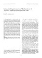
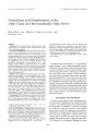
Show journal i)/ Nt: iiro- Oy) illitili> niliwti! 150): iSfi- W), 1995. © 1995 Lippincott- Raven Publishers, Philadelphia Vasculature and Morphometry of the Optic Canal and Intracanalicular Optic Nerve Ping- I Chou, M. D., Alfredo A. Sadun, M. D., Ph. D., and Hwa Lee, M. D. Abstract: Objectives: To study tlie bony structure of the optic canal and the vasculature of the intracanalicular optic nerve in human cadavers. Materials and Methods: Gross and microscopic examinations were performed in 25 optic canals from 13 cadavers to study the pattern of vascular supply of the intracanalicular optic nerve. Neoprene latex was injected through the most proximal part of the ophthalmic artery in seven optic canals. The intracanalicular branches from the ophthalmic artery were carefully identified and quantified. Quantitative measurements of the canal length, canal thickness, canal transverse area, optic nerve transverse area, and subdural space were done for the other 18 canals by means of semiautomated morphometry analysis system. Each canal was divided into anterior, middle, and posterior parts for better visualization and measurement. Results: The ophthalmic artery gives off three branches that supply the intracanalicular optic nerve: medial collateral branch, lateral collateral branch, and ventral branch. Each branch pierces the dura and then supplies the nerve through the pia mater. The middle medial wall was the thinnest bony part of the canal ( 0.31 ± 0.06 mm). The optic canal, optic nerve, and subdural space transverse area varied at different transection levels. The narrowest space was in the middle part of the optic canal. The mean subdural cross- sectional space was only 1.84 mm2. This, multiplied by the average canal length ( 11.79 mm), can be considered the potential space for hemorrhage, optic nerve edema, or hematoma. Conclusions: The vasculature within the bony canal is extremely delicate. Due to the limitation of this space, even a tiny amount of blood or swelling of the nerve ( 21.69 mm") may cause optic nerve compression. It ap- Manuscript received May 18, 1995. From the Department of Ophthalmology ( P. I. C, H. L.), Tri- Service General Hospital, National Defense Medical Center, Taipei, Taiwan; Department of Ophthalmology and Neurosurgery ( A. A. S.), University of Southern California School of Medicine, Dolieny Eye Institute, Los Angeles, California, U. S. A. Supported in part by a grant from the National Science Council, Republic of China, grant NSC 83- 0412- B- 016- 125. Address correspondence and reprint requests to Dr. P, I. Chou, Department of Ophthalmology, Tri- Service General Hospital, No. 42, Sec. 3, Ting Chou Rd., Taipei, Taiwan. pears that these vessels could easily be disrupted in closed head injury by a shearing or concussive force, leading to ischemic infarction of the optic nerve. Since the narrowest portion of the canal is in the middle portion, it is the middle part of the optic canal that is most critical in doing an optic canal decompression. Key Words: Optic canal- Ophthalmic artery- Optic nerve trauma. Traumatic optic neuropathy is a vision- threatening disorder. Several pathophysiologic mechanisms have been proposed, the most likely being a vascular disruption of the intracanalicular optic nerve produced by either a shearing or concussive force after blunt head trauma or direct compression from a bony fragment. Extracranial, microsurgical decompression of the optic canal has been demonstrated as a sometimes effective method in the treatment of traumatic optic neuropathy ( 1- i). Surgery of the optic canal region requires detailed knowledge of this area. In the present study, gross and microscopic dissections of the optic canal were performed to study the relationship between the optic canal structure and intracanalicular optic nerve vasculature. MATERIALS AND METHODS Twenty- five fresh optic canals, including nerves, were dissected completely en bloc from 13 human cadavers. All of the specimens were obtained from the Department of Medicine, University of Southern California, with the approval of the institutional review board. The specimen was placed immediately into heparin water ( 3,000 units heparin/ ml) to prevent coagulation of blood within the vessels and to Iyse the red cells. After carefully rinsing with running water, Neoprene latex ( No. 571, Dupont Co.) was injected through the most 186 VASCULATURE AND MORPHOMETRY OF OPTIC CANAL AND NERVE 187 proximal part of the ophthalmic artery in seven optic canals. The injected specimen was then immersed in concentrated hydrochloric acid overnight to digest the surrounding tissues and delineate a detailed vascular tree. The intracanalicular branches from the ophthalmic artery were carefully identified and quantified. Eighteen other optic canals were fixed in 2% paraformaldehyde- 2% glutaraldehyde for 24 h immediately after dissection and then divided into anterior, middle, and posterior parts. Each specimen was paraffin embedded, sectioned, and stained with Masson trichrome. Under stereomi-croscopy, measurements were made of the length and thickness of the optic canal, and the branching pattern of the ophthalmic artery was studied. The thickness, diameter, transverse area, and subdural space of the optic canals and nerves were also measured by means of a computer assisted semiauto-mated image analysis system ( Fig. 1). RESULTS The ophthalmic artery derived from the internal carotid artery in all of the 25 canals studied. At the cranial end, the ophthalmic artery was located mainly on the inferior medial side and rotated to the inferior lateral side at the orbital end ( Table 1). The ophthalmic artery was observed to contribute three small branches of vessels to supply the intracanalicular optic nerve: the medial collateral, lateral collateral, and ventral branches. Occasionally, one or even two of the branches would be missing, and double ventral branches were noted in one case. In the optic canal, the ophthalmic artery ran TABLE 1. Locations of ophthalmic artery at orbital and cranial ends Cranial end Orbital end Inferior medial ( 14/ 25) Inferior ( 6/ 25) Inferior lateral ( 5/ 25) Interior lateral ( 18/ 25) Inferior ( 4/ 25) Inferior medial ( 3/ 25) intradurally, then penetrated the dura and became epidural at the orbital end. By means of Neoprene injection, each of these small branches could always be traced back to the ophthalmic artery. Instead of supplying the optic nerve directly, these branches first ran into the dura, in which they proliferated a capillary network that penetrated through the subdural space to the optic nerve. The vasculature within the optic canal was extremely delicate. In the dura, the blood vessels, including the ophthalmic artery, were mainly located in the inferior quadrant. These vessels entered the pia through an arachnoid plexus and then supplied the optic nerve by a capillary network surrounding the optic nerve. As the vessels came to the pia, they distributed themselves evenly around the nerve. Our quantitative measurements showed that the thickness of the bony canal wall varied not only from medial to lateral, but also from the anterior ( orbital) to posterior ( cranial) end ( Table 2). The thick anterior medial wall was formed by the sphenoethmoidal junction. The thinnest wall was located in the middle segment on the medial side ( 0.3f ± 0.06 mm). The posterior superior wall was not a real bony wall, but a falciform ligament FIG. 1. Histopathological section of the middle optic canal with intracanalicular optic nerve ( arrow). The medial wall ( double arrow) is the thinnest part of the bony canal ( H& E, 10 x 10). 1 Neun>- Oi> ltt) uilti, vl. Vol. 15, No. 3, 1995 188 P. l. CHOUETAL TABLE 2. Thickness of canal walls ( millimeters) at different levels and positions Anterior Middle Posterior Medial " 2.63 ± 0.24 0.31 ± 0.06 0.87 ± 0.10 Lateral 1.53 ± 0.10 3.82 ± 0.75 3.54 ± 0.56 Superior 1.59 ± 0.35 0.76 ± 0.15 0.38 ± 0.01 Inferior 2.36 ± 0.48 3.64 ± 0.68 3.60 ± 0.47 formed by a fold of the dura. The length of the optic canal varied also ( Table 3). The medial wall was the longest and is formed by the posterior ethmoid sinus and anterior sphenoid sinus walls. The shortest wall was the lateral wall, and it is formed by the optic strut. At the orbital end, the optic canal opening was a vertical ovoid, the horizontal and vertical diameters being 4.34 mm and 5.59 mm, respectively. In the middle part, the ca-naJ was round, the diameter being 4.55 mm. At the cranial end, the optic canal was a horizontal ovoid, the horizontal and vertical diameters being 6.73 mm and 4.64 mm, respectively. The relationships between canal transverse area, nerve transverse area, and subdural transverse area are listed in Table 4. The middle of the canal is the narrowest part, being only 16.22 ± 5,15 mm2 in cross- sectional area. The canal opens up at both ends, and the nerve transverse area gradually increases from anterior to posterior. The subdural transverse areas were 1.68 ± 0.67 mm2, 1.52 ± 0.10 mm2, and 2,31 ± 0.36 mm2 from anterior to posterior, with a mean subdural space of 1.84 mm2. This space was occupied by the cerebral spinal fluid only. The area occupied by the meninges was excluded during measurement. These data, multiplied by the average length of the canal ( 11.79 mm), could be considered as the potential space for optic nerve edema or hemorrhage ( 21.69 mnv5). However, this was not a closed space. Both at the orbital end and the cranial end the intracanalicular subarachnoid space communicated freely with the intracranial subarachnoid space. DISCUSSION Francois first studied the vascularization of the optic nerve pathway by means of Neoprene injection ( Latex 572), microangiography, and serial mi- TABLE 3. Average length of each canalicular wall Length ( mm) Medial wall 13.63 ± 2.05 Lateral wall 6.78 ± 1.56 Superior wall 12.17 ± 4.05 Inferior wall 12.58 ± 3.98 ; Nciiro- Qphtlititmol, Vol. 15. Nti. 3. 1995 croscopic sections ( 5- 7). He found that the optic nerve was supplied by capillaries from the pia mater throughout its length; these capillaries deriving from branches from the ophthalmic artery, anterior cerebral artery, internal carotid artery, lacrimal artery, middle meningeal artery, and long posterior ciliary arteries. For the intracanalicular course, branches from the internal carotid, anterior cerebral, and anterior communicating artery also made contributions that formed the peripheral vascular supply to the optic nerve ( 5). The proximal ophthalmic artery gave off a central optic nerve artery that formed the axiaJ nutritional system of the optic nerve ( 6,7), However, this scheme was later revised by Hayreh, who believed that the intracanalicular part of the optic nerve was entirely supplied by the ophthalmic artery ( 8- 12). Several small branches ( usually one to three in number) arose from the ophthalmic artery near the apex of the orbit. These branches reached the optic nerve by transversing the connective tissue bands that bind the optic nerve to the surrounding dural sheath ( 11,12). No axial vascular system was seen in Hayreh's study. Our present study is in concurrence with Hayreh's findings. In the present study, we always found the origin of the ophthalmic artery to be from the internal carotid artery. As an unusual variant, it has been described to arise from the middle meningeal artery ( 8). The intracanalicular course of the ophthalmic artery varied from the inferior medial position in the optic canal at the cranial end to the inferior lateral position at the orbital end ( Table 1). Throughout its course, we found the intracanalicular ophthalmic artery to be almost entirely embedded in the dura. It then pierced the dural sheath of the optic nerve at the orbital end and became epidural in location. Under gross dissection, the intracanalicular ophthalmic artery seemed exposed under the dura. However, by careful microscopic examination, a thin layer of dura was always seen to wrap about the ophthalmic artery. Our finding differed from Hayreh's report that the ophthalmic artery arises in the subdural space and pierces the dura on its inferior aspect ( 8). The delicacy of the intracanalicular vasculature could be visualized well under the microscope. The optic nerve can be damaged by either concus-sive or compressive mechanisms, and in some cases both conditions can occur in the same patient ( 13- 16). Interruption of the vascular supply to the intracanalicular optic nerve, either from a closed head injury that causes shearing of the pial network or from direct compression by a bony frag- VASCULATURE AND MORPHOMETRY OF OPTIC CANAL AND NERVE 189 TABLE 4. Canal, nerve, and subdural cross- sectional areas at different levels Anterior Middle Posterior Canal transverse area ( mm2) 20.75 ± 4.10 16,22 ± 5.15 18.16 ± 4,50 Nerve transverse area ( mm2) 6.16 ± 1,24 6.66 ± 1.05 7.71 ± 1,07 Subdural transverse area ( mma) 1.68 ± 0.67 1.52 ± 0.10 2.31 ± 0.36 merit or hematoma, is the most widely accepted explanation of visual loss in traumatic optic neuropathy ( 7,11,16). Walsh, in his clinical- pathological correlations, found hemorrhages in the nerve, dura, and sheath space; tears in the nerve or chiasm; and contusion necrosis of the optic nerve to be the primary lesions in indirect trauma to the optic nerve ( 17), Similar reports can also be found elsewhere in the literature ( 18,19). These reports indicate the vulnerability of the intracanalicular capillary network to traumatic disruption and agree with our findings. Habal and Maniscalco performed quantitative measurements of the optic canal ( 20,21). The postnatal growth of the optic canal and its relation to paranasal sinuses were described by Lang ( 22). Great variations existed in the measurement of this structure. This may partly reflect the inappropriate attempt to express the thickness or length of the optic canal by only one value, since the length and thickness of the optic canal varies not only from anterior to posterior, but also from medial to lateral. In the present study, the optic canal was totally removed en bloc from cadavers and then dissected under a stereomicroscope. Each of three levels of the canal was considered separately for measurement. Extremely thin walls, such as the medial wall and falciform ligament, were measured by means of a computerized semiautomated image analysis system and accompanying high- power microscopy. The middle medial wall was the thinnest part of the optic canal. In cases of traumatic optic neuropathy, this part might be most vulnerable to traumatic fracture. Twelve of 58 cases ( 21%) had a fracture of the medial wall of the optic canal, as seen by computed tomographic scan ( 23). In another report, however, five of 379 patients who suffered blindness from optic canal fracture were found to have lesser wing fracture by computed tomogram ( 24). The length of the falciform ligament varied from 1.80 mm to 6.30 mm, with an average of 3.83 mm, which was very close to the measurement of Renn and Rhoton ( 25). The sharp margin of this ligament can severely pinch the optic nerve when optic nerve edema occurs after head injury ( 20,21). The equivalent diameters of the subdural space ( Ds), calculated from the subdural transverse areas, were 1.46 mm, 1,39 mm, and 1.72 mm from anterior to posterior, respectively [ Ds = 2 x ( subdural transverse area/ 3.14) 1' 2]. The middle part of the optic canal was the narrowest passage for the optic nerve and hence a likely site of injury in cases of edema or subarachnoid hemorrhage. This suggests an important consideration in the surgical management of traumatic optic neuropathy patients. For complete and adequate decompression of the optic canal, removal of much of the medial, and sometimes inferior, wall is necessary. Maniscalco and Habal emphasized the importance of removing the optic ring in the distal ( orbital) portion of the optic canal ( 21). However, since the narrowest portion of the canal is in the middle portion, we emphasize the importance of opening this area in optic canal decompression. REFERENCES 1. Fukado Y. Results in 400 cases of surgical decompression of the optic nerve. Moil Prohl Ophthalmol 1975; 14: 474- 81. 2. Kennerdell JS, Amsbaugh GA, Myers EN. Transantral-cthmoidal decompression of optic canal fracture. Arch Ophthalmol 1976; 94: 1040- 3. 3. Nilio S, Yasuda K, Sato T, Sugita S, Murayama K, Ogino N. Decompression of the optic canal by the transethmoidal route. Am I Ophthalmol 1961; 51: 659- 65. 4. Niho S, Niho M., Nino K, Decompression of the optic canal by the transethmoidal route and decompression of the superior orbital fissure. Can j Opfitlialnuil 1970; 5-. 22- 40, 5. Francois J, Neetens A. Vascularization of the optic pathway. 1. Lamina cyibrosa and optic nerve. Br j Ophthalmol 1954; 38: 472- 88. 6. Francois ], Neetens A, Collette JM. Vascular supply of the optic pathway, II. Further studies by micro- arteriography of the optic nerve. Br j Ophthalmol 1955; 39: 220- 32. 7. Francois ], Neetens A, Vascularization of the optic pathway- 111. Study of intra- orbital and intracranial optic nerve by serial sections, Br j Ophthalmol 1956; 40: 45- 52. 8. Hayreh SS, Dass R. The ophthalmic artery. I. Origin and intracranial and intiacanalicular course. Br ) Ophthalmol 1962; 46: 65- 98. 9. Hayreh SS, Dass R, The ophthalmic artery. II. intra- orbital course, Br j Ophthalmol 1962; 46: 165- 85, 10. Hayreh SS. The ophthalmic artery. ( II, branches. Br j Ophthalmol 1962; 46: 212- 47. 11. Hayreh SS. Blood supply and vascular disorders of the optic nerve. An lust Barraquer 1963; 4: 7- 109. 12. Hayreh SS. Blood supply and vascular disorders of the optic nerve. In: Cant JS, ed. The optic nerve. St. Louis: CV Mosby, 1972: 59- 67. 13. Matsuzakt H, An experimental study on indirect injuries of the intracanalicular portion of the optic nerve, Nenro- Ophthalmoi 1986; 6: 23- 8. / Nmtro- Otftfluifatol, Vol. 15, No. .3, 1995 190 P. I. CHOUETAL. 14. Rodger FC. Unilateral involvement of the optic nerve in head injuries. Br j Ophthalmol 1943; 27: 23- 33. 15. Savino PJ, Harbour R. Neuro- ophthalmic manifestations of trauma. In: Smith BC, Delia Rocea RC, Nesi FA, Lisman RD, eds. Ophthalmic plastic and reconstructive surgery. St. Louis: CV Mosby, 1987: 311- 26. 16. Anderson RL, Panje WR, Gross CE. Optic nerve blindness following blunt forehead trauma. Ophthalmology 1982; 89: 445- 55. 17. Walsh FB. Pathological- clinical correlations: 1. Indirect trauma to the optic nerves and chiasm. II. Certain cerebral involvements associated with defective blood supply. Invest Ophthalmol 1966; 5: 433^ 19. 18. Crompton MR. Visual lesions in closed head injury. Brain 1970; 93: 785- 92. 19. Pringle JH. Atrophy of the optic nerve following diffused violence to the skull. Br Med j 1922; 2: 1156- 7. 20. Habal MB, Maniscalco JE, Rhoton A, Microsurgical anatomy of the optic canal; Correlates to optic nerve exposure. ; Surg Res 1977; 22: 527- 33. 21. Maniscalco JE, Habal MB. Microanatomy of the optic canal. / Neurosurg 1978; 48: 402- 6. 22. Lang ], Optic nerve, topographic anatomy. In: Samii M, Jannetta PJ, eds. The cranial nerves. Berlin: Spring- Verlag, 1981: 77- 84. 23. Chou PI, Sadun AA, Chen YC, Su WY, Lin SZ, Lee CC. Clinical experiences in the management of traumatic optic neuropathy. Neuro- Ophthahnol ( in press). 24. Manfredi SJ, Raji MR, Sprinkle PM, Weinstein GW, Minardi LM, Swanson TJ. Computerized tomographic scan findings in facial fractures associated with blindness. Plast Reconstr Surg 1981; 10: 479- 90, 25. Renn WH, Rhoton AL Jr. Microsurgical anatomy of the sellar region. / Neurosmg 1975; 43: 288- 98, / Neuro- Ophtltalmol, Vol. 15, No. 3, 1995 |