| OCR Text |
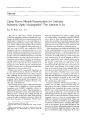
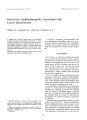
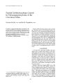
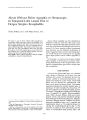
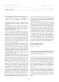
Show journal of Neuro- Ophthalmology 15( 2): 95- 97, 1995. 1995 Raven Press, Ltd., New York Restrictive Ophthalmopathy Associated with Linear Scleroderma William W. Campbell, M. D., and Frank J. Bajandas, M. D. t A patient with a coup de sabre lesion of the forehead developed progressive ipsilateral limitation of ocular motility, primarily involving adduction and depression. Investigation disclosed no other explanation for the ocular motility disturbance, which we suspect represents restrictive myopathy maximally involving ocular muscles immediately subjacent to the area of linear scleroderma. Key Words: Localized scleroderma- Morphea- Ocular myopathy- Restrictive ophthalmopathy. From the Department of Neurology, Medical College of Virginia, Richmond, Virginia, and Department of Ophthalmology, University of Texas Health Science Center, San Antonio, Texas, U. S. A. Address correspondence and reprint requests to Dr. W. W. Campbell, Department of Neurology, Box 599, MCV Station, Richmond, VA 23298, U. S. A. t Deceased. A variety of neurologic, neuromuscular, and neuro- ophthalmic abnormalities may occur in association with linear scleroderma. We investigated a woman with a coup de sabre lesion of the forehead and an ipsilateral restrictive ocular myopathy. CASE REPORT For 2 years, an otherwise healthy 26- year- old woman noted a slowly enlarging, tender patch of discoloration and change in skin texture of the left forehead, accompanied by loss of the hair of the lateral portion of the left eyebrow and loss of the lashes of the left upper lid. Clinically the lesion was a typical coup de sabre lesion of linear morphea, and skin biopsy was consistent with linear scleroderma. A similar, smaller patch developed over the left occipital area. For 3 to 4 months, she had noted blurred vision on gaze to either side, without frank diplopia. Monocular occlusion eliminated the visual blurring. As the blurring became progressively more severe, she noted the need to turn her head in the direction of gaze to maintain clear vision. General physical examination was unremarkable except for a linear streak of atrophic hyperpig-mented skin extending across the left forehead from the hairline to the lateral aspect of the left eyebrow, with alopecia of the lateral brow and upper eyelashes ( Fig. 1). Except for abnormal eye movements, the formal neurologic examination was normal. Opthalmological examination revealed corrected visual acuity of 20/ 20 OU. External examination, Hertel exophthalmometry, corneal sensation, slit-lamp examination, intraocular pressure, Gold-mann perimetry, and dilated fundus examination were all normal. Pupils were normal. Gaze was full in all directions in the right eye. In the left eye, 95 96 W. W. CAMPBELL AND F. /. BA] ANDAS FIG. 1. Coup de sabre lesion on left forehead, with depigmented streak running from hairline to brow. Note also loss of lashes on the temporal aspect of brow and upper eyelid. there was marked limitation of adduction and depression, with mild limitation of abduction and elevation. In primary gaze, there were 2 prism diopters of exotropia and 2 prism diopters of left hypertropia ( Fig. 2). In left gaze, there were 11 prism diopters of exotropia and 4 of left hypertropia. In right gaze, there were 22 prism diopters of exotropia and 7 of left hypertropia ( Fig. 3). In downgaze there were 10 prism diopters of exotropia and 6 of left hypertropia ( Fig. 4). In upgaze there were 4 prism diopters of exotropia and 3 of left hypertropia ( Fig. 5). Forced ductions of the left eye demonstrated restriction of movement primarily medially and temporally, less so inferotempo-rally. Saccadic and pursuit movements were normal. Lid position and movement were normal. The following were normal or negative: hemo-gram, sedimentation rate, urinalysis, VDRL, prothrombin time, partial thromboplastin time, electrolytes, blood urea nitrogen, creatinine, calcium, phosphorus, total protein, albumin, bilirubin, creatine kinase, transaminases, lactate dehydrogenase, alkaline phosphatase, and uric acid. Skull FIG. 2. In primary gaze, muscle was XT2, LHT2. ; Neum- Ophthalmol, Vol. 15, No. 2, 1995 FIG. 3. In gaze right, there is marked limitation of adduction OS. and orbital apex films were normal. Computerized tomographic scan of brain and orbits was normal. All thyroid functions were completely normal, including T3, T4, T3 by radioimmunoassay, free thyroxin index, and radioactive iodine uptake. An 8- day cytomel- suppression test showed normal thyroid suppressability. Because the limitation of adduction was not accompanied by limitation of movement in the other fields of gaze of the third cranial nerve, or by ptosis or pupillary abnormality, and in view of marked limitation to passive movement, a diagnosis of restrictive myopathy, primarily involving the medial and lateral recti and related to the overlying linear scleroderma, was judged most plausible. DISCUSSION A variety of ophthalmological abnormalities may occur in association with juxtaposed linear scleroderma, including changes in eyelid shape, loss of eyelashes, ptosis, eyelid retraction, paresis of extraocular muscles in varying combinations, ocular myopathy, loss of corneal sensation, iridoplegia, iris atrophy, iritis, heterochromia iris, " pseudo-oculomotor palsy," and a fundus picture reminiscent of central retinal vein thrombosis ( 1- 4). The patient reported by Serup et al. had progressive paralysis of the levator, superior rectus, and inferior oblique thought clinically to represent chronic progressive external ophthalmoplegia but associated with typical linear scleroderma of the ipsilat- FIG. 4. Downgaze photo reveals moderate limitation to depression OS. OPHTHALMOPATHY IN SCLERODERMA 97 FIG. 5. Essentially full upgaze. Muscle balance, XT4, LHT3. eral forehead ( 2). Extraocular muscle electromyography was most consistent with a myopathy. Their case is reminiscent of our case as well as Cords' case 2 ( 4). Myopathy in association with linear scleroderma has been recognized for some years. Reported abnormalities of muscle underlying cutaneous lesions of linear scleroderma have included plasma cell fasciitis with severe localized atrophy of types 1 and 2 fibers and thickening of the capillary basal lamina with intramitochondrial inclusions ( 5,6). It is not clear whether these changes have a neuropathic or myopathic basis ( 6). Most current evidence favors a primary disorder of muscle as the etiology of progressive ocular myopathies, although neurogenic abnormalities have been reported as well ( 7). Given the disproportionate restriction of adduction and depression and the location of the coup de sabre lesion, our patient seems to have developed a restrictive myopathy of the extraocular muscles immediately subjacent to the area of linear scleroderma, for reasons unclear. REFERENCES 1. Tang RA, Mewis- Christmann L, Wolf J, Wilkins RB. Pseudo- oculomotor palsy as the presenting sign of linear scleroderma. / Pediatr Ophthalmol Strabismus 1986; 23: 236- 8. 2. Serup J, Serup L, Sjo O. Localized scleroderma " en coup de cabre" with external eye muscle involvement at the same time. Clin Exp Dermatol 1984; 9: 196- 200. 3. Segal P, Jablonska S, Mrzyglod S. Ocular changes in linear scleroderma. Am ] Ophthalmol 1961; 51: 807- 13. 4. Cords R. Strichformige gesichtsatrophie und auge. Bericht Dtsch Ophthalmol Ges 1928; 47: 53- 9. 5. Schwartz RA, Tedesco AS, Stern LZ, Kaminska AM, Har-aldsen JM, Grekin DA. Myopathy associated with sclero-dermal facial hemiatrophy. Arch Neurol 1981; 38: 592- 4. 6. Stern LZ, Payne CM, Alvarez JT, Hannapel AB. Myopathy associated with linear scleroderma. Neurology 1975; 25: 114- 119. 7. Tome FMS, Fardeau M. Ocular myopathies. In: Engel AG, Banker BQ, eds. Myology. New York: McGraw- Hill, 1986: 1327^ 17. / Neuro- Ophthalmol, Vol. 15, No. 2, 1995 |