| OCR Text |
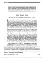
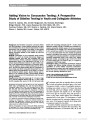
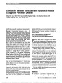
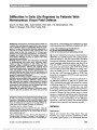
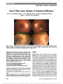
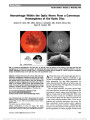
Show Paraneoplastic Syndromes in Neuro-Ophthalmology Lynn K. Gordon, MD, PhD Background: Paraneoplastic syndromes that affect the visual pathways and present with neuro-ophthalmologic signs or symptoms may involve the afferent or efferent systems. Afferent syndromes may involve the optic nerve or retina and, in some cases, these may be associated with systemic neurologic disease. Efferent symptoms typically affect eye movements and may involve the neuromuscular junction or involuntary eye movements. Evidence Acquisition: Literature review and personal clini-cal and research experience. Results: Diagnosis of paraneoplastic syndromes relies on clinical and laboratory evaluations. In the appropriate clinical setting, the presence of specific antibodies may help confirm the diagnosis. Conclusions: In some cases, the visual pathway distur-bance precedes a diagnosis of malignancy. Astute obser-vation and selective evaluation and management are critical to establish the correct diagnosis and institute therapeutic approaches that can be sight or life saving. Journal of Neuro-Ophthalmology 2015;35:306-314 doi: 10.1097/WNO.0000000000000280 © 2015 by North American Neuro-Ophthalmology Society Paraneoplastic syndromes may affect the afferent and efferent visual pathways and involve the retina, optic nerve, or eye movement systems (1-6). In some cases, the neuro-ophthalmic consequences are the first indication of a malignancy, and in others, the visual pathways are affected in patients with known malignancies. The challenges are to recognize when a symptom or sign may be associated with an underlying malignant disease, to understand the diag-nostic challenges in identifying an underlying paraneoplas-tic cause for the observed visual pathway deficits, and to determine the optimal strategy for therapeutic intervention. It is important to understand the spectrum of signs and symptoms that can result from a tumor-stimulated immune process to suspect, diagnose, and treat these diseases. Clinical Disease Spectrum When should the clinician consider a diagnosis of paraneo-plastic syndrome? (1,6-11). In terms of afferent symptoms, an unexplained, painless, progressive vision loss is typical. With retinal involvement, there may be photopsias, night blindness, or ring scotomas. In the optic neuropathies, there is most commonly bilateral disc swelling often accompanied by vitri-tis. Efferent symptoms include myasthenic-like presentation or the presence of opsoclonus/myoclonus syndrome (OMS). Associated systemic neurologic symptoms, such as encephali-tis, cerebellar degeneration, myelitis, or sensory neuropathies, increase suspicion for a paraneoplastic syndrome. Pertinent negatives include lack of alternative explanation for the symp-toms such as a known genetic condition, history of ocular surgery, infection, trauma, mass lesion, or toxic exposures. This review does not focus on syndromes associated with anti-Hu, anti-Ma, and anti-Yo as these typically manifest with a variety of brainstem and cerebellar findings. A history of cancer may heighten the suspicion for a paraneoplastic syn-drome, but the real challenge is to diagnose a potentially treat-able cancer in patients who do not already carry that diagnosis. Afferent Symptoms Autoimmune Paraneoplastic Retinopathy Three types of autoimmune paraneoplastic retinopathy syn-dromes have been described: cancer-associated retinopathy (CAR), melanoma-associated retinopathy (MAR), and bilateral diffuse uveal melanocytic proliferation (BDUMP) (12-19). CAR was the first of these syndromes to be recognized, seems to be the most common of the paraneoplastic retinopathies, and remains a significant diagnostic and therapeutic challenge (12). Visual dysfunction in CAR typ-ically involves a bilateral, progressive, and painless loss of vision with photopsias (14,20) Patients complain of a rapid onset of night blindness and flickering lights associated with progressive vision loss over weeks to months. The signs and symptoms depend on whether rod or cone function is Jules Stein Eye Institute, Department of Ophthalmology, David Geffen School of Medicine at UCLA, Los Angeles, California. The author reports no conflicts of interest. Address correspondence to Lynn K. Gordon, MD, PhD, Jules Stein Eye Institute, David Geffen School of Medicine at UCLA, 100 Stein Plaza, Los Angeles, CA 90095; E-mail: lgordon@mednet.ucla.edu 306 Gordon: J Neuro-Ophthalmol 2015; 35: 306-314 State-of-the-Art Review Section Editors: Valérie Biousse, MD Steven Galetta, MD Copyright © North American Neuro-Ophthalmology Society. Unauthorized reproduction of this article is prohibited. primarily disrupted. In rod disease, there is often constric-tion of the visual field with impaired dark adaptation (Fig. 1). In contrast, when there is primarily cone dysfunction, central scotomas, dyschromatopsia, glare, and loss of visual acuity are more prominent. There may be an associated uveitis involving the anterior segment or vitreous, and this may also include retinal vasculitis and cystoid macular edema (9). Retinal findings may be unexceptional. Alterna-tively, arteriolar narrowing, thinning or mottling of the retinal pigment epithelium (RPE), or pallor of the optic disc may be observed in these patients. Electroretinography (ERG) confirms photoreceptor dysfunction but is not pathognomonic for CAR. ERG shows reductions in the amplitude of the scotopic and photopic a- and b-waves, and in some cases, a negative waveform is observed. Fluo-rescein angiography may reveal cystoid macular edema or retinal vasculitis. Optical coherence tomography (OCT) may demonstrate macular atrophy with photoreceptor FIG. 1. Paraneoplastic retinopathy. An 81-year-old woman complained of decreased vision, impaired light adaptation, and progressive visual field loss. She had a history of breast cancer treated 4 years earlier with lumpectomy, chemotherapy, and radiation therapy and was cancer free at the time of evaluation. On examination, visual acuity was 20/20 in the right eye and 20/50 in the left eye with constricted visual fields. Ophthalmoscopy was unremarkable (A and B), but optical coherence tomography shows cystoid macular edema in the right eye (C) and vitreomacular traction in the left eye with possible early lamellar hole formation (D). ERG showed a significantly truncated b-wave consistent with downstream dysfunction (E). Anti-retinal antibody testing was positive on Western blot for aldolase antibodies and, on immunohistochemistry, for antibodies against bipolar cells and the inner plexiform layer. This case highlights the importance of ERG evaluation in patients despite the presence of other retinal abnormalities such as cystoid macular edema or vitreomacular traction in the diagnosis of retinal dysfunction (Courtesy of Michael Gorin, MD, PhD). ERG, electroretinography. Gordon: J Neuro-Ophthalmol 2015; 35: 306-314 307 State-of-the-Art Review Copyright © North American Neuro-Ophthalmology Society. Unauthorized reproduction of this article is prohibited. thinning and loss of inner/outer segment junction. CAR may occur in patients with known malignancies, but it also may occur before the diagnosis of cancer. Thus, prompt recogni-tion may lead to early diagnosis and cure for patients with specific types of malignancies. Although the most common tumor associated with CAR is small cell lung carcinoma, other tumors originating in many organs including the prostate, bladder, colon, thymus, ovary, endometrium, breast, and cer-vix also have been implicated in CAR (Table 1). Visual symptoms in MAR have rapid onset and include loss of visual acuity, presence of photopsias, and central or paracentral scotomas (21,22). Vision loss is usually mild with the majority of patients having a best-corrected visual acuity in the 20/60 range. Ophthalmoscopy initially may be normal but may show optic atrophy, retinal vascular attenuation, or loss of RPE. ERG abnormalities typically include an electronegative pattern with a normal dark-adapted a-wave and attenuated or absent b-wave (Fig. 2). In a large review of more than 60 patients with MAR, vitreous cells were present in 30% of affected patients (22). However, the fundus evaluation was initially normal in more than 40%. Similar to CAR, disc pallor and retinal vascular attenuation may be observed. Patients with MAR usually have a diagnosis of malignant melanoma that typically predates the retinal disease by several years. Cases of MAR have been primarily associated with cutaneous melanoma but have also been observed in patients with ocular or mucosal sites for the primary tumor, thus neces-sitating a thorough systemic evaluation for tumors in those patients without a known melanoma. BDUMP is rare but distinctive in its proliferation of melanocytes in the uveal tract along with bilateral loss of vision. These patients experience sudden and bilateral visual loss and may have exudative retinal detachment and progressive cataracts. Patients with BDUMP typically have a short life span, less than 15 months after diagnosis on average. Visual disturbance may precede diagnosis of malignancy, and although multiple types of cancers have been identified in association with BDUMP, the most common are lung, colon, pancreas, or gynecologic. OCT is helpful in identifying subretinal fluid. Areas of RPE atrophy and adjacent regions of RPE hypertrophy are typical and seen well on fluorescein angiography. Paraneoplastic Optic Neuropathy Paraneoplastic optic neuropathy (PON) is an unusual but serious cause of bilateral painless loss of vision. Vision loss is typically subacute, occurring over days to weeks. In 2003, TABLE 1. Paraneoplastic syndromes of neuro-ophthalmic interest Syndrome Disease- Associated Antigen Autoreactivity Against Specific Cell Type Suspected Disease-Associated Antigen/Cells Neoplasm Cancer-associated retinopathy Recoverin alpha-enolase Transducin, carbonic anhydrase II, arrestin, TULP1, neurofilament, HSP70, TRPM1, Müller cell specific Lung, prostate, bladder, colon, thymus, breast, gynecologic Melanoma-associated retinopathy Retinal bipolar cells TRPM1, rhodopsin, recoverin, arrestin, transducin, mitofilin, titin Müller cell Melanoma Bilateral diffuse uveal melanocytic proliferation Lung, gynecologic, colon, rectum, pancreas, melanoma Paraneoplastic optic neuropathy Collapsin response-mediating protein-5 22 kDa protein Lung, kidney, thymus, colon, thyroid, breast, uterus, neuroendocrine Opsoclonus/ myoclonus syndrome Purkinje cells Ri (Anna-2), NMDA receptor, Hu (ANNA-1), Yo (PCA1), Ri (ANNA-2). amphiphysin, neuronal calcium channels Lung, breast, neuroblastoma Lambert-Eaton myasthenis syndrome Voltage-gated calcium channels: P/Q, L, N Lung, prostate, thymus, lymphoproliferative 308 Gordon: J Neuro-Ophthalmol 2015; 35: 306-314 State-of-the-Art Review Copyright © North American Neuro-Ophthalmology Society. Unauthorized reproduction of this article is prohibited. a cohort of 16 patients was identified as being positive for the antibody against collapsin response-mediating protein 5 (CRMP-5) (11). Similar to patients with CAR and MAR, photopsias may be present. Ophthalmoscopic findings include optic disc edema and vitreous cells, but patients may present with optic atrophy as well. Retinitis also may be present, and in these patients, the ERG may show scoto-pic or photopic abnormalities. Vitreous cells appear small, without clumping, and without evidence for an intermediate uveitis, and are typically pleomorphic lymphocytes. One of the original 16 patients also had anterior chamber cells, an unusual finding in this disease. The histopathology of the optic nerve in affected cases demonstrates inflammation of the optic nerve with lymphocytic infiltrates. In one case the infiltrating cells were primarily CD8+ T cells and in another case they were identified as both T cells (CD3+) and B cells (CD20+) (11,23). In addition, disorders of eye movement including vertical gaze disturbance, internuclear ophthalmo-plegia, and opsoclonus have been observed in affected patients. Systemic neurologic symptoms are present in essentially all patients during their illness and may include seizures, cognitive abnormalities, dementia, cerebellar findings, and a wide variety of motor and sensory abnormalities (24). Evaluation of the spinal fluid in patients with PON may reveal lymphocytosis or elevated protein (11). The most common cancer in these patients is small cell lung carci-noma but also includes neuroendocrine tumors, nasopha-ryngeal carcinoma, and colon cancer, among others (25- 28). One reported patient with unilateral sudden loss of vision due to an optic neuropathy had a choroid meningi-oma. There was immune seroreactivity against optic nerve sections from multiple species and reversal of the optic neuropathy after tumor resection, suggesting a cause and effect relationship with this tumor (29). POEMS syndrome (polyneuropathy, organomegaly, endocrinopathy, monoclonal gammopathy, and skin changes syndrome) should be included when considering the paraneoplastic afferent syndromes that present with neuro-ophthalmologic signs or symptoms. Major criteria include a monoclonal plasma cell dyscrasia and polyneur-opathy, initially sensory but ultimately both sensory and motor. Bilateral optic disc edema is observed in about half of the patients with POEMS, and this may be asymptom-atic (29% of patients) or alternatively may be associated with symptoms of blurred vision, pain, or double vision (9,30,31). Evaluation of the cerebrospinal fluid (CSF) often shows an increased opening pressure and elevated protein. The mechanism of ophthalmologic signs and symptoms in POEMS is likely distinct from the other paraneoplastic diseases and may involve high levels of inflammatory cyto-kines or vascular endothelial growth factor. Specific immune mechanisms, including cellular or humoral immu-nity, leading to this syndrome have not been implicated. Efferent Symptoms Opsoclonus/Myoclonus Syndrome Although abnormalities of eye movement associated with paraneoplastic disease include disorders of saccades and smooth pursuit, opsoclonus/myoclonus, and nystagmus, the best characterized is the opsoclonus/myoclonus syndrome (OMS). Myoclonus is defined by short sudden involuntary jerking movements, which may involve lower or upper extremities, trunk, or face and may be induced or worsened by a change in posture or stress (32-37). Opsoclonus is the ocular equivalent of myoclonus and is characterized by rapid, involuntary, horizontal, and vertical conjugate eye movements without intersaccadic delay (A video of a patient with paraneoplastic opsoclonus can be viewed on the NOVEL Web site at http://content.lib.utah.edu/cdm/ singleitem/collection/ehsl-shw/id/92/rec/12). In children, up to 43% of cases of OMS are associated with a paraneo-plastic syndrome linked to neuroblastoma (38). However of all patients with neuroblastoma, only 2%-4% will develop OMS. Other etiologies during childhood include ovarian teratoma, hepatoblastoma, or infections including myco-plasma pneumonia and hepatitis C. In 2012, a series of 21 adults with OMS was published in conjunction with a review of all reported cases at that time of adult OMS (n = 116) (35). The age range for newly reported patients was 27-78 years, and symptoms consisted FIG. 2. Melanoma-associated retinopathy. A 76-year-old man noted vibrating and sparkling visual symptoms associated with reduced night vision. His history was significant for a malignant melanoma removed approximately 5 years earlier. ERG tracings show the photopic (A) and scotopic (B) responses and demonstrate the characteristic decrease in the b-wave under scotopic conditions (label 2 in [B]) (Courtesy of John Keltner, MD). ERG, electroretinography. Gordon: J Neuro-Ophthalmol 2015; 35: 306-314 309 State-of-the-Art Review Copyright © North American Neuro-Ophthalmology Society. Unauthorized reproduction of this article is prohibited. of dizziness/balance difficulties in 67%, nausea/vomiting in 48%, and abnormalities of vision from opsoclonus in 28%. Neuroimaging was normal in all except one patient with metastatic disease. Evaluation of CSF revealed abnormalities in more than 50% of patients including elevated protein and/or increased white cells (lymphocytes); in about one-third of cases, there may be an increased IgG index or oligoclonal bands (32,35). Cancer was identified in about half of the reported cases, and although small cell lung cancer was the most common, other malignancies included non-small cell lung cancer, breast adenocarcinoma, and ovarian carcinoma. Several adult patients with OMS were found to have thymic carcinomas, one in whom the OMS significantly improved after surgical removal of the tumor (39). Ocular flutter also has been associated with lung and breast cancer and, therefore, a thorough neoplastic evalua-tion is indicated in these patients (40). The majority of nonmalignant causes of OMS include parainfectious etiol-ogies such as infection with HIV. Lambert-Eaton Myasthenic Syndrome In the Lambert-Eaton myasthenic syndrome (LEMS), there is a decrease in acetylcholine release leading to decreased activity of the neuromuscular junction. Although LEMS is typically characterized by decreased deep tendon refluxes, autonomic dysfunction, and proximal muscle weakness, symptoms of diplopia and ptosis are common (5,7). Auto-nomic symptoms develop in about 90% of affected patients within the first 3 months of disease onset and are very helpful in establishing the diagnosis as LEMS. To distinguish LEMS from myasthenia gravis (MG), clinical findings, such as slight increases in strength on prolonged effort, and electromyog-raphy, showing facilitation in repetitive stimulation testing, are helpful when present. Ocular symptoms generally occur after onset of other disease symptoms such as generalized weakness and ultimately occur in about half of the patients with LEMS. Ocular findings reported in patients with LEMS, that are not generally seen in MG include involun-tary lid closure, decreased ptosis with prolonged upgaze, dilated pupils, and poorly reactive pupils (5). Ductions are typically full in LEMS but may be limited in MG patients. More than 50% of patients with LEMS not only have cancer, commonly lung cancer, but also lymphoproliferative diseases as well as cancers of the prostate or thymus may be present. PATHOPHYSIOLOGY The paraneoplastic syndromes often involve antibodies against normal proteins that are also typically expressed in the tumor. Some syndromes are also associated with tissue infiltration of inflammatory cells. To date, a virtual alphabet soup of proteins have been identified as potential immune targets, and in some cases, these proteins have been validated as playing a significant role in the pathophysiology of the disease (Table 1). For other candidates, the patho-physiological role played by the immune response against a particular autoantigen is not entirely clear. Recoverin, a 23 kDa protein, was the first antigenic target identified in patients with CAR. The protein is widely expressed in the majority of lung cancer samples, irrespective of retinopathy. Serum autoantibodies against recoverin may be present in a small subset of patients with lung cancer, but only a small fraction of those develop CAR. Alpha-enolase, another common protein that is found both in lung cancer and the retina, elicits an antibody in a larger percentage of patients with lung cancer (estimated at 13%- 65%); however, only a small subset of these patients will develop CAR (1,41,42), Alpha-enolase antibodies also are found in many other diseases, including autoimmune hep-atitis, rheumatoid arthritis, and mixed cryoglobulinemia. In addition, these antibodies may be found in normal individ-uals; thus, their presence alone does not indicate disease or causation of symptoms. How are these antibodies associated with disease path-ophysiology? It is believed that high-titer of antibodies may traverse the blood-retinal barrier, leading to exposure to retinal cells. There is also some evidence for different anti-genic epitopes within a protein with differential consequen-ces with regards to pathology. In addition, the immunologic phenomenon of epitope spreading may be associated with differences in pathogenic sequelae. In the retina, there is some evidence that the antibody may be engulfed through an endocytic mechanism into retinal cells (43). Once inter-nalized, the antibody engages its corresponding antigen, and this binding leads to downstream signaling resulting in apo-ptotic cell death. This observation was initially made in vitro using retinal cells in culture but has also been replicated in vivo using either intravitreal or intravenous injections of antibodies against recoverin. Apoptosis was activated through caspase 3 and caspase 9 (44). It is believed that an increase in free Ca2+ precedes apoptosis, a mecha-nism that was also observed in studies using antibodies against enolase. In studies using anti-enolase antibodies, there is a decrease in glycolytic adenosine triphosphate with resultant increase in the intracellular Ca2+. This mechanism is a plausible common pathway leading to cell death. How-ever, additional studies will be required to definitively understand the disease pathophysiology. Many other poten-tial antigens also have been identified, but it is yet unknown whether the antibodies against these antigens play an impor-tant role in disease pathogenesis. In LEMS, there is a well-characterized antibody against P/Q voltage-gated calcium channels (VGCC), present on the presynaptic nerve terminal at the neuromuscular junction. This antibody reproduced the disease in animal models, and the disease can be transmitted passively from mother to child. It is believed that the antibody causes a loss in the VGCC, leading to a decrease in Ca2+ internalization and decrease in release of acetylcholine containing vesicles, with less available acetylcholine at the neuromuscular junction. 310 Gordon: J Neuro-Ophthalmol 2015; 35: 306-314 State-of-the-Art Review Copyright © North American Neuro-Ophthalmology Society. Unauthorized reproduction of this article is prohibited. PATIENT EVALUATION For the well-characterized antigens in disease pathophysiology, such as CRMP-5 and recoverin, the presence of antibodies is helpful in making the diagnosis. The challenges are primarily 1) interpreting and identifying possible disease-associated antibodies against other possible antigens and 2) understand-ing the sensitivity and specificity for the known antigens in making the diagnosis of a paraneoplastic disorder. Laboratory evaluations for the presence of antibodies are performed using multiple techniques including immunofluorescence against tissues, immunofluorescence against cultured or purified cells, Western blot against proteins extracted from tissues or cells, and enzyme-linked immunosorbant assay (ELISA) testing against purified proteins. The potential pitfalls of testing include the tissue or cells of origin, the way that the proteins are extracted from the tissue or cells, the dilution and exposure time to the patient serum, techniques to enhance specificity, and the size of the antigen of interest. In all of these tests, it is important to understand the limitations of the testing strategy and the false-positive and false-negative potential for a partic-ular technique. Antibodies against self-proteins are fairly common. Using Western blot analysis, the majority of normal individuals have immunologic reactivity against at least 1 protein in whole retinal extracts (45). In a review of the literature about anti-retinal antibodies, it was pointed out that standardization among laboratories was lacking and results were not always concordant between different testing sites (46). This was confirmed in a report of 14 patients who were diagnosed with autoimmune retinopathy (47). Their serum was sent to 2 different laboratories for evalu-ation of the presence of anti-retinal antibodies by Western blot. One laboratory used human retina extract with a pos-itive control, and the other used pig retinal extract with a panel of normal controls (without anti-retinal activity). Anti-retinal antibodies were detected in 9 patients by both laboratories. In the remaining 5 patients, 4 were positive for anti-retinal antibodies as detected by one laboratory and one was positive as detected by the other laboratory. Further-more, for the 9 who were positive, there was only a single patient whose sera gave the same results (within 1 kDa of size of the band) from both laboratories. What Antibodies Are Known to be Disease Associated? Disease-associated validated antigenic targets of antibod-ies have been described in several of the paraneoplastic neuro-ophthalmic syndromes (Table 1). However, there are other antigens that have also been identified as poten-tial immunologic targets, but the pathologic significance of these antibodies is uncertain. What testing is recom-mended when you encounter a patient with a suspected paraneoplastic syndrome with neuro-ophthalmologic symptoms? If you suspect CAR, then antibodies against retinal proteins initially should be tested with Western blot and/or immunofluorescence. As noted above, however, it might be important to send materials to different laboratories for investigation. In one large series of CAR patients, only 61% had antibodies against defined retinal proteins, of which the antigenic targets were alpha-enolase in about half of the patients, followed in descending order by transducin, carbonic anhydrase II, and recoverin (1). Antibodies against recoverin, the best characterized antibody, were found in only 10% of the patients. If the clinical suspicion for CAR remains high, but the antibody testing is negative, then one might try empirical therapy for treatment of CAR, in par-ticular in the setting of known malignancy, to determine whether there is clinical improvement. If the testing is negative, consider the possibilities that the patient is negative for anti-retinal antibodies or that the technique used was not sufficiently sensitive to uncover the reactivity. If antibody testing is positive, you need to determine whether the results fit the observed clinical picture before making the diagnosis. For example, anti-recoverin antibodies, the best-studied antibody associated with CAR, have been observed in 1% of patients with retinitis pigmentosa (12). Multiple anti-retinal antibodies may also be found in the same patient (48). Patients with MAR most commonly produce antibodies that react with retinal bipolar cells, as visualized by immunofluorescence. However, specific antigenic targets are not well-defined although several candidate antigens have been identified (22) MAR patients serum may also exhibit immunoreactivity against other retinal layers. There-fore, MAR is primarily a clinical diagnosis, based on the presenting signs and symptoms, a history of melanoma, and ERG findings that are consistent with the disease. If there is no history of melanoma, but your clinical suspicion is high for MAR, then the patient should be referred for dermato-logic evaluation and possibly additional testing, such as body position emission tomography (PET) imaging. In cases of PON, antibodies against the CRMP-5 antigen are detectable by immunofluorescence studies or Western blot (11,49). Pediatric patients with the OMS generally do not have positive paraneoplastic antibody evaluations in the serum or CSF (35). Antibodies against ANNA-2, NMDA receptor, nuclear antigens, and neuronal calcium channels have all been detected in subsets of adult patients with OMS (32,38,50,51). Antibodies against Purkinje cells may be responsible for disease pathogenesis in some patients (32). If testing is warranted, then serum, and perhaps CSF, should be sent for a full paraneoplastic evaluation that will typically use immunofluorescence for reactivity against brain and ELI-SA for reactivity against specific antigenic targets. In some patients, paired evaluations of antibodies in the serum and CSF are preferred (52). Given that an antibody is typically not identified, evaluation for occult malignancy is required in all patients with OMS. Patients with neuroblastoma- Gordon: J Neuro-Ophthalmol 2015; 35: 306-314 311 State-of-the-Art Review Copyright © North American Neuro-Ophthalmology Society. Unauthorized reproduction of this article is prohibited. associated OMS may have tumors that are small, located in the paravertebral regions, and may be challenging to identify on neuroimaging, which must be done using thin sections or ultrasound. Identification of an underlying neuroblastoma may sometimes be made on the basis of elevated catechol-amine metabolites (33). Specific testing for antibodies against the VGCC should be performed in patients who are suspected to have LEMS. These studies are generally available as ELISA tests with good specificity. Antibodies against the P/Q-type VGCC are present in 85%-90% of patients with LEMS but may also be seen in up to 4% of patients with small cell lung cancer in the absence of neurologic disease. In addition, LEMS patients may have antibodies against N-type VGCC (in up to 30%-40%) or L-type VGCC (in up to 25%) (35). What Systemic Evaluation Should be Performed for an Occult Malignancy? CT of the chest, abdomen, and pelvis are essential. Breast examination and mammography should be performed. Pelvic and abdominal CT may reveal many other cancers that are associated with CAR, and pelvic examination, prostate examination, and colonoscopy should be consid-ered in the appropriate patients. Serologic testing for specific cancer markers may also have a role in the evaluation. These tests are best ordered through a collabo-rative care of the patient with a hematologist-oncologist. Whole-body PET using fluorodeoxyglucose (FDG-PET) can be performed in patients in whom other testing was not revealing (53). Hematologic malignancies have been associated with the paraneoplastic syndromes, and these may require special testing including serum protein electro-phoresis and bone marrow biopsy. Management A common thread among all of the paraneoplastic syn-dromes is that treatment of the underlying malignancy may be beneficial to the neuro-ophthalmological symptoms and signs. Immunologic treatment targeting the antibody-mediated paraneoplastic syndrome and symptomatic ther-apy also are of benefit. In addition to cytoreduction of tumor, the mainstay for treatment for CAR is use of oral or intravenous corticosteroids or a combinatorial therapy of cyclosporine, azathioprine, and prednisone with a reported response rate of up to 70% (54). Newer therapies have included rituximab (55). Many types of treatments have been used with variable success in MAR. In general, corticosteroids are of little benefit, whereas intravenous immunoglobulin (IVIg) alone or in combination with cytoreduction of tumor burden has proven more effective (22). Successful treatment also has been reported with combination therapy such as oral prednisone, plasmapheresis, azathioprine, and gabapentin or the combination of intravenous steroids with plasmaphe-resis. The total number of affected patients with MAR is small and, therefore, best practice is not yet determined for these patients. BDUMP also is rare, but there have been reports of improvement with plasma exchange with or with-out systemic corticosteroids (56). In some cases of PON, use of intravitreal triamcinolone was associated with improvement in vision (57). In OMS, adult patients generally respond favorably to immunotherapy treatment with intravenous corticosteroids and/or IVIg (35) in addition to the treatment of the primary tumor (surgery, chemotherapy, radiation). A wide variety of other agents have been used for symptomatic control in small numbers of patients with some success, including clonazepam, valproic acid, or gabapentin (35). Many pedi-atric patients with this syndrome demonstrate long-term neuropsychological morbidity, but this is believed to be ameliorated by early immunosuppressive therapy (58). A multicenter clinical trial is currently in progress to try to provide evidence-based therapeutic recommendations for OMS in pediatric patients (ClinicalTrials.gov Identifier: NCT01868269). Suggested therapy for LEMS includes 3,4- diaminopyridine, IVIg, plasma exchange, steroids, and immunosuppressive agents (59-62). According to a Co-chrane publication that reviewed trials through 2010, there is good evidence for the use of 3,4-diaminopyridine, an agent that increases the release of acetylcholine, and a single trial that demonstrated benefit from IVIg (59). The use of cortocosteroids and azathioprine in addition to 3,4- diaminopyridine may be beneficial, but this is not based on prospective clinical trials (61). A new calcium channel agonist, with selectivity for both the N-type and P/Q-type VGCC, was used in an experimental model of LEMS, with promising results (62). If these results can be replicated in humans, then this would give an additional therapeutic option. In conclusion, the paraneoplastic disorders in neuro-ophthalmology may predate identification of a malignancy. These patients require careful discussion about the possi-bility of an underlying disease such as cancer and also will require periodic follow-up. Treatment consists of control of tumor itself followed by symptomatic relief and use of immunosuppressant and immunomodulatory drugs. REFERENCES 1. Adamus G. Autoantibody targets and their cancer relationship in the pathogenicity of paraneoplastic retinopathy. Autoimmun Rev. 2009;8:410-414. 2. Bataller L, Dalmau J. Neuro-ophthalmology and paraneoplastic syndromes. Curr Opin Neurol. 2004;17:3-8. 3. Chan JW. Paraneoplastic retinopathies and optic neuropathies. Surv Ophthalmol. 2003;48:12-38. 4. Jacobson DM. Paraneoplastic disorders of neuro-ophthalmologic interest. Curr Opin Ophthalmol. 1996;7:30-38. 5. Ko MW, Dalmau J, Galetta SL. Neuro-ophthalmologic manifestations of paraneoplastic syndromes. J Neuroophthalmol. 2008;28:58-68. 312 Gordon: J Neuro-Ophthalmol 2015; 35: 306-314 State-of-the-Art Review Copyright © North American Neuro-Ophthalmology Society. Unauthorized reproduction of this article is prohibited. 6. Ling CP, Pavesio C. Paraneoplastic syndromes associated with visual loss. Curr Opin Ophthalmol. 2003;14:426-432. 7. Wray SH, Dalmau J, Chen A, King S, Leigh RJ. Paraneoplastic disorders of eye movements. Ann N Y Acad Sci. 2011;1233:279-284. 8. Rosenfeld MR, Titulaer MJ, Dalmau J. Paraneoplastic syndromes and autoimmune encephalitis: five new things. Neurol Clin Pract. 2012;2:215-223. 9. Rahimy E, Sarraf D. Paraneoplastic and non-paraneoplastic retinopathy and optic neuropathy: evaluation and management. Surv Ophthalmol. 2013;58:430-458. 10. Khan N, Huang JJ, Foster CS. Cancer associated retinopathy (CAR): an autoimmune-mediated paraneoplastic syndrome. Semin Ophthalmol. 2006;21:135-141. 11. Cross SA, Salomao DR, Parisi JE, Kryzer TJ, Bradley EA, Mines JA, Lam BL, Lennon VA. Paraneoplastic autoimmune optic neuritis with retinitis defined by CRMP-5-IgG. Ann Neurol. 2003;54:38-50. 12. Braithwaite T, Vugler A, Tufail A. Autoimmune retinopathy. Ophthalmologica. 2012;228:131-142. 13. Dobson R, Lawden M. Melanoma associated retinopathy and how to understand the electroretinogram. Pract Neurol. 2011;11:234-239. 14. Keltner JL, Thirkill CE, Tyler NK, Roth AM. Management and monitoring of cancer-associated retinopathy. Arch Ophthalmol. 1992;110:48-53. 15. Murphy MA, Thirkill CE, Hart WM Jr. Paraneoplastic retinopathy: a novel autoantibody reaction associated with small-cell lung carcinoma. J Neuroophthalmol. 1997;17: 77-83. 16. Shildkrot Y, Sobrin L, Gragoudas ES. Cancer-associated retinopathy: update on pathogenesis and therapy. Semin Ophthalmol. 2011;26:321-328. 17. Thirkill CE, FitzGerald P, Sergott RC, Roth AM, Tyler NK, Keltner JL. Cancer-associated retinopathy (CAR syndrome) with antibodies reacting with retinal, optic-nerve, and cancer cells. N Engl J Med. 1989;321:1589-1594. 18. Thirkill CE, Roth AM, Keltner JL. Cancer-associated retinopathy. Arch Ophthalmol. 1987;105:372-375. 19. Volpe NJ, Rizzo JF III. Retinal disease in neuro-ophthalmology: paraneoplastic retinopathy and the big blind spot syndrome. Semin Ophthalmol. 1995;10:234-241. 20. Thirkill CE. Cancer-induced, immune-mediated ocular degenerations. Ocul Immunol Inflamm. 2005;13:119-131. 21. Lu Y, Jia L, He S, Hurley MC, Leys MJ, Jayasundera T, Heckenlively JR. Melanoma-associated retinopathy: a paraneoplastic autoimmune complication. Arch Ophthalmol. 2009;127:1572-1580. 22. Keltner JL, Thirkill CE, Yip PT. Clinical and immunologic characteristics of melanoma-associated retinopathy syndrome: eleven new cases and a review of 51 previously published cases. J Neuroophthalmol. 2001;21:173-187. 23. Sheorajpanday R, Slabbynck H, Van De Sompel W, Galdermans D, Neetens I, De Deyn PP. Small cell lung carcinoma presenting as collapsin response-mediating protein (CRMP) -5 paraneoplastic optic neuropathy. J Neuroophthalmol. 2006;26:168-172. 24. Thambisetty MR, Scherzer CR, Yu Z, Lennon VA, Newman NJ. Paraneoplastic optic neuropathy and cerebellar ataxia with small cell carcinoma of the lung. J Neuroophthalmol. 2001;21:164-167. 25. Slamovits TL, Posner JB, Reidy DL, Thirkill CE, Keltner JL. Pancreatic neuroendocrine paraneoplastic optic neuropathy: confirmation with antibody to optic nerve and hepatic metastasis. J Neuroophthalmol. 2013;33:21-25. 26. Hoh ST, Teh M, Chew SJ. Paraneoplastic optic neuropathy in nasopharyngeal carcinoma-report of a case. Singapore Med J. 1991;32:170-173. 27. Chao D, Chen WC, Thirkill CE, Lee AG. Paraneoplastic optic neuropathy and retinopathy associated with colon adenocarcinoma. Can J Ophthalmol. 2013;48:e116-e120. 28. Yu Z, Kryzer TJ, Griesmann GE, Kim K, Benarroch EE, Lennon VA. CRMP-5 neuronal autoantibody: marker of lung cancer and thymoma-related autoimmunity. Ann Neurol. 2001;49:146-154. 29. Nakano S, Kanamori A, Nakamura M, Mizukawa K, Negi A. Paraneoplastic optic neuropathy associated with cerebellar choroid meningioma. Eye (Lond). 2013;27:1220-1221. 30. Kaushik M, Pulido JS, Abreu R, Amselem L, Dispenzieri A. Ocular findings in patients with polyneuropathy, organomegaly, endocrinopathy, monoclonal gammopathy, and skin changes syndrome. Ophthalmology. 2011;118:778-782. 31. Lally DR, Moster ML, Foroozan R. Muscle cramping over the diagnosis. Surv Ophthalmol. 2014;59:128-131. 32. Jen JC, Lopez I, Baloh RW. Opsoclonus: clinical and immunological features. J Neurol Sci. 2012;320:61-65. 33. Hero B, Schleiermacher G. Update on pediatric opsoclonus myoclonus syndrome. Neuropediatrics. 2013;44:324-329. 34. Laroumagne S, Elharrar X, Coiffard B, Plojoux J, Dutau H, Breen D, Astoul P. "Dancing eye syndrome" secondary to opsoclonus-myoclonus syndrome in small-cell lung cancer. Case Rep Med. 2014:545490. 35. Klaas JP, Ahlskog JE, Pittock SJ, Matsumoto JY, Aksamit AJ, Bartleson JD, Kumar R, McEvoy KF, McKeon A. Adult-onset opsoclonus-myoclonus syndrome. Arch Neurol. 2012;69:1598-1607. 36. Scarff JR, Iftikhar B, Tatugade A, Choi J, Lippmann S. Opsoclonus myoclonus. Innov Clin Neurosci. 2011;8:29-31. 37. Sahu JK, Prasad K. The opsoclonus-myoclonus syndrome. Pract Neurol. 2011;11:160-166. 38. Brunklaus A, Pohl K, Zuberi SM, de Sousa C. Investigating neuroblastoma in childhood opsoclonus-myoclonus syndrome. Arch Dis Child. 2012;97:461-463. 39. Yamaguchi Y, Wada M, Tanji H, Kurokawa K, Kawanami T, Ohtake H, Kato T. Marked improvement in opsoclonus and cerebellar ataxia after the surgical removal of a squamous cell carcinoma of the thymus: a case report. J Neurol Sci. 2013;325:156-159. 40. Kruger JM, Yonekawa Y, Skidd P, Cestari DM. Ocular flutter as the presenting sign of lung adenocarcinoma. Digit J Ophthalmol. 2014;20:4-6. 41. Dot C, Guigay J, Adamus G. Anti-alpha-enolase antibodies in cancer-associated retinopathy with small cell carcinoma of the lung. Am J Ophthalmol. 2005;139:746-747. 42. Weleber RG, Watzke RC, Shults WT, Trzupek KM, Heckenlively JR, Egan RA, Adamus G. Clinical and electrophysiologic characterization of paraneoplastic and autoimmune retinopathies associated with antienolase antibodies. Am J Ophthalmol. 2005;139:780-794. 43. Shiraga S, Adamus G. Mechanism of CAR syndrome: anti-recoverin antibodies are the inducers of retinal cell apoptotic death via the caspase 9- and caspase 3-dependent pathway. J Neuroimmunol. 2002;132:72-82. 44. Adamus G. Autoantibody-induced apoptosis as a possible mechanism of autoimmune retinopathy. Autoimmun Rev. 2003;2:63-68. 45. Shimazaki K, Jirawuthiworavong GV, Heckenlively JR, Gordon LK. Frequency of anti-retinal antibodies in normal human serum. J Neuroophthalmol. 2008;28:5-11. 46. Forooghian F, Macdonald IM, Heckenlively JR, Heon E, Gordon LK, Hooks JJ, Detrick B, Nussenblatt RB. The need for standardization of antiretinal antibody detection and measurement. Am J Ophthalmol. 2008;146:489-495. 47. Faez S, Loewenstein J, Sobrin L. Concordance of antiretinal antibody testing results between laboratories in autoimmune retinopathy. JAMA Ophthalmol. 2013;131:113-115. 48. Saito M, Saito W, Kanda A, Ohguro H, Ishida S. A case of paraneoplastic optic neuropathy and outer retinitis positive for autoantibodies against collapsin response mediator protein-5, recoverin, and alpha-enolase. BMC Ophthalmol. 2014;14:5. 49. Margolin E, Flint A, Trobe JD. High-titer collapsin response-mediating protein-associated (CRMP-5) paraneoplastic optic neuropathy and Vitritis as the only clinical manifestations in a patient with small cell lung carcinoma. J Neuroophthalmol. 2008;28:17-22. Gordon: J Neuro-Ophthalmol 2015; 35: 306-314 313 State-of-the-Art Review Copyright © North American Neuro-Ophthalmology Society. Unauthorized reproduction of this article is prohibited. 50. Blaes F, Fuhlhuber V, Preissner KT. Identification of autoantigens in pediatric opsoclonus-myoclonus syndrome. Expert Rev Clin Immunol. 2007;3:975-982. 51. Kirsten A, Beck S, Fuhlhuber V, Kaps M, Kreutz T, Korfei M, Schmitt S, Preissner KT, Blaes F. New autoantibodies in pediatric opsoclonus myoclonus syndrome. Ann N Y Acad Sci. 2007;1110:256-260. 52. McKeon A, Pittock SJ, Lennon VA. CSF complements serum for evaluating paraneoplastic antibodies and NMO-IgG. Neurology. 2011;76:1108-1110. 53. Schoenberger SD, Kim SJ, Lavin P. Paraneoplastic optic neuropathy from cutaneous melanoma detected by positron emission tomographic and computed tomographic scanning. Arch Ophthalmol. 2012;130:1223-1225. 54. Ferreyra HA, Jayasundera T, Khan NW, He S, Lu Y, Heckenlively JR. Management of autoimmune retinopathies with immunosuppression. Arch Ophthalmol. 2009;127:390-397. 55. Or C, Collins DR, Merkur AB, Wang Y, Chan CC, Forooghian F. Intravenous rituximab for the treatment of cancer-associated retinopathy. Can J Ophthalmol. 2013;48:e35-e38. 56. Jaben EA, Pulido JS, Pittock S, Markovic S, Winters JL. The potential role of plasma exchange as a treatment for bilateral diffuse uveal melanocytic proliferation: a report of two cases. J Clin Apher. 2011;26:356-3561. 57. Pulido J, Cross SA, Lennon VA, Pulido J, Swanson D, Muench M, Lachance DH. Bilateral autoimmune optic neuritis and vitreitis related to CRMP-5-IgG: intravitreal triamcinolone acetonide therapy of four eyes. Eye (Lond) 2008;22:1191- 1193. 58. Brunklaus A, Pohl K, Zuberi SM, de Sousa C. Outcome and prognostic features in opsoclonus-myoclonus syndrome from infancy to adult life. Pediatrics. 2011;128:e388-e394. 59. Keogh M, Sedehizadeh S, Maddison P. Treatment for Lambert Eaton myasthenic syndrome. Cochrane Database Syst Rev. 2011;2:CD003279. 60. Titulaer MJ, Lang B, Verschuuren JJ. Lambert-Eaton myasthenic syndrome: from clinical characteristics to therapeutic strategies. Lancet Neurol. 2011;10:1098-1107. 61. Maddison P. Treatment in Lambert-Eaton myasthenic syndrome. Ann N Y Acad Sci. 2012;1275:78-84. 62. Tarr TB, Malick W, Liang M, Valdomir G, Frasso M, Lacomis D, Reddel SW, Garcia-Ocano A, Wipf P, Meriney SD. Evaluation of a novel calcium channel agonist for therapeutic potential in Lambert-Eaton myasthenic syndrome. J Neurosci. 2013;33:10559-10567. 314 Gordon: J Neuro-Ophthalmol 2015; 35: 306-314 State-of-the-Art Review Copyright © North American Neuro-Ophthalmology Society. Unauthorized reproduction of this article is prohibited. |