| OCR Text |
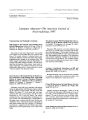
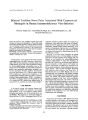
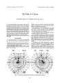
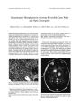
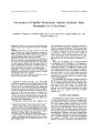
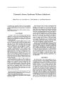
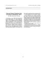
Show Journal of Neuro- Ophthalmology 19( 2): 128- 130, 1999. © 1999 Lippincott Williams & Wilkins, Inc., Philadelphia The Perils of a Sneeze Francine B. Wein, M. D., and Mark S. Gans, M. D., F. R. C. S. A 51- year- old woman had a 3- day history of severe left supraorbital pain associated with blurred vision of the left eye. Examination revealed visual acuity of 20/ 20 OD and 20/ 100 OS. A left relative afferent pupillary defect was present. Neu-roimaging revealed a large intra-, supra-, and parasellar mass that had eroded through the sphenoid sinus into the maxillary sinus. Secondary pneumocephalus was present. Pathologic examination of the tissue revealed a pituitary adenoma of the null cell type. To the best of our knowledge, there is only one other case in the literature in which a spontaneous pneumatocele represents the initial manifestation of a pituitary adenoma. Key Words: Null cell adenoma- Pituitary adenoma- Pneumocephalus. CASE REPORT A 51- year- old woman had severe left supraorbital pain. She described the pain as lancinating in quality and Manuscript received July 13, 1998; accepted February 17, 1999. From the Department of Ophthalmology, Montreal General Hospital, McGill University, Montreal, Quebec, Canada. Address correspondence to Mark Gans, M. D., Department of Ophthalmology, Montreal General Hospital, 1650 Cedar La- 211, Montreal, Quebec, H3G 1AA, Canada. related its onset to a sneeze 3 days earlier. She subsequently noted blurring of vision in the left eye. Ocular history was otherwise unremarkable. Her medical history was noncontributory. One week before onset of ocular symptoms she had a nonproductive cough associated with shortness of breath and was treated with amoxicillin for " bronchitis." Current medications included nortriptyline, triazolam, clonazepam, sertraline, and amoxicillin. On examination, the patient was found to be in significant discomfort, with her hand clutched over her left eye. Visual acuity measured 20/ 20 OD and 20/ 100 OS. Pupils measured 3.5 mm OD and 4.0 mm OS; a left relative afferent pupillary defect was present. Findings in the remainder of the neuro- ophthalmic examination were normal. A Goldmann visual field demonstrated a superior bitemporal hemianopia and a small paracentral scotoma in the left eye ( Fig. 1). Computed tomography ( CT) and magnetic resonance imaging ( MRI) of the orbits and sella revealed a large intra-, supra-, and parasellar mass displacing the third ventricle and optic chiasm superiorly i i i i i i i i FIG. 1. The Goldmann visual field shows defect with a central scotoma of the left eye. 128 PERILS IN SNEEZING 129 FIG. 2. The thin arrows indicate the margins of the lesion in a coronal computed tomographic scan. The wide arrows delineate the edge of the pneumatocele. ( Figs. 2 and 3). The lesion had eroded through the sphenoid sinus roof and floor into the nasopharynx. Secondary pneumocephalus was present, with a collection of air between the mass and the medial aspect of the temporal lobe. The patient underwent transsphenoidal subtotal removal of the tumor. Pathologic analysis revealed a pituitary macroadenoma of the null cell type. At her 1- month postoperative visit, the patient's left eye visual acuity had improved to 20/ 40 with a persistent left afferent pupillary defect. At 6 months visual acuity increased to 20/ 25, and there was no relative afferent pupillary defect. Goldmann visual fields returned to normal in each eye. DISCUSSION Null cell adenomas are clinically nonfunctioning and are thus usually diagnosed at the macroadenoma stage when mass effects become evident. Our patient's visual loss was precipitated abruptly by the mass effect of a pneumocephalus secondary to an expanding adenoma with sinonasal involvement. To the best of our knowledge there has been been only one report in the literature documenting spontaneous pneumocephalus as the initial manifestation of a pituitary adenoma ( 1). That patient, however, had a 3- year history of recurrent headaches and a 2- month history of progressive visual loss. Pneumocephalus refers to an intracranial gas collection. The largest review of pneumocephalus was conducted by Markham in 1967 ( 2). He stated that the most common causes of this entity, comprising 74% of cases, were trauma and craniofacial surgery. Mass lesions, most frequently sinus osteomas, were responsible for 13% of cases. The ophthalmologist is most likely to encounter pneumocephalus in the setting of orbital roof fractures. Involvement of the sinonasal tract by a pituitary adenoma is estimated to be 2% ( 3). Patients may have frontal headache, recurrent epistaxis, nasal obstruction, postnasal drip, and intermittent nasal discharge. Our patient's " bronchitis" symptoms may have been associated with the pneumatocele. The manner in which air enters the brain has been likened to a ball- valve mechanism and an " inverted soda FIG. 3. T1- weighted coronal magnetic resonance image shows a large intra-, supra-, and parasellar mass displacing the optic chiasm superiorly ( black arrow) and encasing the left internal carotid artery ( solid white arrow). There is a collection of air between the mass and the temporal lobe ( white double arrowhead). J Neuro- Ophthalmol Vol. 19. No. 2. 1999 130 F. B. WEIN AND M. S. GANS pop bottle" phenomenon ( 4). The former explanation was well illustrated in our patient. Her pituitary adenoma eroded through arachnoid, dura, and the sphenoid sinus roof and floor, thus establishing a communication between the intracranial compartment and the nasopharynx. A ball- valve mechanism allowed the air to enter, but not to escape through the " valve" of eroded bone and dura. The volume of air could progressively increase with repeated coughing, sneezing, or straining. Similar to the air trapped in an inverted soda bottle, intracranial air rises and is trapped by the soft tissue below. Eventually, this volume occupies enough space to cause symptoms of increased intracranial pressure. Our patient may have had pneumocephalus before her symptoms appeared, but when she sneezed, the volume of the pneumatocele became large enough to cause tension and, thus, pain. In summary, we present a patient whose pituitary adenoma initially manifested with periorbital pain related to a pneumatocele that probably expanded with her perilous sneeze. REFERENCES 1. Sage MR, McAllister VL. Spontaneous intracranial " aerocele" with chromophobe adenoma. Br J Radiol 1974; 47: 727- 9. 2. Markham JW. The clinical features of pneumocephalus based on a survey of 284 cases with report of 11 additional cases. Acta Neu-rochir 1967; 15: 1- 78. 3. Luk ISC, Chan JKC, Chow SM, Leung S. Pituitary adenoma presenting as sinonasal tumor: pitfalls in diagnosis. Hum Pathol 1996; 27: 605- 9. 4. Wanamaker JR, Mehle ME, Wood BG, Lavertu P. Tension pneumocephalus following cranio- facial resection. Head and Neck 1995; 17: 152- 6. J Neuro- Ophthalmol, Vol. 19, No. 2, 1999 |