| OCR Text |
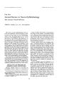
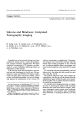
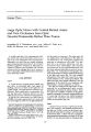
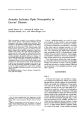
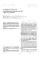
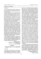
Show Journal of Neuro- Ophthalmology 14( 3): 170- 174, 1994. © 1994 Raven Press, Ltd., New York Anterior Ischemic Optic Neuropathy in Graves' Disease Andre Dosso, M. D., Avinoam B. Safran, M. D., Gordana Sunaric, M. D., and Albert Burger, M. D. Optic neuropathy occurred in two patients suffering from Graves' disease with marked limitation of eye movement. Optic nerve changes were moderate. They consisted of parapapillary flame- shaped hemorrhages, swelling of the disc, and bundle defects in the visual field on the involved side. This clinical pattern suggested that the optic neuropathy was anterior and ischemic in nature. In one patient, symptoms of optic neuropathy were noted 3 days after starting stretching exercises with the ocular muscles, performed following a friend's advice in an attempt to prevent increase in restrictive myopathy. In patients with Graves' disease, it is conceivable that mild optic neuropathy occasionally occurs as a result of elevation in intraocular pressure, and stretching exercises of the ocular muscles might consequently favor such ischemic events. In the mechanisms of optic nerve involvement associated with Graves' disease, the role of ischemia should be considered in addition to the widely accepted role of optic nerve compression by enlarged extraocular muscles, at the level of the orbital apex. Key Words: Myopathy- Graves' disease- Anterior ischemic optic neuropathy. Graves' ophthalmopathy can result in optic nerve damage. In this condition, optic neuropathy is generally considered to be caused by a posteriorly located compressive process that is, crowding at the orbital apex by enlarged muscles ( 1,2). We observed two patients suffering from Graves' disease, who presented with mild anterior ischemic optic neuropathy ( AION). In both patients, ocular motility showed marked restriction. Interestingly, in one patient reduction in vision was noted 3 days after starting intensive eye movement exercises. These observations indicate that optic neuropathy associated with Graves' disease is occasionally anterior and ischemic in nature, and may possibly result from elevation in ocular pressure following eye movements, when markedly restrictive myopathy is present. CASE REPORTS From the Neuro- ophthalmology Unit, Department of Ophthalmology ( A. D., A. B. S., G. S.), and the Division of Endocrinology ( A. B.), Geneva University Hospital, Geneva, Switzerland. Address correspondence and reprint requests to Dr. A. B. Safran, Neuro- ophthalmology Unit, Department of Ophthalmology, Geneva University Hospital, 1211 Geneva 4, Switzerland. Case 1 A 50- year- old woman was referred for evaluation of bilateral exophthalmos. Fifteen years earlier she had shown clinical evidence of thyrotoxicosis, which was treated with carbimazole. Visual acuity was found to be 20/ 30 in both eyes. Automated perimetry showed in both eyes large areas of relative loss in sensitivity, which were considered to result from bundle defects. In addition, in the right eye, a small absolute scotoma was observed at the inferior border of the visual field area, which theoretically includes the blind spot, and which is not tested with this examination program ( Fig. 1). The lids and conjunctiva were swollen, and the horizontal rectus insertions showed redness. Values measured with Hertel exophthalmometry were 18- 19- 110. A slight relative afferent pupillary defect was noted on the right. Ocular motility presented 270 OPTIC NEUROPATHY IN GRAVES' DISEASE 171 tNTKMXUC OCTOV08 200011 Mauro- Opfathalao log l e a l rrograa HI ( MlMMd July 1991 ) C E N T R A L n » l l > P ( D I F F E R K H C 1) • • • t •••• FIG. 1. Patient 1. Right visual field ( central 30 degrees), evaluated using Octopus automated perimetry, demonstrating, at the border of the unchecked area of the field, a small absolute scotoma, which represents the displaced blind spot, and areas with relative loss of sensitivity. the following changes. The right eye showed marked downward and nasal deviation, with no possible elevation; abduction was markedly impaired. The left eye showed only moderate limitation in elevation, abduction, and adduction. When the patient was instructed to gaze straight ahead with no effort, intraocular pressure was 23 mmHg in both eyes. Upon attempting to upgaze, however, the pressure rose to 45 mmHg. In the right eye, fundus examination revealed pronounced ex-cyclotorsion, moderate venous dilatation, blurring of the margins of the optic disc, and a flame-shaped hemorrhage at the temporal border of the optic disc ( Fig. 2). Torsion of the right eye, observed in the fundus, suggested that the small absolute scotoma observed in the corresponding visual field represented the displaced blind spot ( Fig. 1). In the opposite eye, the fundus was within normal limits and cup/ disc ratio was about 0.4. A computed tomography ( CT) scan showed pronounced posterior enlargement of the extraocular muscles on both sides. Case 2 A 57- year- old woman, suffering for 1 year with Graves' hyperthyroidism, treated with carbima-zole, was referred for diplopia, which had progressively developed over the previous months. Visual acuity was normal, and the visual fields, evaluated using Octopus automated perimetry ( Program Nl), were unremarkable. The eyelids were minimally swollen, and the insertions of the horizontal recti showed little hyperemia. Motility, however, was markedly impaired in both eyes: elevation was virtually nil, abduction was limited to 25 degrees in the right eye, and 20 degrees in the left eye, and adduction was, respectively, 30 degrees in the right eye and 35 degrees in the left eye; depression, however, was only mildly limited. Moderate upper lid retraction was also observed. Values measured with Hertel exophthalmometry were 14- 14- 105. No relative afferent pupillary defect was FIG. 2. Patient 1. Excyclotorsion is evident in the fundus of the right eye. In addition, the optic disc appears swollen, with flame- shaped hemorrhages and moderate venous dilatation. / Neuro- Ofihthalmol. Vol. 14, No. 3. 1994 172 A. DOSSOETAL. noted. Mechanical components in the limitation of ocular motility were demonstrated in both eyes by an elevation of intraocular pressure from 15mmHg in primary gaze to more than 35 mmHg when attempting to upgaze. The fundi were normal, showing a 0.45 cup: disc ratio on both sides. A CT scan revealed marked posterior enlargement of the extraocular muscles in both orbits. Six weeks later, following a friend's advice, the patient started intensive eye movement exercises in the hope of preventing progression of the restrictive muscular disorder. Three days after starting, however, she noted a sudden loss of vision in the left eye. Examination showed a visual acuity of 20/ 25 in the left eye, and a moderate left relative afferent pupillary defect. Mild optic disc edema and a small parapapillary flame- shaped hemorrhage were noted in the left fundus ( Fig. 3). In addition, repeated automated perimetry showed a small, relative scotoma at the border of the presumed area of the blind spot ( Fig. 4). Ophthalmologic examination, including ocular pressure, showed no other recent changes. The patient was then lost to follow- up for 3 months. When she was next examined, the parapapillary hemorrhage in the right eye had disappeared and torsion of the eye had occurred, resulting in displacement of the blind spot in the visual field. ( The displacement of the blind spot, and diagnostic difficulties resulting from this phenomenon, have been discussed in another paper ( 3).) Discussion Our patients showed optic nerve disorders in association with Graves' ophthalmopathy. This as- C E N T R A L F I B L D ( D i r r K H I H C l) •• lift M M M M M M •••• •••• •••• 111! M M M M M M Difference in % 0- 10 11- 22 23- 34 35- 46 47- 58 59- 70 71- 82 83- 94 95- 100 FIG. 4. Patient 2. Left visual field ( central 30 degrees), evaluated using Octopus automated perimetry, showing a small inferior paracecal relative scotoma. sociation reportedly occurs in about 5% of patients suffering from Graves' disease ( 4). In the second patient, visual changes occurred without significant exophthalmos, a clinical picture often observed in patients with optic neuropathy due to Graves' disease. ( 5). However, out patients were unique in that they showed morphologic features in the fundi, indicating that the optic nerve lesions were anterior and ischemic in nature. The anterior location of the optic nerve lesions which were shown by our patients, contrasted with the generally held belief that, as a rule, optic nerve involvement in Graves' FIG. 3. Patient 2. Fundus photography of left eye, showing a flame-shaped hemorrhage at the border of the optic disc. ] Neuro- Ophthalmol, Vol. 14, No. .3, 1994 OPTIC NEUROPATHY IN GRAVES' DISEASE 173 disease results from compression by enlarged extraocular muscles at the level of the orbital apex ( 1,2). Reportedly, in this condition, the optic discs appear normal in 50%> of cases, moderately swollen in 21% of cases, markedly swollen in 14% of cases, and pale in 15% ( 6). We are unaware of any previous description of anterior ischemic optic neuropathy ( AION) occurring in patients with Graves' disease. Nor have we found any previous reports of flame- shaped hemorrhages at the borders of affected optic discs, which are characteristically associated with AION ( 7). In a large series devoted to optic nerve disorders in Graves' disease, parapapillary hemorrhages were not listed among the morphologic features observed in the fundi. In a paper by Trobe and colleagues ( 6), on optic nerve disorders associated with this condition, we noted, in a photograph, the presence of a small round parapapillary hemorrhage in the fundus. Based on the literature, an association between AION and Graves' disease appears to be rare. It is possible, however, that this condition is not unusual but rather that it is frequently overlooked, owing to the smallness of the functional and morphologic changes that occur. In our first patient, visual changes were minimal, and they were moderate in the second patient. This clinical pattern is a form of discreet AION, which frequently escapes attention ( 8). Moreover, the morphologic changes in the fundus may be minute and ephemeral, and therefore missed during follow- up examinations. In the fundus of our second patient, for example, only a splinter parapapillary hemorrhage was noted, which lasted for a few weeks, and then disappeared. Such lesions frequently occur in isolation in the fundi of patients with glaucoma ( 9). When unassociated with retinopathy and hemor-rages elsewhere in the fundus, small linear hemorrhages at the optic nerve head suggest microinfarctions of the anterior part of the optic nerve ( 10), but they rarely result in field defects that are detectable by routine, nonautomated perimetry ( 11). It is well established that, in subjects with AION, the cup: disc ratio is generally lower than that found in normal individuals ( 12- 14). In our patients, the cup: disc ratio was not small. However, a small ratio is not a prerequisite for the development of ischemic lesions in the anterior part of the optic nerve. A study by Feit and colleagues ( 13) showed that in 2 of 30 patients with AION, the cup. disc ratio was greater than 0.45, that is, greater than the ratio found in our patients. In the paper by Beck and colleagues ( 14) 5 of 126 affected patients showed a ratio of 0.45 or more. Therefore, the cup appearance in the discs of our patients is not inconsistent with a diagnosis of AION. It is conceivable that, in the patients reported here, the relatively large cup: disc ratio contributed to limiting the functional and morphologic changes induced by ischemia in the optic nerve head. Average ocular pressure in patients presenting with AION is higher than normal ( 15). It has therefore been suggested that raised ocular pressure might play a significant role in the development of AION. Now, limitation of ocular movements in Graves' disease is characteristically associated with elevation of ocular pressure during excentric gaze ( 16). In our patients, this factor may have played a decisive role in the development of AION. In our second patient, the fact that the appearance of AION coincided with the beginning of intensive stretching exercises of the ocular muscles suggests that the elevation of intraocular pressure plays a role in this condition, even though a cause and effect relationship between exercises and optic neuropathy cannot be proven. It is also conceivable that, in patients with Grave's disease, a reduced orbital venous outflow ( 17) plays a role in the development of optic disc ischemia. References 1. Neidel JM, Rootman J, Belkin RI, Nugent RA, Drance SM, Beattie CW, Spinelli JA. Dysthyroid optic neuropathy: the crowded orbital apex syndrome. Ophthalmology 1988; 95: 1515- 21. 2. Kennerdel JS, Rosenbaum AE, EI- Hoshy M. Apical optic nerve compression of dysthyroid optic neuropathy on computed tomography. Arch Ophthalmol 1981; 99: 807- 9. 3. Safran AB, Sunaric G, Burger A. The trap of the displaced blind spot in automated perimetry. Br / Ophthalmol 1993; 77: 274- 5. 4. Werner SC, Ingbar SH, The thyroid, 3rd Ed. New- York: Harper & Row; 1971: 536. 5. Feldon SE, Muramatsu S, Weiner JM. Clinical classification of Graves' ophthalmopathy. Arch Ophthalmol 1984; 102: 1469- 72. 6. Trobe JD, Glaser SJ, Laflamme P. Dysthyroid optic neuropathy. Arch Ophthalmol 1978; 96: 1199- 209. 7. Miller NR. Walsh and Hoyt's clinical neuro- ophthalmology. Vol. 1. Baltimore: William & Wilkins, 1982: 214. 8. Beri M, Klugman MR, Kohler JA, Hayreh SS. Anterior ischemic optic neuropathy. VII. Incidence of bilaterally and various influencing factors. Ophthalmology 1987; 94: 1020- 8. 9. Gloster J. Incidence of optic disc haemorrhages in chronic simple glaucoma and ocular hypertension. Br / Ophthalmol 1981; 65: 452- 6. 10. Susanna R, Drance MS, Douglas GD. Disc hemorrhages in patients with elevated intraocular pressure: occurrence with and without field changes. Arch Ophthalmol 1979; 97: 284- 5. 11. Airasksinen PJ, Mustonen E, Alanko HI. Optic disc hemorrhages precede retinal nerve fiber layer defects in ocular hypertension. Acta Ophthalmol 1981; 59: 627- 41. / Neuro- Ophthalmol, Vol. 14, No. 3, 1994 174 A. DOSSO ET AL. 12. Beck RW, Savino PJ, Repka MX, Schatz NJ, Sergott RC. Optic disc structure in anterior ischemic optic neuropathy. Ophthalmology 1984; 91: 1334- 7. 13. Feit RH, Tomsak RL, Ellenberger C. Structural factors in the pathogenesis of ischemic optic neuropathy. Am / Ophthalmol 1984; 98: 105- 8. 14. Beck RW, Servais GE, Hayreh SS. Anterior ischemic optic neuropathy. IX. Cup- to- disc ratio and its role in pathogenesis. Ophthalmology 1987; 94: 1503- 8. 15. Katz B, Weireb RN, Wheeler DT, Klauber MR. Anterior ischemic optic neuropathy and intraocular pressure. Br ] Ophthalmol 1990; 74: 99- 102. 16. Gamblin GT, Gelentine PG, Eil C. Intraocular pressure and thyroid disease. In: Gorman CA, Waller RR, and Dyer JA, eds. The eye and orbit in thyroid disease. New York: Raven Press; 1984: 155- 66. 17. Hudson HL, Levin L, Feldon SE. Graves's exophthalmos unrelated to extraocular muscle enlargement: superior rectus muscle inflammation may induce venous obstruction. Ophthalmology 1991; 98: 1495- 9. / Neuro- Oplithahmtl, Vol. 14. No. 3, 1994 |