| OCR Text |
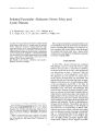
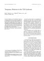
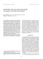
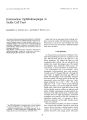
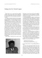
Show journal of Nemo- Ophthalmology 14( 1): 34- 37, 1994. © 1994 Raven Press, Ltd., New York Usher's Syndrome and Multiple Sclerosis Review of an Individual with Usher's Syndrome with a Multiple Sclerosis- like Illness Sharon G. Lynch, M. D., Kathleen Digre, M. D., and John W. Rose, M. D. We describe an individual with Type I Usher's syndrome and a multiple sclerosislike illness. MRI scan showed vermian atrophy on Tl- weighted images and multiple white matter lesions in the periventricular areas on T2- weighted images. Although MRIs demonstrating increased signal intensity on weighted images are reported in some individuals with Usher's syndrome, the cerebrospinal fluid findings are not described in these cases. In the present case, oligoclonal bands were present in the spinal fluid. The possibility of a linkage between the two diseases is raised. Key Words: Multiple sclerosis- Usher's syndrome- Retinitis pigmentosa- Congenital deafness- MRI. Usher's syndrome is an autosomal recessive disorder characterized by congenital neurosensory deafness and retinitis pigmentosa ( 1). Associations with vestibular abnormalities, cerebellar ataxia, and psychiatric problems are found in many individuals with Usher's syndrome ( 2,3). Cranial magnetic resonance imaging ( MRI) and computed tomography ( CT) scans demonstrate cerebral and cerebellar atrophy ( 4- 6). In addition, multiple white matter lesions are seen by MRI ( 5,6). We describe the clinical, MRI, and retinal features of an individual with Usher's syndrome and a multiple sclerosis- like illness. MRI of the brain showed multiple white matter lesions consistent with multiple sclerosis ( MS) and cerebrospinal fluid results were positive for oligoclonal bands. From the Department of Neurology ( S. G. L.), University of Kansas Medical Center, Kansas City, Kansas; Departments of Neurology ( K. D., J. W. R.), and Ophthalmology ( K. D.), University of Utah School of Medicine, and the Neurovirology Research Laboratory ( J. W. R.), VA Medical Center, Salt Lake City, Utah, U. S. A. This work was supported by Research Grant 1929- A- l from the National Multiple Sclerosis Society. SGL was supported by Post- Doctoral Fellowship Award FG 796- A- l from the National Multiple Sclerosis Society and KD was supported in part by an unrestricted grant from Research to Prevent Blindness, University of Utah. Address correspondence and reprint requests to Dr. Sharon G. Lynch, Department of Neurology, University of Kansas Medical Center, 3901 Rainbow Boulevard, Kansas City, KA 66160- 7314, U. S. A. CASE SUMMARY A 32- year- old white male presented to our clinic with a 9- year history of MS. He was diagnosed after the subacute onset of lower extremity weakness at age 23. The method of diagnosis was uncertain. He had been deaf since birth. There was no family history of deafness, and the cause had never been determined. He had been functioning as a T- 12 paraplegic for the past 5 years. He also had severe upper extremity ataxia and oscillopsia for several years, treated with clonazepam with some success. He had a remote history of optic neuritis. He had been treated with cyclophosphamide in 1986 and with methylprednisolone therapy in 1990, with partial return of motor function after therapy. At the time of admission, he had a 1- week history of increasing weakness and ataxia of his upper extremities, causing difficulty with his transfers 34 USHER'S SYNDROME AND MS 35 and increasing difficulty with his vision, causing trouble with reading. Neurological examination was remarkable for the following: The patient was deaf and used American sign language to communicate. This was difficult for him because ataxia made his movements imprecise. The interpreters sometimes had difficulty interpreting his signs. He was able to speak one or two words with difficulty. The speech was excessively loud and severely dysar-thric. He had severe upper extremity and truncal ataxia. He was paraplegic with spasticity of his lower extremities, and had 4/ 5 strength in his upper extremities. He had decreased vibratory sensation in all four extremities, but normal pinprick and temperature sensation. He was hyperreflexic with Babinski signs present bilaterally. Ophthalmic examination revealed best correct visual acuity of 20/ 80 OD, 20/ 200 OS. Color vision was decreased ( on 2/ 6- isochromatic plates). There was no relative afferent pupillary defect. Slit- lamp examination revealed no cataract. Extraocular muscle examination revealed gaze evoked and primary pendular nystagmus. Funduscopic examination showed bone spicules and degeneration typical of retinitis pigmentosa ( Fig. 1) The macula was normal. Electroretinogram ( ERG) demonstrated minimal scotopic response. Photopic response FIG. 1. Funduscopic view of patient's retina demonstrates bone spicules and degeneration of retinitis pigmentosa. showed 10 microvolt amplitude response. Visual evoked potentials were not detectable. Optic nerves were pale. An MRI of the brain was performed. Structurally, the patient had a magna cisterna magna and cerebellar vermian hypoplasia ( Fig. 2A) On T2- weighted imaging, nonspecific white matter abnormalities were seen in the periventricular region, right pons, and posterior limbs of the internal capsule ( Fig. 2B). MRI of the thoracic spine was normal. A lumbar puncture was performed. Abnormal results in the cerebrospinal fluid ( CSF) included an IgG index of 0.96 ( normal range: 0.35- 0.65), IgG synthesis of 10.0 ( normal range: 5.0- 5.0 mg/ day) and the presence of oligoclonal bands. The patient was admitted to the University of Utah hospital for treatment with methylpredniso-lone. Some increase in his motor strength was seen, but no improvement in his ataxia. DISCUSSION The combination of retinitis pigmentosa and hearing loss was first described in 1858 by von Graefe ( 7), and was further defined by Usher in 1914 ( 1). It is usually an autosomal recessive disorder with complete penetrance, although cases of an X- linked recessive inheritance have been described ( 8). Four subtypes have been identified ( 8). Type I is characterized by profound static hearing loss, vestibular dysfunction, and nyctalopia with onset in the first decade of life. Type II individuals present with moderate to severe hearing loss, normal vestibular dysfunction, and the development of retinitis pigmentosa in the second and third decades. Type III is similar to type II, except the individuals have progressive hearing loss. Type IV is an X- linked recessive variant ( 8). Retinitis pigmentosa is rarely seen in individuals with Usher's syndrome at birth, but appears over time ( 3). The amount of visual loss in individuals with Usher's syndrome is progressive and varies widely ( 3,9). The patient we describe had the profound hearing loss of Type I individuals. CT scans and MRI have been performed on a limited number of individuals with Usher's syndrome ( 4- 6). Both CT and MRI studies have shown cerebellar atrophy, and occipital lobe atrophy ( 4- 6). MRI studies have also shown high signal-intensity lesions ( 5,6). In one study, MRI showed the posterior fossa in Type II individuals ( 6). Two of seven had high signal- intensity lesions. One patient had a single lesion in the left cerebral peduncle. The second had multiple foci present in the cerebellum and midbrain. In a single case report by / Neuro- Ophthalmol, Vol. 14, No. 1, 1994 36 S. G. LYNCH ET AL. FIG. 2. ( A) T1- weighted MRI ( TR 400, TE 20) of the posterior fossa, showing vermian hypoplasia and mega cisterna magna. ( B) T2- weighted MRI ( TR 2333, TE30) of the cerebral hemispheres, showing increased signal intensity at multiple sites in the periventricular white matter. Koizumi and coworkers ( 5) a patient with Type II Usher's syndrome, who had psychiatric disturbances and ataxia, had prominent cortical and cerebellar atrophy and diffuse periventricular high-intensity lesions on T2- weighted axial MRI. The manifestations of Usher's syndrome are quite varied, making a diagnosis of any other neurologic condition difficult in the presence of this disorder. Cerebellar and vestibular signs and abnormalities are common, as are behavioral abnormalities ( 2,3). Paraplegia and sensory abnormalities are not described in this syndrome. In our patient, several features seemed more consistent with a second diagnosis of MS than with Usher's syndrome alone. The patient had a history of an exacerbating- remitting course at the time of his original diagnosis, with some response to immunosuppresive therapy. He had paraplegia with spasticity and a neurogenic, flaccid bladder. His sensory examination was also abnormal, with loss of vibratory and position sensation, but preservation of pinprick and temperature. Visual loss in Usher's syndrome is quite variable ( 10), but generally 89% preserve acuity better than 20/ 80; optic nerve pallor is not a prominent feature. In our case viaual aciity was decreased more than expected for his age or the severity of the retinitis pigmentosa. His optic pallor was greater than expected for Usher's syndrome. Again, these findings underscore a secondary diagnosis of MS. Further, the spinal fluid showed an increased IgG index and oligoclonal bands. These abnormalities have not been previously described in Usher's syndrome. The MRI results in this case could not alone suggest a diagnosis of MS, since a previous patient with Usher's syndrome had similar findings by MRI ( 5). In addition to our patient, 3 of 8 patients with Usher's syndrome who had cranial MRI performed had high signal- intensity lesions in the white matter ( 5,6). The significance of this finding is uncertain. Perhaps the other individuals with Usher's syndrome who had white matter lesions also had MS or a subclinical demyelinating disease. If this is the case, a high percentage of individuals with Usher's syndrome may also have demyelinating disease. Further, MRI and pathological studies are needed to clarify this issue. In view of the recent interest in an MS susceptibility gene ( 11,12), perhaps the possibility of a genetic linkage between demyelinating disease and Usher's syndrome should be pursued. REFERENCES 1. Usher CH. On the inheritance of retinitis pigmentosa, with notes of cases. R Lond Ophthalmol Hosp Rep 1914; 19: 130- 256. / Neuw- Ophthalmol, Vol. 14, No. 1, 1994 USHER'S SYNDROME AND MS 37 2. Hallgren B. Retinitis pigmentosa combined with congenital deafness: with vestibulo- cerebellar ataxia and mental abnormality in a proportion of cases: a clinical and genetico-statistical study. Acta Psychiatr Neurol Scand 1959; 34( Suppl 138): 5- 101. 3. Vernon M. Usher's syndrome- deafness and progressive blindness: clinical cases, prevention, theory, and literature survey. / Chron Dis 1969; 22: 133- 51. 4. Bloom TS, Fishman GA, Mafee MF. Usher's syndrome: CNS defects determined by computed tomography. Retina 1983; 3: 108- 13. 5. Koizumi J, Ofuku K, Sakuma K, et al. CNS changes in Usher's syndrome with mental disorder: CT, MRI, and PET findings. / Neurol Neurosurg Psychiatry 1988; 51: 987- 90. 6. Piazza L, Fishman G, Kaplan R, et al. Magnetic resonance imaging of central nervous system defects in Usher's syndrome. Retina 1987; 7: 241- 5. 7. von Graefe A. Exceptionelles Verhalten des Gesichtfeldes bei Pigmententartung der Netzhaut. Arch Exp Ophthalmol 1858; 4: 250- 3. 8. Davenport SLH, Omenn GS. The heterogeneity of Usher's syndrome [ Abstract]. In: Littlefield JW, Ebling FJG, Henderson IW, eds. Fifth international conference on birth defects. Amsterdam: Excerpta Medica, 1977: 87- 8. 9. Matthews TW, Poliquin J, Mount J, MacFie D. Is there genetic heterogeneity in Usher's syndrome? / Otololaryngol 1987; 16: 61- 6. 10. Piazza L, Fishman GA, Forder M, et al. Visual acuity in patients with Usher's syndrome. Arch Ophthalmol 1986; 104: 1336- 9. 11. Ebers GC, Bulwen DE Sadovnik AD, et al. A population based study of multiple sclerosis in twins. N Engl J Med 1986; 315: 1638- 42. 12. Sadovnik AD, Baird PA, Ward RH. Multiple sclerosis: updated risks for relatives. Am ] Med Genet 1988; 29: 533- 41. / Neuro- Ophthalmol, Vol 14, No. 1, 1994 |