| Title |
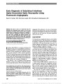
Diagnostic Algorithm for Patients With Suspected Giant Cell Arteritis |
| Creator |
Mays A. EI-Dairi; Lan Chang; Alan D. Proia; Thomas J. Cummings; Sandra S. Stinnett; M. Tariq Bhatti |
| Abstract |
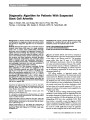
BACKGROUND: To identify clinical and laboratory factors contributing to the diagnosis of giant cell arteritis (GCA) and develop a diagnostic algorithm for the evaluation of GCA. METHODS: Retrospective review of 213 consecutive cases of temporal artery biopsy (TAB) seen at a single academic center over a 10-year period (2000-2009). Pathologic specimens were re-reviewed and agreement between the original and second readings was assessed. A composite clinical suspicion score was created by adding 1 point for each of the following criteria: anterior extracranial circulation ischemia, new onset headache, abnormal laboratory results (erythrocyte sedimentation rate, C-reactive protein (CRP), or platelet count), jaw claudication, abnormal or tender superficial temporal artery, constitutional symptoms, and polymyalgia rheumatica; one point was subtracted if a comorbid condition could explain a criterion. RESULTS: Of the 204 TABs analyzed, pathologic findings were confirmatory in 49 (24.0%) and suggestive in 12 (5.9%). TAB-positive patients were more likely to be older (age 75.2 7.8 vs 69.7 11.0 years, P = 0.0002), complain of jaw claudication (relative-risk = 3.26, P = 0.0014), and have thrombocytosis (relative-risk = 3.3, P = 0.0072) and elevated CRP (relative-risk = 1.8, P = 0.037). None of the patients with a clinical score less than 2 had a positive TAB. Diabetes mellitus and kidney disease were often the explanation for the symptoms and abnormal clinical finding(s) that led to a negative TAB. CONCLUSIONS: We propose a clinical algorithm that is highly predictive for a positive TAB and can be valuable in the evaluation process of suspected cases of GCA. |
| Subject |
Adult; Older people; Older people, 80 and over; Algorithms; Blood Sedimentation; C-Reactive Protein; Diagnosis, Differential; Diagnostic Techniques, ; Ophthalmological; Female; Giant Cell Arteritis; Humans; Longitudinal Studies; Male; Middle Older people; Platelet Count; ROC Curve |
| Format |
application/pdf |
| Publication Type |
Journal Article |
| Collection |
Neuro-Ophthalmology Virtual Education Library: Journal of Neuro-Ophthalmology Archives: https://novel.utah.edu/jno/ |
| Publisher |
Lippincott, Williams & Wilkins |
| Holding Institution |
Spencer S. Eccles Health Sciences Library, University of Utah |
| Rights Management |
© North American Neuro-Ophthalmology Society |
| Setname |
ehsl_novel_jno |
| ID |
227740 |
| Reference URL |
https://collections.lib.utah.edu/ark:/87278/s62j9hxc/227740 |