| OCR Text |
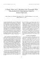
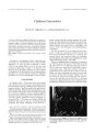
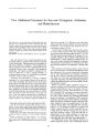
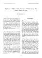
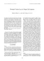
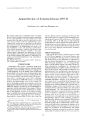
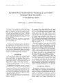
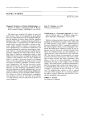
Show Journal of Neiim- Ophthalmology 18( 2): 81- 83, 1998. & 1998 LippincoU- Ravcn Publishers, Philadelphia A Family With Leber's Hereditary Optic Neuropathy With Mitochondrial DNA Heteroplasmy Related to Disease Expression Akiko Tanaka, M. D., Motohiro Kiyosawa, M. D., Ph. D., Yukihiko Mashima, M. D., Ph. D., and Takashi Tokoro, M. D., Ph. D., A Japanese family has members with Leber's hereditary optic neuropathy resulting from the heteroplasmic 11778 mutation and the homoplasmic 4216 mutation. Quantitative determination of heteroplasmy was performed by a combination of polymerase chain reaction and single- strand conformation polymorphism analysis. The association between heteroplasmy and clinical features was determined. Eleven people from the maternal side of the family, including four affected and seven unaffected members, showed heteroplasmy of the mtDNA mutation ranging from 5% to more than 95%. Four possibly affected patients had more than 90% of the mutant mtDNA. Seven unaffected people had mutant mtDNA ranging from 5% to 77%. A recovery episode of visual acuity was noted in the history of three of the four patients. Heteroplasmy is likely to be a factor in the expression of disease in this family. Key Words: Heteroplasmy- Leber's hereditary optic neuropathy- Mitochondrial 11778 mutation- Visual recovery. Leber's hereditary optic neuropathy ( LHON) is a maternally inherited form of optic neuropathy characterized by an acute or subacute bilateral loss of central vision that mainly affects young men. Several point mutations of mitochondrial DNA ( mtDNA) are primarily or secondarily associated with LHON ( 1,2). Approximately 40% to 90% of LHON families have an mtDNA mutation at position 1 1778 of the nicotinamide adenine di-nucleotide dehydrogenase subunit 4 ( ND4) gene ( 1,2). In families carrying inherited disorders with point mutations of mtDNA, some maternal relatives demonstrate a mixture of mutant and wild- type mtDNA, a condition known as heteroplasmy. Heteroplasmic people are more likely to remain asymptomatic than those with a homoplasmic mutant ( 3- 6). In LHON, heteroplasmy with Manuscript received August 18, 1997; accepted February 18, 1998. From the Department of Ophthalmology , Tokyo Medical and Dental University ( A. T., M. K., T. T), and the Department of Ophthalmology, Keio University School of Medicine ( Y. M.), Tokyo, Japan. Address correspondence and reprint requests to Motohiro Kiyosawa M. D., Ph D., Department of Ophthalmology, School of Medicine, Tokyo Medical and Dental University, 1- 5- 45, Yushima, Bunkyo- ku, Tokyo, Japan. the 11778 mutation can be detected in about 14% of families ( 7). In heteroplasmic LHON families, the percentage of mutant mtDNA tends to increase in successive generations ( 7- 13). A correlation between the extent of heteroplasmy and the risk of developing optic atrophy or the severity of the disease has also been reported in some pedigrees of LHON ( 7- 15). However, it has been thought that heteroplasmic people who become symptomatic do not differ clinically from symptomatic patients who exhibit the homoplasmic mutant ( 7). We report on an LHON family of three generations that carries the 11778 mutation with mtDNA heteroplasmy. In this family, disease expression was related to the percentage of mutant mtDNA. Recovery of visual acuity had occurred in the past in some members of this family. PATIENTS AND METHODS We surveyed 11 members on the maternal side in one Japanese family spanning three generations ( Fig. 1). The proband was a 25- year- old man ( III- 10 in Fig. 1) who experienced a sudden, painless loss of vision in the left eye in November 1995. Within 4 weeks, vision in the right eye also began to decrease, and he was referred to Tokyo Medical and Dental University Hospital. Eight months later, he was able to see only hand movement in both eyes. Examination by Goldmann perimetry showed bilateral central scotomata with some constriction of the peripheral fields. Both optic discs were pale, and no sign of visual recovery was seen. There was a family history of maternally inherited optic neuropathy, and LHON was suspected in this patient. Molecular genetic analysis of mtDNA identified a mutation at nucleotide position ( np) 11778, confirming the diagnosis. The medical histories in this patient's family included subjective onset of visual disturbance, with and without recovery of visual acuity. Subsequent ophthalmic examinations included visual acuity, slit lamp biomicroscopy, kinetic and static visual field examination, and fundus examination. 81 82 A. TANAKA ET AL. 40 Yo 37 YD 33Yo 31 Yo 7 4% 5 % 28 Yo 25 Yo QO 1 9 0% 9 5%? FIG. 1. Family exhibiting Leber's hereditary optic neuropathy with heteroplasmy of the 11778 mutation. Percentages and Yo indicate the proportion of mutated mtDNA and present age, respectively. Solid circles or squares indicate affected patients. Patients II- 2, II- 5, and II- 6 reported visual recovery. All members with percentages underwent ophthalmic examination. Analysis of mtDNA Blood samples were taken from the maternal members of the proband's family, including 4 visually affected and 7 unaffected people, after they gave informed consent. Total cellular DNA was extracted from peripheral blood leukocytes by the standard method. MtDNA from the 11 people was screened for three primary LHON mutations at np 3460, np I 1778, and np 14484, and at nine secondary LHON mutations at np 3394, np 4216, np 4917, np 5244, np 7444, np 9438, np 13708, np 15257, and np 15812. To determine the level of mtDNA heteroplasmy, we used the technique of single- strand conformation polymorphism analysis, as described previously ( 15). In brief, the oligonucleotide primers for detecting the 11778 mutation were 5'- GGCGCAGTCATTCACTCATAAT- 3' ( nt 11690- 11709 of the mtDNA sequence) and 5'- AAGTAGGAGAGTGATATTTG- 3' ( nt 11925- 11944). The 5' ends of primers were labeled with [ 7- 32P] adenosine triphosphate. Labeled polymerase chain reaction products were obtained by cycling amplification and then were diluted, denatured, and electrophorescd on a 6% polyacrylamide gel for single- strand conformation polymorphism analysis. The percentage of mutant mtDNA was calculated using an image analyzer ( BAS 300011; Fujifilm, Tokyo, Japan). REPORT OF CASES I- 1: An 84- year- old woman had blurred vision caused by senile cataract. A local ophthalmologist reported that her visual acuity had been 20/ 20 in both eyes before she had cataract. Optic disc atrophy was not reported. II- 1: A 63- year- old man had no visual problems. His visual acuity was 20/ 20 in both eyes, and he had normal visual fields. II- 2: A 59- year- old man had loss of central vision in the right eye in 1958, when he was 22 years old. He reported that his visual acuity recovered within a year. He developed retinal branch vein occlusion in the right eye in 1991, which was treated with laser photocoagulation. He had a cerebral infarction in 1995. In 1996 his visual acuity was 20/ 80 in the right eye and 20/ 20 in the left eye. both optic discs were partially pale. Goldmann perimetry showed left homonymous hemianopia and a local defect in the right eye. These visual field defects were attributed to the brain infarction and retinal branch vein occlusion. II- 3: A 56- year- old woman had no ocular symptoms. Her visual acuity was 20/ 20 bilaterally. II- 4: A 54- year- old man had no ocular symptoms. His visual acuity was 20/ 20 bilaterally. II- 5: A 50- year- old woman experienced bilateral visual loss and was unable to read letters on a blackboard when she was 10 years old. She was treated with vitamins in a university hospital, and some visual recovery occurred in 6 months. Her visual acuity was 20/ 200 in the right eye and 20/ 60 in the left eye in 1996. She had optic disc atrophy bilaterally. II- 6: A 48- year- old man had loss of vision to finger counting in the right eye when he was 18 years old in 1966. Within a few months, the vision in the left eye began to decrease. He was treated with vitamins, and visual recovery occurred in 3 months. In 1996, his visual acuity was 20/ 20 in the right eye and 20/ 40 in the left eye. Both optic discs were partially pale. Examination results with Goldmann perimetry were normal in the right eye, and the left eye had an enlarged Mariotte's scotoma with relative central scotoma. III- 5: A 33- year- old man had no ocular symptoms. His visual acuity was 20/ 20 in both eyes. His visual fields tested by Goldmann and octopus perimetry were almost normal. III- 6: A 31- year- old man had no ocular symptoms. His visual acuity was 20/ 20 in both eyes, and results of the Goldmann visual fields test were normal. III- 9: A 28- year- old woman had no ocular symptoms. Results of Goldmann visual fields examination were normal and fundus was normal. She had received a diagnosis of systemic lupus erythematosus 10 years before. RESULTS Figure 2 shows results of the polymerase chain reaction- single- strand conformation polymorphism analysis in this family exposed to radiographic film. Two separate bands represent the wild- type ( lower) and mutant strands ( upper). We calculated the sets of the wild- type and mutant bands for quantification. The percentages of mutant mtDNA in this family are shown in Figure 1. The people in this family who had LHON carried at least 90% of the mutant mtDNA. This family also carried the secondary mtDNA mutation at np 4216, with homoplasmy. ./ Neiim- Ophlluilnml, Vol. IX, No. 2, 1998 LEBER'S HEREDITARY OPTIC NEUROPATHY WITH HETEROPLASMY 83 J 31 III 1 1 2 : i 5 5 5 S S 10 FIG. 2. Single- strand conformation polymorphism analysis of polymerase chain reaction- amplified mtDNA fragments exposed to radiographic film in the family shown in Figure 1. Two separate bands represent the wild- type ( lower) and mutant strands ( upper). DISCUSSION Because most patients with LHON and their unaffected maternal relatives carry mutant mtDNA in ho-moplasmy, the percentage of mutant mtDNA and the incidence of disease development do not always show a close association. However, a correlation between the extent of heteroplasmy and the risk of development of optic atrophy has been reported in some LHON families ( 7- 15). Regarding the I 1778 mutation of mtDNA, Smith et al. ( 7) estimated that at least 76% of the mtDNA in circulating leukocytes must be mutant for LHON to develop. Mashima et al. ( 15) suggested that people with less than 60% of mutant mtDNA in circulating leukocytes were at a reduced risk for development of LHON. In the current family, people who were thought to have LHON had more than 90% of mutant mtDNA, whereas people who had mutant mtDNA in percentages of 5% to 77% did not have LHON. Our findings suggest that the percentage of mutant mtDNA in circulating leukocytes is a probable factor in disease expression. Heteroplasmy is one of the significant factors in disease expression. However, the percentage of mutated mtDNA in peripheral blood is not always the same as that in the retinal or optic nerve tissue, or both. Different tissues of a heteroplasmic person may have different proportions of mutant mtDNA molecules ( 12,16). However, a high proportion of mutated mtDNA in leukocytes is reportedly correlated with a high proportion of mutated mtDNA in other tissues ( 11,17). Although there is limited applicability of data on leukocyte heteroplasmy to relevant target tissue ( retinal or optic nerve tissue), examination and quantification of mutant mtDNA in peripheral blood may provide helpful information for genetic counseling of families affected by LHON with heteroplasmy. The visual prognosis of LHON patients with the 11778 mutation is usually poor ( 18- 20). Clinical futures of heteroplasmic patients are reportedly similar to those of homoplasmic mutant patients once visual symptoms occur ( 7,18). Two LHON patients with the heteroplasmic 11778 mutation, however, reportedly had spontaneous recovery of partial vision ( 14,21). Although detailed medical records at the time of onset were not available, the current family showed a high frequency of visual recovery. We found no evidence that heteroplasmy correlated with visual recovery in this family, because all affected patients had more than 90% of mutant mtDNA. Other factors may have contributed to promote recovery of vision in this family. The 4216 mutation, expressed in homoplasmic manner as a secondary LHON mutation ( 1,2), was also identified in this family. However, the correlation of this mutation and disease expression in this family is not clear. REFERENCES 1. Newman NJ. Leber's hereditary optic neuropathy. New genelie considerations. Arch Neurol 1993; 50: 540- 8. 2. Brown MD, Wallace DC. Spectrum of mitochondrial DNA mutations in Leber's hereditary optic neuropathy. Clin Neurosci 1994; 2: 138- 45. 3. Sato W, Hayasaka K, Komatsu K, et al. Genelie analysis of three pedigrees of mitochondrial myopathy, encephalopathy, lactic acidosis, and stroke like episodes ( MELAS). Am J Hum Genet 1992; 50: 655- 7. 4. Shoffner JM, Lott MT, Lezza AMJ, et al. Myoclonic epilepsy and ragged- red fiber disease ( MERRF) is associated with a mitochondrial DNA tRNA Lys mutation. Cell 1990; 61: 931- 7. 5. Holt IJ, Harding AE, Petty RK, Morgan HJ. A new mitochondrial disease associated with mitochondrial DNA heteroplasmy. Am J Hum Genet 1990; 46: 428- 33. 6. Tatuch Y, Christodoulou J, Feigenbaum A, el al. Heteroplasmic mtDNA mutation ( T- G) at 8993 can cause Leigh disease when the percentage of abnormal mtDNA is high. Am J Hum Genet 1992; 50: 852- 8. 7. Smith KH, Johns DR, Heher KL, Miller NR. Heteroplasmy in Leber's hereditary optic neuropathy. Arch Ophthalmol 1993; 11 1: 1486- 90. 8. Holt IJ, Miller DH, Harding AE. Genelie heterogeneity and mitochondrial DNA heteroplasmy in Leber's hereditary optic neuropathy. J Med Genet 1989; 26: 739- 43. 9. Vilkki J, Savontaus M- L, Nikoskelainen EK. Segregation of mitochondrial genomes in a heteroplasmic lineage with Leber hereditary optic neuroretinopathy. Am J Hum Genet 1990; 47: 95- 100. 10. Bolhuis PA, Bleeker WE, Ponne NJ, et al. Rapid shift in genotype of human mitochondrial DNA in a family with Leber's hereditary optic neuropathy. Biochem Biophys Rex Commun 1990; 170: 994- 7. I 1. Barboni P, Mantovani V, Montagna P, et al. Mitochondrial DNA analysis in Leber's hereditary optic neuropathy. Ophthalmic Pae-diatr Genet 1992; 13: 219- 26. 12. Howell N, Xu M, Halvorson S, Bodis Wl, Sherman J. A heteroplasmic LHON family: tissue distribution and transmission of the 11778 mutation. Am J Hum Genet 1994; 55: 203- 6. 13. Black GC, Morten K, Laborde A, Poulton J. Leber's hereditary optic neuropathy: heteroplasmy is likely to be significant in the expression of LHON in families with Ihe 3460 ND1 mutation, Br J Ophthalmol 1996; 80: 915- 7. 14. Zhu DP, Economou EP, Antonarakis SE, Maumcnee IH. Mitochondrial DNA mutation and heteroplasmy in type I Leber hereditary optic neuropathy. Am J Med Genet 1992; 42: 173- 9. 15. Mashima Y, Saga M, Hiida Y, et al. Quantitative determination of heteroplasmy in Leber's hereditary optic neuropathy by single-strand conformation polymorphism. Invest Ophthalmol Vis Set 1995; 36: 1714- 20. 16. Lott MT, Voljavec AS, Wallace DC. Variable genotype of Leber's hereditary optic neuropathy patients. Am J Ophthalmol 1990; 109: 625- 31. 17. Juvonen V, Nikoskelainen E, Lammincn T, et al. Tissue distribution of the ND4/ 1I778 mutation in heteroplasmic lineages with Leber hereditary optic neuropathy. Hum Mutal 1997; 9: 412- 7. 18. Newman NJ, Lott MT, Wallace DC. The clinical characteristics of pedigrees of Leber's hereditary optic neuropathy with the I 1778 mutation. Am J Ophthalmol 1991 ; 1 11: 750- 62. 19. Hotta Y, Fujuki K, Hayakawa M, et al. Clinical features of Japanese Leber's hereditary optic neuropathy with I 1778 mutation of mitochondrial DNA. Jpn J Ophthalmol 1995; 39: 96- 109. 20. Stone EM, Newman NJ, Miller NR, et al. Visual recovery in patients with Leber's hereditary optic neuropathy and the I 1778 mutation. ./ Clin Neuro- Ophthalmol 1992; 12: 10- 4. 21. Isashiki Y, Nakagawa M. Clinical correlation of mitochondrial DNA heteroplasmy and Leber's hereditary optic neuropathy. Jpn .1 Ophthalmol 1991 ; 35: 259- 67. ./ Neiiro- Oplilhalniol, Vol. IS, No. 2, I99H |